Proanthocyanidins Suppresses Lipopolysaccharide-stimulated Inflammatory Responses via Heme Oxygenase-1 Induction in RAW264.7 Macrophages
Hye-Jin Cheon1, Sun Young Park1, Hee-Ji Jang1, Da-Young Cho1, Jiwon Jung2, Gimin Park2, Kyeong Mi Jeong2 and Jin-Kyung Kim1*
1Department of Biomedical Science, Daegu Catholic University, 38430 Gyeongsan-si, Korea
2Yeongcheon Girl’s High School, 38841 Yeongcheon-si, Korea
Received January 22, 2019 /Revised February 19, 2019 /Accepted February 20, 2019
Proanthocyanidins are naturally occurring polyphenolic compounds abundant in many vegetables, plant skins (rind/bark), seeds, flowers, fruits, and nuts. Numerous in vitro and in vivo studies have demonstrated myriad effects potentially beneficial to human health, such as antioxidation, immun- omodulation, DNA repair, and antitumor activity. Among immune cells, macrophages are crucial players in a variety of inflammatory responses to environmental conditions. However, it has been widely reported that macrophages cause chronic inflammation and are involved in a variety of dis- eases, such as obesity, diabetes, metabolic syndrome, and cancer. In this study, we report the sup- pressive effect of proanthocyanidins via the heme oxygenase-1 (HO-1)-related system, on the immune response of the LPS-stimulated mouse macrophage cell line RAW264.7. Increased HO-1 expression at mRNA and protein levels were found in proanthocyanidins-treated RAW264.7 cells. Further, proan- thocyanidins enhanced nuclear factor-erythroid 2-related factor 2 translocation into the nucleus.
RAW264.7 cells were treated with lipopolysaccharide (LPS) with or without proanthocyanidins, and inflammatory mediator expression levels were assessed. Proanthocyanidins treatment resulted in the attenuation of nitric oxide production and inducible nitric oxide synthase expression in LPS-stimulated RAW264.7 cells. In addition, mRNA and protein expression of proinflammatory cytokines, such as tu- mor necrosis factor-α and interleukin-6, was inhibited by proanthocyanidins treatment in LPS-stimu- lated RAW264.7 cells. These findings support proanthocyanidins as a promising anti-inflammatory agent.
Key words : Heme oxygenase-1, inducible nitric oxide synthase, inflammation, macrophages, proan- thocyanidins
*Corresponding author
*Tel : +82-53-850-3774, Fax : +82-53-850-3774
*E-mail : [email protected]
This is an Open-Access article distributed under the terms of the Creative Commons Attribution Non-Commercial License (http://creativecommons.org/licenses/by-nc/3.0) which permits unrestricted non-commercial use, distribution, and reproduction in any medium, provided the original work is properly cited.
Journal of Life Science 2019 Vol. 29. No. 4. 484~491 DOI : https://doi.org/10.5352/JLS.2019.29.4.484
Introduction
Among immune cells, macrophages are one of the most widely studied pleiotropic cells in the immune system.
Macrophages play critical and plastic roles in host defense, immune regulation, and wound healing, adapting to their local environments and adopting diverse phenotypes [20].
When the sophisticated balance of macrophage activity col- lapses, macrophages can cause various diseases [20]. For ex- ample, several reports have indicated the important relation- ship between the particular steps of colon cancer develop- ment and inflammation due to obesity, which is mediated
by inflammatory cytokines, such as interleukin-6 (IL-6) and tumor necrosis factor-α (TNF-α) secreted by macrophages [6, 15]. Therefore, it is important to prevent abnormal activation and to keep macrophages within their proper range of activ- ity to avoid lifestyle-related diseases caused by macrophages.
One of the antioxidant enzymes, heme oxygenase (HO), is a rate-limiting enzyme catalyzing the degradation of heme to biliverdin, free iron, and carbon monoxide (CO). In partic- ular, HO-1 is one of three mammalian HO isozymes and is a stress-responsive protein induced by various stimuli in- cluding oxidative stress, ischemia-reperfusion, heavy metals, and cytokines [6, 15]. It is generally assumed that these heme-related catabolites have antioxidant activity [6, 15, 17].
For example, CO mediates potent anti-inflammatory effects [27]. Otterbein et al., revealed that CO at low concentrations inhibited the expression of the lipopolysaccharide (LPS)-in- duced inflammatory cytokines TNF-α, IL-1β, and others [14].
Proanthocyanidins are a group of polyphenolic bio- flavonoids that are naturally occurring in many vegetables,
flowers, fruits, and nuts [25]. A number of pharmacological effects have been reported for proanthocyanidins, such as antiviral, antimicrobial, anti-HIV, anti-oxidative and anti- tumor-promoting properties, as well as cardiotonic and an- ti-arteriosclerotic activities [1, 11, 16, 19, 22]. In this study, we examined the effects of proanthocyanidins on lip- opolysaccharide (LPS)-induced inflammatory responses in murine RAW264.7 macrophages. LPS is a component of the outer membrane of gram-negative bacteria. Since LPS is widely used in studies of inflammation and chronic in- flammation can be modeled by administration of LPS in vivo [12], we used LPS to study inflammation in an in vitro model.
Materials and Methods
Chemicals and reagents
All reagents were from obtained Sigma-Aldrich (St. Louis, MO) unless otherwise indicated. Proanthocyanidins was ob- tained from Avention (Incheon, Korea). Dulbecco’s modified Eagle’s medium (DMEM), fetal bovine serum (FBS), pen- icillin and streptomycin were obtained from Hyclone (Logan, UT, USA). TNF-α and IL-6 ELISA kits were purchased from eBioscience (San Diego, CA, USA). PRO-PREP™ Protein Extraction Solution and NE-PER™ Nuclear and Cytoplasmic Extraction Reagents were obtained from iNtRON Biotech- nology (Seongnam, Korea) and Thermo Scientific™ (Waltham, MA, USA), respectively. Antibodies against inducible nitric oxide synthase (iNOS) and HO-1 were purchased from BD Biosciences (San Jose, CA) and Santa Cruz Biotechnology (Santa Cruz, CA), respectively.
Cell culture and cell viability assay
Murine RAW264.7 macrophages were obtained from Korea Cell Bank (Seoul, Korea) and cultured in DMEM con- taining 10% FBS, 100 U/ml penicillin, and 100 μg/ml strep- tomycin at 37℃ in 5% CO2. A D-Plus™ CCK cell viability assay kit (Dongin Biotech, Seoul, Korea) was used for the cell viability assay. A defined number of cells were centri- fuged after collection, resuspended with fresh complete me- dium and then seeded into 96-well plates at a density of 2×10⁴ cells per well. Proanthocyanidins was added to each plate at indicated concentrations. After 24 hr incubation, the number of viable cells was counted according to the manu- facturer's instructions.
Measurement of nitrite
The amount of nitric oxide (NO) produced by the mouse macrophages was measured in the RAW264.7 cell culture supernatant. RAW264.7 cells were plated at a density of 2.5×10⁵ cells in a 48-well cell culture plate with 500 μl of culture medium and incubated for 12 hr. They were then treated with indicated concentrations of proanthocyanidins plus LPS (500 ng/ml) and incubated for another 24 hr. The amount of nitrite produced was measured using the Griess reagent system (Promega, Madison, WI, USA).
Cytokine measurement
The amount of IL-6 and TNF-α in the cell culture super- natant was measured using an ELISA kit. RAW264.7 cells were plated in a 48-well cell culture plate at a density of 2.5×10⁵ cells and incubated with indicated concentrations of proanthocyanidins in 500 ng/ml LPS for 24 hr. The culture supernatant was collected and assayed according to the manufacturer’s instructions to determine the amount of IL-6 and TNF-α released from the cells.
Quantitative real-time reverse-transcription polymerase chain reaction (qRT-PCR)
Total RNA was isolated from RAW264.7 cells using Trizol Reagent (Invitrogen, Carlsbad, CA, USA). DNA was elimi- nated from total RNA using RNA Qualified RNase-Free DNase (Promega) and cDNA was synthesized by GoScript™ Reverse Transcription System (Promega). qRT-PCR assay was carried out with LightCycler (Roche Diagnostics, Basel, Switzerland) using LightCycler FastStart DNA Master SYBR Green I (Roche Diagnostics). All the experiments were re- peated twice in triplicate each time. Transcripts of glycer- aldehyde-3-phosphate dehydrogenase (GAPDH) as a house- keeping gene were quantified as endogenous RNA of refer- ence to normalize each sample. Relative quantities were esti- mated by the -ΔΔCt method. The primers used in this study corresponded to mouse iNOS: F 5'-CCT CCT CCA CCC TAC CAA GT-3', R 5'-CAC CCA AAG TGC TTC AGT CA-3', mouse IL-6: F 5'-CAT CCA GTT GCC TTC TTG GGA-3', R 5'-CTG AAG GAC TCT GGC TTG TC-3', mouse TNF-α:
F 5'-TGT CTC AGC CTC TTC TCA TT-3' R 5'-AGA TGA TCT GAG TGT GAG GG-3', mouse HO-1: F 5'-GCT TGT TGC GCT CAT TCT CC-3’ R 5'- GCC ACC AAG GAG GTA CAC ATA -3' and mouse GAPDH: F 5'-TCT TGC TCA GTG TCC TTG C-3'. R 5'-CTT TGT CAA GCT CAT TTC CTG G-3'
A B
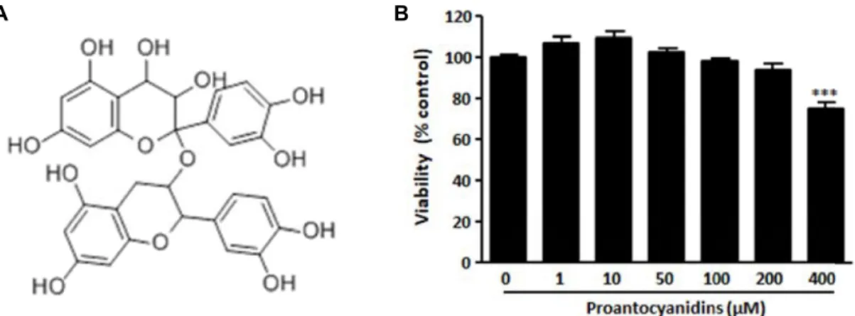
Fig. 1. Effect of proanthocyanidins on cell viability in RAW264.7 macrophages. (A) The chemical structure of proanthocyanidins.
(B) The viability of RAW264.7 cells were tested by CCK-8 assay after 24 hr incubation in medium containing indicated concentration of proanthocyanidins. Triplicate experiments were used to evaluate the data, and the mean value is expressed as ± SEM. Significant compared to 0 μM proanthocyanidins-treated cells, ***p<0.001.
Western blotting analysis
Whole cell, cytosolic or nuclear extracts were separated by 10% sodium dodecyl sulfate-polyacrylamide gel electro- phoresis (SDS-PAGE). The separated proteins were electro- phoretically transferred to nitrocellulose membranes. The membranes were incubated with the indicated Abs and spe- cific bands were visualized using an enhanced chem- iluminescence kit (Amersham Biosciences, Piscataway, NJ, USA).
Data analysis
The data were presented means ± SEM. The values were evaluated by one-way ANOVA with Bonferroni multiple comparison range tests using GraphPad Prism 4.0 software (GraphPad Software Inc, San Diego, CA). Statistically sig- nificant differences were considered at a P-value < 0.05.
Results
Cell viability of proanthocyanidins-treated RAW 264.7 macrophages
Murine RAW264.7 macrophages were chosen for use in an investigation of the anti-inflammatory effects of proan- thocyanidins. We first analyzed the effects of proanthocyani- dins on the viability of RAW 264 macrophages by D-Plus™ CCK cell viability assay. Cells were seeded and incubated with medium containing 0, 10, 50, 100, 200, 400 μM proan- thocyanidins for 24 hr. No notable cytotoxicity was observed when the cells were exposed up to 200 μM proanthocyani- dins (Fig. 1B). Since proanthocyanidins showed no cytotox- icity with concentrations up to 200 μM in RAW264.7 macro-
phages, we used up to 200 μM proanthocyanidins for the rest of the experiments.
HO-1 protein expression analysis of proanthocyanidins- treated RAW264.7 macrophages
We next examined whether proanthocyanidins induces HO-1 mRNA and protein expression in RAW264.7 cells.
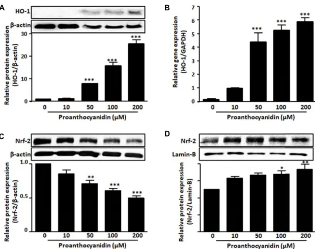
Cells were challenged with different proanthocyanidins con- centrations and the levels of HO-1 mRNA and protein were quantified by qRT-PCR and Western blotting analysis, respectively. HO-1 mRNA and protein expression levels of the proanthocyanidins-added group were significantly upre- gulated compared with those of the control group in a dose-dependent (Fig. 2A, Fig. 2B). These results suggest that proanthocyanidins addition induces HO-1 expression at mRNA and protein levels without noticeable cell damage in RAW264.7 cells.
The transcription of HO-1 is mainly regulated by tran- scription factor nuclear factor-erythroid 2-related factor 2 (Nrf-2) [6, 15]. Under resting condition, the cytosolic Nrf-2 is combined with Kelch-like ECH-associated protein 1 (Keap1), a repressor of Nrf-2, followed by Nrf-2 degradation through proteasome. Upon activation, Nrf-2 is dissociated from Keap1, leading to translocation into the nucleus, where it activates the transcription of phase II genes including HO-1 by binding to the antioxidant response element (ARE) sequence in their promoter regions of target genes [6, 15].
Therefore, we examined the nuclear translocation of the Nrf-2 protein in proanthocyanidins-treated RAW264.7 cells.
To determine whether proanthocyanidins induced the nu- clear translocation of Nrf-2 in macrophages, RAW264.7 cells
A B
C D
Fig. 2. Proanthocyanidins induced HO-1 protein expression. RAW264.7 cells were treated with indicated concentration of proanthocya- nidins for 18 hr or 8 hr to measure (A) HO-1 protein and (B) mRNA expression levels, respectively. After cells were treated with indicated concentrations of proanthocyanidins for 2 hr, Nrf-2 was measured both cytoplasmic (C) and nuclear fraction (D). β-actin and lamin B1 were used as loading controls of cytoplasmic and nuclear protein, respectively. Triplicate experiments were used to evaluate the data, and the mean value is expressed as ± SEM. Significant compared to 0 μM proanthocyani- dins-treated cells, *p<0.05, **p<0.01, ***p<0.001.
were exposed to proanthocyanidins for different concen- trations, and then Western blot analysis was performed us- ing nuclear and cytoplasmic extracts. As shown in Fig. 2C and Fig. 2D, the Nrf-2 nuclear accumulation increased by proanthocyanidins treatment, whereas the cytosolic accumu- lation of Nrf-2 was decreased. These results suggest that proanthocyanidins activates the Nrf-2 nuclear translocation that mediate HO-1 expression.
NO production and iNOS expression in RAW264.7 macrophages under LPS stimulation with proantho- cyanidins
Since we evaluated the effect of proanthocyanidins alone on HO-1 and nuclear translocation of Nrf-2, we investigated the effect of proanthocyanidins on the activity of the RAW 264.7 cells. NO, a free radical produced by iNOS, has been shown to have a number of important biological functions,
including tumor cell killing and host defense against intra- cellular pathogens [10]. iNOS is generally not present in in- active cells but is induced by various stimuli such as LPS [10]. Therefore, NO releases and iNOS expression of RAW 264.7 macrophages under LPS stimulation with proantho- cyanidins was performed to assess the effect of proanthocya- nidins on the immune response.
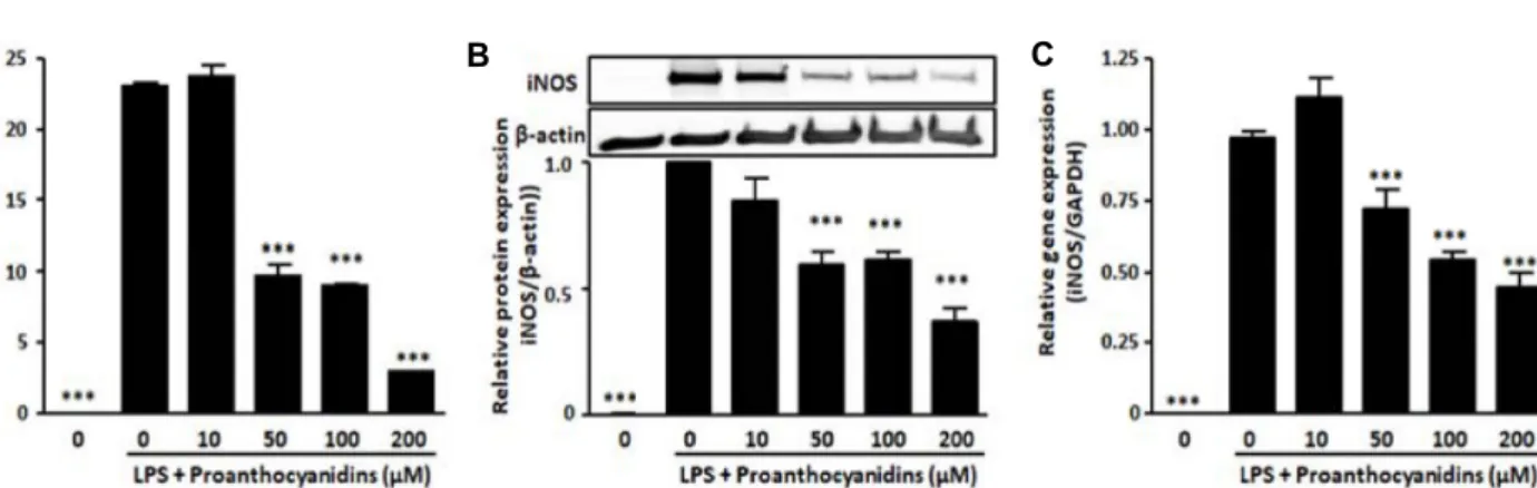
To evaluate the effects of proanthocyanidins on the pro- duction of NO, Griess reagent was used to detect the level of LPS-induced NO release in the supernatant media of RAW 264.7 cells. The results showed that LPS treatment caused significant increase of NO release compared to blank, while proanthocyanidins dose-dependently inhibited LPS-in- duced NO production in RAW264.7 cells as shown in Fig.
3A. Subsequently, to investigate whether these effects of proanthocyanidins are related to iNOS expression of the gene and protein, we used qRT-PCR and Western blot analy-
A B C
Fig. 3. Proanthocyanidins significantly reduced LPS-induced NO production and iNOS expression. RAW264.7 cells was incubated in 0.5 μg/ml LPS condition with or without proanthocyanidins for 24 hr. (A) After incubation, the concentrations of nitrite were measured as described in Materials and Methods. (B) iNOS protein and (C) mRNA levels were analyzed by Western blotting and qRT-PCR analysis, respectively. β-actin was used as control. Triplicate experiments were used to evaluate the data, and the mean value is expressed as ± SEM. Significant compared to LPS-treated cells, ***p<0.001.
sis, respectively. The protein levels of iNOS significantly in- creased in response to LPS treatment, whereas proanthocya- nidins inhibited this effect in a dose-dependent manner (Fig.
3B). Our results also showed that proanthocyanidins sig- nificantly reduced the expression of iNOS gene induced by LPS (Fig. 3C). These data demonstrate proanthocyanidins can restrain the production of NO by down-regulating the expression of iNOS.
Proinflammatory cytokine production in RAW264.7 macrophages under LPS stimulation with proantho- cyanidins
In addition to NO production investigation, we further analyzed the action of proanthocyanidins in the production of proinflammatory cytokines, such as TNF-α and IL-6, known to be induced by LPS stimulation. After LPS stim- ulation with or without proanthocyanidins, the mRNA ex- pression levels of TNF-α and IL-6 were assessed by qRT-PCR analysis (Fig. 4A, Fig. 4B). The mRNA levels of each of the proinflammatory cytokines were upregulated by LPS treat- ment. In contrast, these LPS-inducible cytokines were sig- nificantly reduced with proanthocyanidins addition com- pared with the LPS-alone condition. In addition, the ELISA analysis of TNF-α and IL-6 showed a significant reduction when RAW264.7 cells were treated with LPS and proantho- cyanidins, compared with the LPS-treated group (Fig. 4C, Fig. 4D). These results are consistent with Otterbein et al.
[14] and they suggested that CO, which is the by-product of heme degradation by HO-1, inhibits the expression of LPS-induced proinflammatory cytokines. Thus, our data in- dicated that the addition of proanthocyanidins leads to HO-1
expression and a downregulative effect on proinflammatory mediators.
Discussion
In this study, we evaluated the functions of proanthocya- nidins-induced expression of HO-1 by macrophage cells un- der LPS treatment. First, we showed that proanthocyanidins addition induces HO-1 mRNA and protein expression with- out obvious cell damage. Second, we revealed that proantho- cyanidins causes changes in several proinflammatory factors induced by LPS stimulation. In particular, proanthocyani- dins treatment induced the attenuation of iNOS expression and NO production and the downregulation of proin- flammatory cytokines, such as TNF-α and IL-6, were shown.
These data indicate that proanthocyanidins, as a HO-1 in- ducer, has promise as a novel anti-inflammatory agent.
HO-1 is well-known as a member of the heat-shock pro- tein group, which is involved in responses to a number of cellular injuries, including thermal and oxidant stresses [6, 15]. HO-1 catalyzes the rate-limiting step in the degradation of cellular heme, producing equimolar quantities of iron, CO, and biliverdin [6, 15]. It is generally assumed that this HO-1 and a bi-product-related system may act as a crucial regulator in inflammatory processes, and regulates the bal- ance between proinflammatory and anti-inflammatory medi- ators [15]. The beneficial effects of HO-1 induction have been attributed to several factors, including the degradation of pro-oxidant heme [24], formation of biliverdin and bilirubin with their antioxidant properties [23], as well as the release of CO, which has anti-apoptotic and anti-inflammatory
A B
C D
Fig. 4. Proanthocyanidins treatment shown significant inhibitory effect on proinflammatory cytokines. RAW264.7 cells were treated with indicated concentration of proanthocyanidins for 6 hr to measure the mRNA levels of (A) TNF-α and (B) IL-6 by qRT-PCR.
RAW264.7 cells was incubated in 0.5 μg/ml LPS condition with or without proanthocyanidins for 24 hr. The production of (C) TNF-α and (D) IL-6 in cell culture medium was measured by ELISA. Triplicate experiments were used to evaluate the data, and the mean value is expressed as ± SEM. Significant compared to LPS-treated cells, *p<0.05, **p<0.01, ***p<0.001.
properties [14, 15, 26, 27]. Although the exact mechanisms involved in anti-inflammatory actions of the HO-1 system have not been fully elucidated, one or more of the HO-1 reaction products have been evaluated as possible factors.
For example, CO has been shown to exert significant anti-in- flammatory effects in several models of inflammatory tissue injury [14, 17, 27]. CO has been shown to decrease the pro- duction of IL-6 [13] and NO [21], and increase the production of IL-10 [9] in LPS-stimulated macrophages. It should also be noted that biliverdin and/or bilirubin are capable of blocking key events in inflammation. Using the well-de- scribed rat model of LPS-induced shock, Otterbein et al. [14]
demonstrated that exposure to biliverdin imparts a potent defense against lethal endotoxemia systemically, as well as in the lungs, and effectively abrogates the inflammatory re- sponse [18]. From the above consideration, we hypothesize the following mechanism of the anti-inflammatory effect of proanthocyanidins. Proanthocyanidins may upregulate heme synthesis, which in turn increases HO-1, which then acts to
degrade heme. At the same time, the catabolites of heme increase and show a suppressive effect on iNOS and other proinflammatory cytokines.
Proanthocyanidins are ubiquitous in fruits, seeds, cereals, bark, flowers, nuts, and vegetables. Proanthocyanidins func- tion as powerful scavengers of oxygen free radicals, with potency comparable to vitamins C and E [2, 3, 9]. In addition, emerging evidence indicates that proanthocyanidins target deleterious signaling pathways activated downstream of free radical production [7, 8]. Therefore, proanthocyanidins are of great interest in nutrition and medicine because of their potent antioxidant capacity and possible protective effects on human health. We believe that our data help to interpret the beneficial effects of proanthocyanidins in reducing the risk of inflammation.
Acknowledgement
This study was supported by the Future Scientist Training
program of the Gyeongsangbuk-do Office of Education.
References
1. Ammar el, S. M., Said, S. A., El-Damarawy, S. L. and Suddek, G. M. 2013. Cardioprotective effect of grape-seed proanthocyanidins on doxorubicin-induced cardiac toxicity in rats. Pharm. Biol. 51, 339-344.
2. Bagchi, D., Bagchi, M., Stohs, S. J., Das, D. K., Ray, S. D., Kuszynski, C. A., Joshi, S. S. and Pruess, H. G. 2000. Free radicals and grape seed proanthocyanidin extract: impor- tance in human health and disease prevention. Toxicology 148, 187-197.
3. Bagchi, D., Garg, A., Krohn, R. L., Bagchi, M., Tran, M. X.
and Stohs, S. J. 1997. Oxygen free radical scavenging abilities of vitamins C and E, and a grape seed proanthocyanidin extract in vitro. Res. Commun. Mol. Pathol. Pharmacol. 95, 179- 189.
4. Braune, J., Weyer, U., Hobusch, C., Mauer, J., Brüning, J.
C., Bechmann, I. and Gericke, M. 2017. IL-6 Regulates M2 Polarization and local proliferation of adipose tissue macro- phages in obesity. J. Immunol. 198, 2927-2934.
5. De Simone, V., Franzè, E., Ronchetti, G., Colantoni, A., Fantini, M. C., Di Fusco, D., Sica, G. S., Sileri, P., Mac Donald, T. T., Pallone, F., Monteleone, G. and Stolfi, C. 2015.
Th17-type cytokines, IL-6 and TNF-α synergistically activate STAT3 and NF-κB to promote colorectal cancer cell growth.
Oncogene 34, 3493-503.
6. Dennery, P. A. 2014. Signaling function of heme oxygenase proteins. Antioxid. Redox. Signal. 20, 1743-1753.
7. El-Shitany, N. A. and Eid, B. 2017. Proanthocyanidin pro- tects against cisplatin-induced oxidative liver damage through inhibition of inflammation and NF-κB/TLR-4 pathway.
Environ. Toxicol. 32, 1952-1963.
8. He, L., Li, P., Yu, L. H., Li, L., Zhang, Y., Guo, Y., Long, M., He, J. B. and Yang, S. H. 2018. Protective effects of proan- thocyanidins against cadmium-induced testicular injury through the modification of Nrf2-Keap1 signal path in rats.
Environ. Toxicol. Pharmacol. 57, 1-8.
9. Lee, T. S. and Chau, L. Y. 2002. Heme oxygenase-1 mediates the anti-inflammatory effect of interleukin-10 in mice, Nat.
Med. 8, 240-246.
10. Lind, M., Hayes, A., Caprnda, M., Petrovic, D., Rodrigo, L., Kruzliak, P. and Zulli, A. 2017. Inducible nitric oxide syn- thase: Good or bad? Biomed. Pharmacother. 93, 370-375.
11. Long, M., Yang, S., Zhang, Y., Li, P., Han, J., Dong, S., Chen, X. and He, J. 2017. Proanthocyanidin protects against acute zearalenone-induced testicular oxidative damage in male mice. Environ. Sci. Pollut. Res. Int. 24, 938-946.
12. Matzneller, P., Strommer, S., Drucker, C., Petroczi, K., Schörgenhofer, C., Lackner, E., Jilma, B. and Zeitlinger, M.
2017. Colistin reduces LPS-triggered inflammation in a hu- man sepsis model in vivo: A randomized controlled trial.
Clin. Pharmacol. Ther. 101, 773-781.
13. Morse, D., Pischke, S. E., Zhou, Z., Davis, R. J., Flavell, R.
A., Loop, T., Otterbein, S. L., Otterbein, L. E. and Choi, A.
M. 2003. Suppression of inflammatory cytokine production by carbon monoxide involves the JNK pathway and AP-1.
J. Biol. Chem. 278, 36993-36998.
14. Otterbein, L. E., Bach, F. H., Alam, J., Soares, M., Tao Lu, H., Wysk, M., Davis, R. J., Flavell, R. A. and Choi, A. M.
2000. Carbon monoxide has anti-inflammatory effects in- volving the mitogen-activated protein kinase pathway. Nat.
Med. 6, 422-428.
15. Paine, A., Eiz-Vesper, B., Blasczyk, R. and Immenschuh, S.
2010. Signaling to heme oxygenase-1 and its anti-inflamma- tory therapeutic potential. Biochem. Pharmacol. 80, 1895-1903.
16. Ravindranathan, P., Pasham, D., Balaji, U., Cardenas, J., Gu, J., Toden, S. and Goel, A. 2018. Mechanistic insights into anticancer properties of oligomeric proanthocyanidins from grape seeds in colorectal cancer. Carcinogenesis 39, 767-777.
17. Rochette, L., Cottin, Y., Zeller, M. and Vergely, C. 2013.
Carbon monoxide: mechanisms of action and potential clin- ical implications. Pharmacol. Ther. 137, 133-152.
18. Sarady-Andrews, J. K., Liu, F., Gallo, D., Nakao, A., Over- haus, M., Ollinger, R., Choi, A. M. and Otterbein, L. E. 2005.
Biliverdin administration protects against endotoxin-induced acute lung injury in rats. Am. J. Physiol. Lung Cell Mol.
Physiol. 289, L1131-L1137.
19. Shahat, A. A., Ismail, S. I., Hammouda, F. M., Azzam, S.
A., Lemière, G., De Bruyne, T., De Swaef, S., Pieters, L. and Vlietinck, A. 1998. Anti-HIV activity of flavonoids and pro- anthocyanidins from Crataegus sinaica. Phytomedicine 5, 133- 136.
20. Shapouri-Moghaddam, A., Mohammadian, S., Vazini, H., Taghadosi, M., Esmaeili, S. A., Mardani, F., Seifi. B., Mo- hammadi, A., Afshari, J. T. and Sahebkar, A. 2018.
Macrophage plasticity, polarization, and function in health and disease. J. Cell. Physiol. 233, 6425-6440.
21. Srisook, K., Han, S. S., Choi, H. S., Li, M. H., Ueda, H., Kim, C. and Cha, Y. N. 2006. CO from enhanced HO activity or from CORM-2 inhibits both O2- and NO production and downregulates HO-1 expression in LPS-stimulated macro- phages. Biochem. Pharmacol. 71, 307-318.
22. Su, X., Howell, A. B. and D'Souza, D. H. 2010. Antiviral effects of cranberry juice and cranberry proanthocyanidins on foodborne viral surrogates--a time dependence study in vitro. Food Microbiol. 27, 985-991.
23. Takeda, T. A., Sasai, M., Adachi, Y., Ohnishi, K., Fujisawa, J. I., Izawa, S. and Taketani, S. 2017. Potential role of heme metabolism in the inducible expression of heme oxygen- ase-1. Biochim. Biophys. Acta Gen. Subj. 1861, 1813-1824.
24. Yang, G., Li, Y., Wu, W., Liu, B., Ni, L., Wang, Z., Miao, S., Wang, L. and Liu, C. 2015. Anti-oxidant effect of heme oxygenase-1 on cigarette smoke-induced vascular injury.
Mol. Med. Rep. 12, 2481-2486.
25. Yang, L., Xian, D., Xiong, X., Lai, R., Song, J. and Zhong, J. 2018. Proanthocyanidins against oxidative stress: From molecular mechanisms to clinical applications. Biomed. Res.
Int. 2018, 8584136.
26. Wang, Y. R., Chen, K. L., Li, C. M., Li, L. and Wang, G.
초록:프로안토시아니딘의 항염증효과
천혜진1․박선영1․장희지1․조다영1․정지원2․박기민2․정경미2․김진경1*
(1대구가톨릭대학교 의생명과학과, 2영천여자고등학교)
프로안토시아니딘(proanthocyanidins)은 식물계에 가장 풍부한 폴리페놀성 화합물로 다양한 고등식물의 뿌리, 잎, 열매, 나무껍질 등에 널리 존재할 뿐만 아니라 이러한 원료로 만들어진 차, 와인, 맥주 등과 같은 식품에도 상당량 함유되어 있다. 세포 및 실험동물을 이용한 다수의 연구보고에 의하면 프로안토시아니딘은 항산화활성 및 면역조절활성, DNA 복구 및 항종양 작용과 같은 인체 건강에 유익한 무수한 효과를 가지고 있는 것으로 밝혀 졌다. 면역 세포 중 대식세포(macrophage)는 염증반응을 매개하는 중요한 세포로 외부 병원체 제거에 중요한 역 할을 수행하고 있다. 그러나 대식세포가 만성 염증을 유발하고 비만, 당뇨병, 대사 증후군 및 암과 같은 다양한 질병에 관여한다는 것 또한 널리 보고되어왔다. 본 연구에서는 마우스의 대식세포주인 RAW264.7세포를 이용하 여 프로안토시아니딘의 항염증활성의 일단이 Heme oxygenase-1 (HO-1)의 유도에 의해서 매개됨을 밝혔다.
RAW264.7세포에 프로안토시아니딘을 처리한 결과 세포독성을 보이지 않은 농도에서 HO-1의 발현을 증강시켰 다. 또한 프로안토시아니딘의 처리는 HO-1의 발현을 조절하는 핵심 전사인자인 Nrf (nuclear factor-erythroid 2-related factor)-2의 핵으로의 이동을 유의적으로 증가시켰다. 프로안토시아닌딘의 처리는 LPS (lipopolysaccharide) 에 의해 유도된 NO (nitric oxide)의 생성 및 iNOS (inducible NO synthase)의 발현과 염증성 사이토카인의 생성 및 발현도 유의적으로 억제 하였다. 이러한 결과는 프로안토시아니딘의 항염증제제로서의 개발 가능성을 제시하 는 결과이다.
L. 2019. Heme oxygenase 1 regulates apoptosis induced by heat stress in bovine ovarian granulosa cells via the ERK1/2 pathway. J. Cell. Physiol. 234, 3961-3972.
27. Zhang, R. G., Pan, K., Hao, Y., Yip, C. Y. and Ko, W. H.
2019. Anti-inflammatory action of HO-1/CO in human bronchial epithelium in response to cationic polypeptide challenge. Mol. Immunol. 105, 205-212.