INTRODUCTION
Lung cancer is the most prevalent and lethal cancer world- wide, and has been reported in many nations, including the Republic of Korea and many Western countries (1). The high mortality rate attributed to lung cancer may be the result of induced multi-drug resistance and a high frequency of me- tastatic occurrence. Despite aggressive treatment with sur- gery, radiation, and chemotherapy, survival rates for lung cancer patients remain low (2). New approaches to the treat- ment of lung cancer are urgently needed. Immunotherapy may represent one such approach, and several different strate- gies for lung cancer immunotherapy have recently been de- veloped (3).
The goal of cancer immunotherapy is to augment the weak- ened host immune response against tumors using specific and/or nonspecific immune stimulants. Treatment may in- clude the administration of cytokines, tumor-specific anti- bodies, and T-cells (3-5). Toxoplasma gondii is an obligate intra- cellular parasite; latent infections with T. gondii are common in human populations throughout the world (6). It has been
reported that the development of spontaneous mammary tumors, leukemia, and 20-methylcholanthrene-autoinduced tumors is suppressed in animal models following injection of Toxoplasma antigen or viable parasites (7, 8). Both Toxo- plasma infection and the cell-free parasite extract are able to reverse the multi-drug resistance of mouse lymphoma and human gastric cancers in vitro (9). Moreover, potent antitu- mor effects are induced by intralesional injection with for- malin-fixed T. gondii organisms in Lewis lung carcinoma (LLC) in Toxoplasma-infected mice (10, 11).
These reports suggest that T. gondii is a powerful agent for cancer immunotherapy, and is useful as a stimulant of the cellular immune responses. Recently, there were many reports that antitumor and antimetastatic actions are associated with inhibition of tumor-induced neovascularization (12-14). How- ever, the immunologic mechanisms of the antitumor activi- ty elicited by T. gondii in lung cancer, as well as the immu- nological characteristics of LLC, are not well documented.
Also, the anti-angiogenic effects of T. gondii have not been determined in the LLC mouse model. In order to determine the antitumor and anti-angiogenic activities of T. gondii in-
Ju-Ock Kim, Sung-Soo Jung, Sun-Young Kim, Tae Yun Kim*, Dae-Whan Shin*,�, Jae-Ho Lee�, Young-Ha Lee*,�
Departments of Internal Medicine, Infection Biology*
and Pediatrics�, College of Medicine, Research Institute for Medical Science�, Chungnam National University, Daejeon, Korea
Address for correspondence Young-Ha Lee, M.D.
Department of Infection Biology, Chungnam National University, College of Medicine, 6 Munhwa-dong, Jung-gu, Daejeon 301-131, Korea
Tel : +82.42-580-8273, Fax : +82.42-583-8216 E-mail : [email protected]
S38
Inhibition of Lewis Lung Carcinoma Growth by Toxoplasma gondii through Induction of Th1 Immune Responses and Inhibition of Angiogenesis
Toxoplasma gondii is an obligate intracellular protozoan parasite that induces anti- tumor activity against certain types of cancers. However, little information is avail- able regarding the immunologic mechanisms that regulate these effects. For this purpose, C57BL/6 mice were administered either the T. gondii Me49 strain orally or Lewis lung carcinoma (LLC) cells intramuscularly. Survival rates, tumor size, histopathology, and immune responses were determined for each group, and angio- genesis was evaluated by in vivo Matrigel plug assay. Toxoplasma-infected (TG- injected) mice survived the entire experimental period, whereas cancer cell-bear- ing (LLC-injected) mice died within six weeks. Mice injected with both T. gondii and cancer cells (TG/LLC-injected group) showed significantly increased survival rates, CD8+T-cell percentages, IFN- mRNA expression levels, serum IgG2a titers, and CTL responses as compared to the LLC-injected mice. In addition, angiogenesis in the TG/LLC-injected mice was notably inhibited. These effects in TG/LCC-injected mice were similar or were increased by the addition of an adjuvant, Quil-A. How- ever, TG/LLC-injected mice showed decreased percentages of CD4+and CD8+T- cells, IFN- mRNA expression levels, and serum IgG1 and IgG2a titers as com- pared to TG-injected mice. Taken together, our results demonstrate that T. gondii infection inhibits tumor growth in the Lewis lung carcinoma mouse model through the induction of Th1 immune responses and antiangiogenic activity.
Key Words : Toxoplasma gondii, Immunotherapy; Lewis Lung Carcinoma; Antitumor; Antiangiogenesis
Received : 3 May 2007 Accepted : 21 July 2007
fection in LLC-bearing mice, C57BL/6 mice were injected with LLC cells alone or in combination with T. gondii. And then, we evaluated the survival rates, tumor size, histopathol- ogy, cell-mediated immune responses, and angiogenesis.
MATERIALS AND METHODS Mice and parasite strains
Female inbred C57BL/6 mice were obtained from the Korea Research Institute of Bioscience and Biotechnology, Daejeon, Korea. All mice used were 6-8 weeks old and doc- umented as specific pathogen-free animals. All animal stud- ies were carried out in a pathogen-free barrier zone at the College of Medicine, Chungnam National University, in accordance with the procedures outlined in the Guide for the Care and Use of Laboratory Animals. Two strains of T.
gondii were used; the RH strain was used to prepare Toxo- plasma lysate antigen (TLA) and mice were orally infected with the Me49 strain to evaluate antitumor activity.
Lewis lung carcinoma (LLC) cell cultures and experimen- tal groups
The LLC cell line was obtained from the American Type Culture Collection (ATCC, Rockville, MD, U.S.A.) and was cultured in a humidified atmosphere of 5% CO2 at 37℃ using Dulbecco’s modified Eagle’s medium (DMEM) (Gib- coBRL Co., Grand Island, NY, U.S.A.) that contained 10%
heat-inactivated fetal bovine serum (FBS; GibcoBRL), 2 mM glutamine, 100 U/mL penicillin, and 100 g/mL strepto- mycin.
Mice were divided into seven experimental groups. Each group was composed of 50 mice; 10 mice to evaluate survi- val rates, 35 mice to evaluate immunologic characteristics, and 5 mice to check angiogenesis. The seven experimental groups included mice that were administered with: LLC cells (LLC-injected group); T. gondii (TG-injected group); both Toxoplasma parasites and LLC cells (TG/LLC-injected group);
Quil-A only (QA-injected group) (Accurate Chemical and Scientific Co., Westbury, NY, U.S.A.); both LLC cells and Quil-A (LLC/QA-injected group); both T. gondii and Quil-A (TG/QA-injected group); and T. gondii, LLC cells and Quil- A (TG/LLC/QA-injected group). To calculate the survival rate for each group, the mice were examined each day for eight weeks following LLC cell injection or T. gondii infec- tion. To evaluate the immunological mechanisms of antitu- mor activity induced by Toxoplasma, mice were sacrificed weekly and blood, muscle, lung, and spleen tissues were col- lected. In addition, we checked the antiangiogenic activity and cytotoxic T-lymphocyte responses one week after infec- tion with T. gondii. Untreated age- and sex-matched control mice were treated with equivalent doses of normal saline.
In vivo evaluation of antitumor activity
According to the experimental protocol, mice were inject- ed with LLC cells, T. gondii parasites or Quil-A. LLC cells were implanted at a density of 1×10⁵viable cells into the femoral muscle and Quil-A (20 g per mouse) was injected intraperitoneally twice weekly for three consecutive weeks.
Mice were orally infected with five cysts of the T. gondii Me49 strain. The extent of tumor growth was measured weekly using sterile metric calipers. Tumor volume was calculated using the following formula (13): tumor volume ( L)=tumor width (mm)2×tumor length (mm)×0.5.
Histopathological analysis
The lung and muscle samples from each mouse were re- moved immediately after anesthesia and placed in 10% bu- ffered neutral formaldehyde (Polyscience Inc., Warrington, PA, U.S.A.). Paraffin-embedded tissues were cut and stained with hematoxylin and eosin (H-E), and the cancer cells were examined using a bright-field microscope.
Enzyme-linked immunosorbent assay (ELISA)
Serum samples were obtained from each mouse, and the IgG subclasses were quantified. TLA was prepared accord- ing to the protocol outlined by Lee et al. (15). Each well of a 96-well plate was coated with TLA (10 g/mL) and incubat- ed overnight at 4℃. After blocking, serum samples were diluted 1:100 in 0.1% bovine serum albumin/phosphate- buffered saline (BSA/PBS) that contained 0.05% Tween-20, and 100 L of sample was added to each well. The plates were incubated for 2 hr, and HRP-conjugated goat anti-mouse IgG1 or IgG2a (Southern Biotechnology Associates Inc., Birm- ingham, AL, U.S.A.) was added. Following the final wash- ing step, freshly prepared O-phenylenediamine dihydrochlo- ride was added, and the reaction was stopped by the addition of 4 N H2SO4. The optical density was read at 492 nm using an automated ELISA reader (Tecan A-5082, Salzburg, Austria).
Splenocyte preparation and flow cytometry
Following removal, the spleens were homogenized and erythrocytes were lysed in Tris-NH4Cl (pH 7.2). Splenocyte extracts that contained 1×10⁶cells were incubated with 50 L of fluorescein isothiocyanate (FITC)-conjugated anti-mou- se CD4+and CD8+monoclonal antibodies or an isotype-spe- cific control (1:100 dilution in 0.1% BSA/PBS; PharMin- gen, San Diego, CA, U.S.A.) for 1 hr at 4℃. The cells were washed three times in 0.1% BSA/PBS with centrifugation, and then fixed with 1% paraformaldehyde and analyzed by flow cytometry (FACScan; Becton Dickinson, Franklin Lakes, NJ, U.S.A.). All data were analyzed using the Cell-Quest program (Becton Dickinson).
RT-PCR assessment of cytokines
Splenic cytokine mRNA expression was assessed accord- ing to the methods of Lee et al. (15). Each sample was pro- cessed for total mRNA isolation using the RNAgent kit (Promega, Madison, WI, U.S.A.). The cDNA was prepared using a starting mixture that contained 5 g of total RNA, 8 L of 5×RT buffer, 4 L of dNTPs, 25 M oligo (dT), and 1 L of avian myeloblastosis virus (AMV) reverse tran- scriptase. PCR was performed using 2-13 L of the cDNA reaction mixture that contained 10×polymerase buffer, 250 M dNTPs, 0.4 M of the 3′-primer and 5′-primer, and 2.5 U Taq polymerase. Each PCR cycle consisted of 94℃for 1 min, 57℃for 1 min, and 72℃for 1 min. The cytokine primer sequences were as follows: for hypoxanthine phospo- ribosyltransferase (HPRT), 3′-GAGGGTAGGCTGGCCT- ATGGCT-5′and 5′-TTGGATACAGGCCAGACTTTGT- TG-3′; for interferon gamma (IFN- ), 3′-CTCATGGAAT- GCATCCTTTTTCG-5′and 5′-ACGCTACACAC-TGCA- TCTTGG-3′; and for interleukin-10 (IL-10), 3′-TGTCTA- GGTCCTGGAGTCCAGCAGACTCAA-5′and 5′-CCA- GTTTTACCTGGTAGAAGTGATG-3′. After 35 PCR cycles, the products were separated by electrophoresis on 2%
agarose gels. Quantification of mRNA was performed with an imaging densitometer (Bio-Rad, Hercules, CA, U.S.A.).
Cytotoxic T lymphocyte assay
One week after T. gondii (or cancer cell) injection, splenic lymphocytes were isolated from each group and cultured in RPMI 1640 for three days. The splenocytes were then co-cul- tured with irradiated LLC cells at various effector to target (E:T) ratios for seven days. One Ci of 51Cr (NEN Research Products, Boston, MA, U.S.A.) was added to each well and the plate was allowed to incubate for 3 hr. The plate was then washed several times with RPMI 1640 until the culture supernatant contained <500 counts per minute (CPM) of
51Cr. The plates were incubated for an additional 4 hr at 37℃, followed by removal of 100 L of each sample, and the CPM were determined by a gamma counter (Beckman, Fullerton, CA, U.S.A.). The percent lysis was calculated as follows: per- cent lysis=(mean CPM of test sample-mean CPM of sponta- neous release)/(mean CPM of maximal release-mean CPM of spontaneous release)×100.
Matrigel angiogenesis assay
Matrigel angiogenesis assay was performed as described by Passaniti et al. (16). One week after T. gondii (or cancer cell) injection, the C57BL/6 mice were injected subcutaneous- ly near the abdominal mid-line using a 25G needle with 0.5 mL Matrigel (BD Bioscience, Bedford, MA, U.S.A.) that contained 300 ng of basic fibroblast growth factor (bFGF;
Sigma Chemical Co., St. Louis, MO, U.S.A.) and 32 U/mL
heparin (Sigma). The injected Matrigel rapidly formed a sin- gle, solid gel and the mice were sacrificed six days later. The underlying skin of the mouse was removed, and the gels were recovered for hemoglobin (Hb) measurement using the Drab- kin reagent kit (Sigma). Hemoglobin concentrations were calculated from a known Hb control, which was run in par- allel.
Statistical analysis
All results are expressed as the mean±standard error (SE) for each group. Kaplan-Meier and Log-Rank tests were per- formed to analyze the differences in survival rates. To evalu- ate differences in tumor sizes, antibody titers, phenotype profiles, cytokine mRNA levels, Hb concentrations, and cytotoxicity, Kruskal-Wallis tests were used to check for sta- tistical differences among the groups. Differences between groups were considered significant at p<0.05.
RESULTS
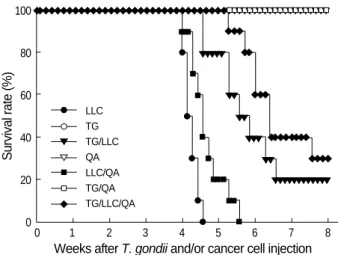
Increased survival rates of cancer-bearing mice by T. gondii infection
Mice were treated with T. gondii parasites, LLC cells, or Quil-A according to our protocol, and survival rates were evaluated over eight weeks. As shown in Fig. 1, the TG-, QA-, and TG/QA-injected mice survived the entire experi- mental period. However, the LLC- and LLC/QA-injected mice began dying 28 days after cancer cell injection, and all of these mice died after 39 days. The survival rate of the TG/
LLC-injected mice was 20%, and the TG/LLC/QA-injected mice had a 30% survival rate at 8 weeks after cancer cell injection, which were significantly higher than the rates for the LLC- and LLC/QA-injected mice (p<0.05).
T. gondii infection inhibits the tumor growth in mice
Mice infected with T. gondii developed brain cysts within two weeks of infection (data not shown). Cancer cells were observed in the muscle tissues within one week in the LLC- and LLC/QA-injected groups (Fig. 2A, B). However, cancer cells were found in the TG/LLC- and TG/LLC/QA-injected mice by two weeks. In addition, cancer cells were found in the lungs three weeks after LLC cell injection in the LLC- and LLC/QA-injected groups (Fig. 2C, D), whereas cancer they were found within five weeks in TG/LLC- and TG/LLC/QA- injected mice. The skin overlying the tumor began to ulcerate three to four weeks after cancer cell injection in the LLC-inject- ed mice (Fig. 2E), whereas ulceration was observed within five weeks in the TG/LLC- and TG/LLC/QA-injected mice.
Tumor masses were palpable by two to three weeks after cancer cell injection in the cancer-bearing LLC-, LLC/QA-,
TG/LLC-, and TG/LLC/QA-injected mice. In the LLC- and LLC/QA-injected mice, tumor masses increased abruptly in size at two to four weeks after cancer cell injection, and the tumor volumes at four weeks were 8,760±465 and 6,740
±360 L, respectively (Fig. 3). The tumor volumes were significantly decreased in the TG/LLC- and TG/LLC/QA- injected mice (2,155±520 L and 1,980±342 L, respec- tively) at four weeks as compared with the LLC- and LLC/
QA-injected mice (p<0.05).
IgG2a titers were increased in T. gondii-infected cancer- bearing mice
Serum samples were assayed for IgG subclasses against TLA by ELISA. The specific IgG1 and IgG2a titers of the LLC-, QA-, and LLC/QA-injected mice were similar to those of control mice. However, the specific IgG1 titers of the TG- and TG/QA-injected mice were significantly increased after one week of T. gondii infection (Fig. 4A). In addition, the IgG1 titers of the TG/LLC- and TG/LLC/QA-injected mice were markedly depressed following cancer cell injection as compared with the TG- and TG/QA-injected mice (p<0.05).
The IgG2a titers of the TG- and TG/QA-injected mice were significantly increased after T. gondii infection as com- pared to the control mice. In the TG/LLC- and TG/LLC/QA- injected mice, the IgG2a titers following cancer cell injec- tion were significantly depressed until four weeks compared with those of the TG- and TG/QA-injected mice, although the IgG2a titers increased abruptly thereafter (Fig. 4B).
Fig. 2. Histopathologic findings for the muscles and lungs of C57- BL/6 mice injected with T. gondii, LLC cells or Quil-A. Paraffin- embedded tissues were stained with hematoxylin and eosin, and visualized under a microscope at ×400 magnification. (A) Nor- mal muscle; (B) cancerous changes in the muscle at cancer cell- injected sites; (C) normal lung; (D) cancerous changes in the lung (cancer metastasis to the lungs); (E) tumor masses of mice in- jected with LLC cells (left and middle) and injected with both T.
gondii and LLC cells (right). Ulceration was observed in cancer- bearing mice (arrow).
A B
C D
E Fig. 1.Survival rates of C57BL/6 mice injected with Toxoplasma
gondii, Lewis lung carcinoma (LLC) cells or Quil-A. Mice were checked daily for eight weeks following cancer cell injection, to measure survival rates. The groups (n=10 per group) were as fol- lows: LLC, injected in the femoral muscle with 1×10⁵viable LLC cells; TG, infected with five cysts of the Me49 strain of T. gondii;
TG/LLC, received both Toxoplasma parasites and LLC cells; QA, received 20 g/mouse of Quil-A twice weekly for 3 weeks; LLC/
QA, received both LLC cells and Quil-A; TG/QA, received both T.
gondii and Quil-A; TG/LLC/QA, received T. gondii, LLC cells, and Quil-A.
0 1 2 3 4 5 6 7 8
Weeks after T. gondii and/or cancer cell injection
LLC TG TG/LLC QA LLC/QA TG/QA TG/LLC/QA
Survival rate (%)
100
80
60
40
20
0
Cancer mass (L)
10,000
8,000
6,000
4,000
2,000
0
0 1 2 3 4 5 6
Weeks after T. gondii and/or cancer cell injection Fig. 3. Sizes of tumors in C57BL/6 mice injected with T. gondii, LLC cells or Quil-A. Tumor growth was measured weekly using sterile metric calipers. Tumor volume ( L)=tumor width (mm)2× tumor length (mm)×0.5. Data are presented as the means±SE of five mice (*, p<0.05 compared with LLC- or LLC/QA-injected group).
LLC TG TG/LLC QA LLC/QA TG/QA TG/LLC/QA
*
*
*
*
*
*
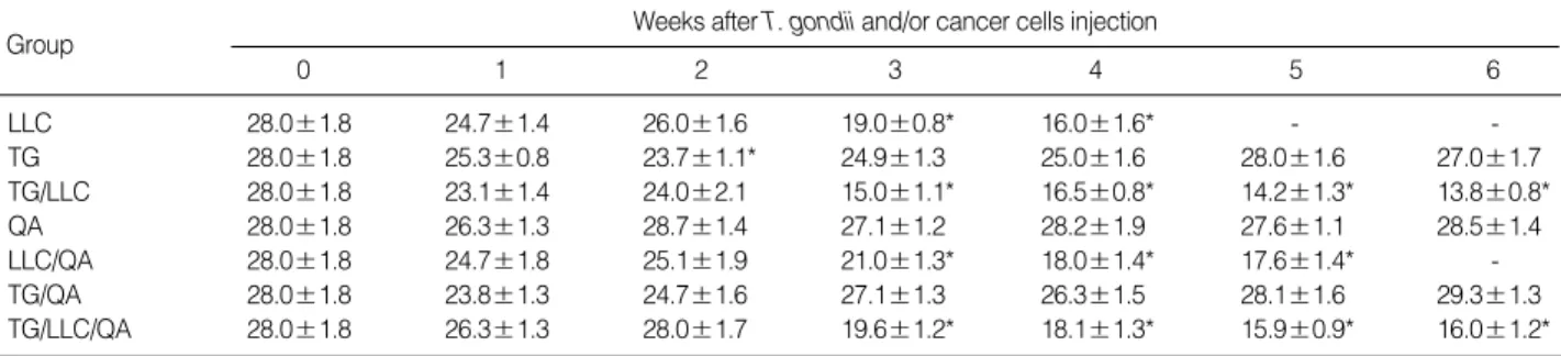
CD4+and CD8+T-cells are decreased in cancer-bearing mice
To evaluate changes in T-cell subtypes following either T.
gondii infection or cancer cell injection, we analyzed the phe- notypic profiles of murine splenocytes. The percentages of
CD4+T-cells in the TG-injected mice were temporarily dec- reased at one to two weeks post-infection compared to those of the control mice, and these levels returned to baseline shortly thereafter (Table 1). The percentages of CD4+T-cells in the LLC-, LLC/QA-, TG/LLC-, and TG/LLC/QA-injected mice were significantly decreased following cancer cell injection
Toxoplasma-specific IgG1 (492 nm)
1.5
1.2
0.9
0.6
0.3
0.0
0 1 2 3 4 5 6
Weeks after T. gondii and/or cancer cell injection
Fig. 4. Serum IgG1 (A) and IgG2a (B) titers of C57BL/6 mice injected with T. gondii, LLC cells or Quil-A, as measured by ELISA. Serum samples were obtained from mice at the indicated time-points, and the IgG subclasses against Toxoplasma lysate antigen were quanti- fied. Data are presented as the mean±SE of five mice (*, p<0.05 compared with LLC- or LLC/QA-injected group; �, p<0.05 compared with TG- or TG/QA-injected group).
LLC TG TG/LLC QA LLC/QA TG/QA TG/LLC/QA
A
Toxoplasma-specific IgG2a (492 nm)
1.2
1.0 0.8
0.6
0.4 0.2
0.0
0 1 2 3 4 5 6
Weeks after T. gondii and/or cancer cell injection
LLC TG TG/LLC QA LLC/QA TG/QA TG/LLC/QA
B
*,�
�
� �
�
*,�
*,�
� �
�
�
�
Group Weeks after T. gondii and/or cancer cells injection
0 1 2 3 4 5 6
LLC 28.0±1.8 24.7±1.4 26.0±1.6 19.0±0.8* 16.0±1.6* - -
TG 28.0±1.8 25.3±0.8 23.7±1.1* 24.9±1.3 25.0±1.6 28.0±1.6 27.0±1.7
TG/LLC 28.0±1.8 23.1±1.4 24.0±2.1 15.0±1.1* 16.5±0.8* 14.2±1.3* 13.8±0.8*
QA 28.0±1.8 26.3±1.3 28.7±1.4 27.1±1.2 28.2±1.9 27.6±1.1 28.5±1.4
LLC/QA 28.0±1.8 24.7±1.8 25.1±1.9 21.0±1.3* 18.0±1.4* 17.6±1.4* -
TG/QA 28.0±1.8 23.8±1.3 24.7±1.6 27.1±1.3 26.3±1.5 28.1±1.6 29.3±1.3
TG/LLC/QA 28.0±1.8 26.3±1.3 28.0±1.7 19.6±1.2* 18.1±1.3* 15.9±0.9* 16.0±1.2*
Table 1.Percentages of CD4+T cell subsets in splenocytes from mice injected with T. gondii, Lewis lung carcinoma cells, or Quil-A
Splenocytes were stained with FITC-conjugated CD4 monoclonal antibody, and then analyzed by FACScan.
*Statistically significant differences compared with the control group (day 0) (p<0.05). The data are represented as the mean±SE of 5 mice.
LLC, Lewis lung carcinama; TG, T. gondii; QA, Quil-A.
Group Weeks after T. gondii and/or cancer cells injection
0 1 2 3 4 5 6
LLC 19.0±1.5 19.2±1.3 17.0±1.2 14.8±0.8* 13.2±1.5* - -
TG 19.0±1.5 17.6±1.2 16.5±0.8 18.0±1.3 22.1±1.5 19.6±1.8 20.8±1.9
TG/LLC 19.0±1.5 19.3±1.0 18.1±1.0 14.0±1.4* 12.0±0.5* 16.2±1.2* 16.5±1.1
QA 19.0±1.5 20.4±0.9 21.7±1.1 22.6±1.7 22.1±1.3 22.8±0.7 22.1±1.4
LLC/QA 19.0±1.5 17.8±1.3 16.5±1.4 15.5±1.0* 14.9±0.8 15.3±1.5* -
TG/QA 19.0±1.5 19.3±1.6 17.3±1.6 20.1±1.3 21.6±1.6 20.9±1.2 21.1±1.6
TG/LLC/QA 19.0±1.5 21.5±1.7 18.8±1.3 15.4±1.3* 14.0±1.7* 16.9±1.3 16.8±1.2 Table 2.Percentages of CD8+T cell subsets in splenocytes from mice injected with T. gondii, Lewis lung carcinoma cells, or Quil-A
Splenocytes were stained with FITC-conjugated CD8 monoclonal antibody, and then analyzed by FACScan.
*Statistically significant differences compared with the control group (day 0) (p<0.05). The data are represented as the mean±SE of 5 mice.
LLC, Lewis lung carcinama; TG, T. gondii; QA, Quil-A.
and remained markedly depressed after three weeks (p<0.05).
As shown in Table 2, the percentages of CD8+T-cells in the LLC-, LLC/QA-, TG/LLC-, and TG/LLC/QA-injected mice were significantly decreased three weeks after cancer cell injection. The LLC- and LLC/QA-injected mice main- tained these low values, whereas the percentage of CD8+T- cells in the TG/LLC- and TG/LLC/QA-injected mice recov- ered slightly at five weeks after T. gondii infection.
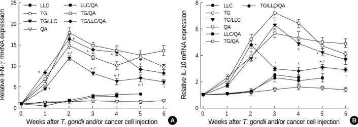
IFN- mRNA expression was significantly increased in T. gondii-infected cancer-bearing mice
As shown in Fig. 5A, the levels of IFN- mRNA expres- sion in the LLC- and LLC/QA-injected mice were increased two- to three-fold at three weeks after cancer cell injection
as compared to the control (p<0.05). However, the levels of IFN- mRNA expression in the TG-, TG/QA-, and TG/
LLC/QA-injected mice were increased immediately after T.
gondii infection, peaked two to three weeks post-infection (14-19-fold increase), and slowly decreased thereafter. How- ever, IFN- mRNA expression in the TG/LLC-injected mice was significantly lower than that in the TG-, TG/QA-, and TG/LLC/QA-injected mice, even though IFN- mRNA expression in the TG/LLC-injected mice was significantly increased by one week post-infection.
As compared to the control mice, the levels of IL-10 mRNA expression in the LLC- and LLC/QA-injected mice increased significantly, by 1.3-2.3-fold, three weeks after cancer cell injection. The levels of IL-10 mRNA expression in the TG-, TG/QA-, and TG/LLC-injected mice increased immediately following T. gondii infection, peaking at three weeks post-in- fection (5.5-7.5-fold increase). However, IL-10 mRNA ex- pression in the TG/LLC/QA-injected mice, which was signif- icantly lower than in the TG-, TG/QA-, and TG/LLC-inject- ed mice from 3 weeks postinfection, peaked at two weeks and subsequently decreased.
T. gondii infection induced tumor cytolysis by splenocytes
An in vitro CTL assay was performed to determine whether the splenic T-cells of Toxoplasma-infected cancer-bearing mice increased cytotoxic activity against LLC cells. As shown in Fig. 6, the LLC-, QA-, and LLC/QA-injected mice did not show CTL responses. However, the splenocyte CTL activi- ties of the TG- and TG/QA-injected mice increased as the E:T ratio increased. The percentages of specific lysis seen in the TG/LLC- and TG/LLC/QA-injected mice were consid- erably higher than those in the LLC- and LLC/QA-injected mice. However, the CTL activities of the TG/LLC- and TG/
LLC/QA-injected mice were significantly lower than those
Relative IFN-mRNA expression
25
20
15
10
5
0
0 1 2 3 4 5 6
Weeks after T. gondii and/or cancer cell injection
Fig. 5. Relative IFN- (A) and IL-10 (B) mRNA expression in splenocytes from C57BL/6 mice injected with T. gondii, LLC cells or Quil-A.
The transcript levels are relative to those in splenocytes obtained from control mice (designated value of 1). Splenocytes were harvested at the time-points indicated and the expression of IFN- and IL-10 mRNAs was assayed by RT-PCR. Data are presented as mean±SE of five mice.
A
Relative IL-10 mRNA expression
8
6
4
2
0
0 1 2 3 4 5 6
Weeks after T. gondii and/or cancer cell injection
LLC TG TG/LLC QA
LLC/QA TG/QA TG/LLC/QA
B
*,�
*,�
*,�
*,�
*,�
*
*
*
* *
*
* *
*
*
�
� �
�
LLC TG TG/LLC QA LLC/QA TG/QA
TG/LLC/QA
*,�
*,�
*,�
*,�
*,�
�
Percent specific lysis
35 30 25 20 15 10 5 0
6.25:1 12.5:1 25.0:1 50.0:1
Effector:target ratio
Fig. 6. Cytotoxic T-cell responses of C57BL/6 mice injected with T. gondii, LLC cells or Quil-A. One week after T. gondii (or cancer cell) injection, splenic lymphocytes were isolated from each mou- se, labeled with 51Cr, and cultured with irradiated LLC cells at dif- ferent cell ratios. Data are presented as mean±SE of five mice.
LLC TG TG/LLC QA LLC/QA TG/QA TG/LLC/QA
of the TG- and TG/QA-injected mice (p<0.05).
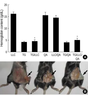
T. gondii infection inhibits tumor angiogenesis in vivo
The Matrigel plugs that formed following subcutaneous implantation of Matrigel alone produced little or no local reaction, nor did they produce an angiogenic response. How- ever, the plugs that formed following the implantation of Matrigel that was supplemented with bFGF and heparin caused an angiogenic reaction (Fig. 7). We measured the Hb concentrations inside the Matrigel plugs in order to quanti- fy the extent of angiogenesis. The Hb concentrations in the LLC-, QA-, and LLC/QA-injected mice ranged from 14.6± 2.0 to 16.2±1.6 g/dL, whereas those in the TG- and TG/
QA-injected mice ranged from 4.1±0.7 to 4.3±0.9 g/dL.
The Hb concentrations in the TG/LLC- and TG/LLC/QA- injected mice were significantly lower than those in the LLC- or LLC/QA-injected mice (p<0.05), but were not significantly different from those in the TG- or TG/QA-injected mice.
DISCUSSION
Tumors frequently interfere with the development and
function of immune responses (3-5). The ability of various infections to suppress neoplastic growth has been well docu- mented. In addition, T. gondii is a nonspecific immune stim- ulant used for cancer immunotherapy (7-11). In the present study, we examined the mechanisms of the antitumor effects of T. gondii infection on lung cancer in vivo. Toxoplasma in- fection of LLC-bearing mice significantly increased the sur- vival rates, serum IgG2a titers, IFN- mRNA expression, CD8+T-cell percentages, CTL responses, and also inhibited angiogenesis. These values were similar or further improved by the addition of an adjuvant, Quil-A. These results indi- cate that T. gondii activates Th1 immune responses and inhi- bits angiogenesis in LLC-bearing mice, leading to the induc- tion of antitumor and antimetastatic activities.
Cancer immunotherapy attempts to harness the exquisite power and specificity of the immune system for the treat- ment of malignancy. Although cancer cells are less immuno- genic than pathogens, the immune system is clearly capable of recognizing and eliminating tumor cells. Lung cancer immunotherapy may represent one new approach that has low toxicity and high specificity, but implementation has been a challenge due to poor antigenic characterization and the ability of lung cancers to escape immune responses (3-5, 17). Several different immunotherapeutic treatment strate- gies and mouse models have been developed. In this study, we utilized LLC cells, which remain highly tumorigenic in C57BL/6 mice and produce primary tumors and lung meta- stases histologically indistinguishable from the original tumor line (18). In the LLC- and LLC/QA-injected mice, tumor masses were initially palpable in the muscle tissues located at the site of cancer cell injection from two weeks after can- cer cell injection, and cancer cells were found in the lungs three weeks after LLC cell injection. Thus, it was confirmed that the LLC cell line is useful for studying the mechanisms of cancer chemotherapeutic agents. After T. gondii infection, tumor volumes were significantly decreased in the TG/LLC- and TG/LLC/QA-injected mice as compared with the LLC- and LLC/QA-injected mice. However, the mechanisms of antitumor activity induced by T. gondii infection are not easi- ly introduced in LLC, so we suggested the mechanisms of antitumor effects in this study.
Infection with T. gondii results in a strong cell-mediated immune response, especially the T-cell-mediated response (19). In the present study, the percentages of CD8+and CD4+ T-cells were significantly decreased in cancer-bearing LLC- and LLC/QA-injected mice. Following infection with T.
gondii, the percentages of CD8+T-cells in TG/LLC- or TG/
LLC/QA-injected mice were increased from 5 weeks post- infection, but those of CD4+T-cells were not. Collectively, these data indicate that T-cell-mediated immune responses were severely suppressed in Lewis lung carcinoma, and T.
gondii alone or in combination with Quil-A may provide an appropriate epitope for MHC-1 presentation (17, 19), and therefore, the percentages of CD8+T-cells were recovered.
Fig. 7. Angiogenesis in C57BL/6 mice injected with T. gondii, LLC cells or Quil-A. One week after T. gondii (or cancer cell) injection, a Matrigel that was supplemented with heparin and basic fibrob- last growth factor was injected into the mice. The gels were recov- ered on day 6 postinjection for hemoglobin measurement using the Drabkin reagent kit. Data are presented as the mean±SE of five mice. (A) Hemoglobin content of each group; (B) Matrigel plugs were removed 6 days after injection and photographed (arrows): LLC-bearing mice (left), Toxoplasma-infected mice (mid- dle), and Matrigel alone (right).
Hemoglobin content (g/dL)
20
15
10
5
0
LLC TG TG/LLC QA LLC/QA TG/QA TG/LLC/
QA A
* *
B
This phenomenon is confirmed by the results of a cytotoxic T lymphocyte assay. Namely, Toxoplasma-infected TG/LLC- and TG/LLC/QA-injected mice demonstrated higher CTL responses of CD8+T-cells than the cancer-bearing LLC- or LLC/QA-injected mice. Previous studies have also demon- strated that strong antitumor responses are induced by either abundant infiltration of both CD4+and CD8+T-cells and tumor-specific CTLs in LLC-bearing mice after treatment with IL-2 and IL-12 (17) or increases in the numbers of apop- totic tumor cells, CD8+T-cells, and natural killer (NK) cells that invade the tumors after injection with ergosterol (14).
Cytokines serve as critical regulators of cell-mediated im- munity by both enhancing and limiting immune responses (20). The CD4+T-cells are divided into two classes on the basis of the range of cytokines produced. Th1 cells produce IL-2 and IFN- , and control the production of IgG2a, where- as Th2 cells produce IL-4, IL-5, and IL-10, and control the production of IgG1 and IgE. Because of these interleukin secretion patterns, Th1 cells promote cell-mediated immu- nity. In contrast, Th2 cells induce B-cell differentiation and inhibit cell-mediated immunity and antitumor responses.
In the present study, we found that IFN- mRNA expres- sion in TG/LLC-injected mice was significantly higher than that in LLC-injected mice and significantly lower than that in TG-injected mice. These results indicate that Th1 immune responses were depressed in Lewis lung carcinoma, and show that the production of cytokines such as IFN- , TNF- , IL- 12, and IL-2 can arrest proliferation of malignant cells and prevent the angiogenesis necessary for tumor growth (3, 5, 17, 21, 22). Similar results were reported in melanoma, liver cancer, and thyroid cancer (22-24). And these phenomena were also confirmed in the titrations of the IgG subclasses.
The IgG2a titer, which is the primary indicator of a Th1 immune response (25), was significantly increased in the TG/LLC- and TG/LLC/QA-injected mice four weeks after infection, while the IgG1 titer, an indicator of the Th2 im- mune response, remained unchanged.
The development of a vascular supply is essential for the growth, maturation, and maintenance of normal tissues. It is also required for wound healing and rapid growth of solid tumors, and is involved in various other pathologic condi- tions. Tumor angiogenesis is regulated by the production of angiogenic stimulators, including members of the fibrob- last growth factor and the vascular endothelial growth fac- tor families (13, 14, 26, 27). In the present study, a combi- nation of bFGF and heparin recruited vessels from the sur- rounding tissues into the Matrigel. The Hb concentrations in T. gondii-infected TG-, TG/QA-, TG/LLC-, and TG/LLC/
QA-injected mice were significantly decreased by three- to four-fold as compared to uninfected QA-, LLC-, and LLC/
QA-injected mice. In a similar study, Hunter et al. (28) show- ed that T. gondii infection is accompanied by strong systemic suppression of angiogenesis. This suppression results in severe hypoxia and avascular necrosis, which are incompatible with
progressive neoplastic growth. Beside this, heyneanol A ob- tained from an Oriental medicinal herb, sodium pyrogluta- mate isolated from Agaricus blazei, and glycosaminoglycan isolated from the African giant snail were also reported as potent antitumor and anti-angiogenic substances in LLC- bearing mice (13, 14, 26, 27).
The antitumor activity of cancer immunotherapy has been explored using advanced molecular and immunological me- thods. Host immunity is likely related to various factors, including the route of challenge, mouse strain, antigen prepa- ration, and adjuvant (29). Our present results suggest that Toxoplasma infection in LLC-bearing mice induces antitumor activity against lung cancer through the induction of a Th1- type immune response and antiangiogenic activity. These effects were similar or further increased by the modulation of Quil-A. Quil-A, a saponin derived from the bark of Quil- laia saponaria Molina, induces antigen presentation, cytokine production, and T-lymphocyte cytotoxicity (29, 30). How- ever, the molecular mechanisms underlying Quil-A activity have not yet been completely described. Infection with T.
gondii leads to the rapid induction of a strong cell-mediated immune response in the experimental murine host. These facts suggest the possibility that new immunostimulating molecules could be found in Toxoplasma organisms.
ACKNOWLEDGMENT
This study was financially supported by Research Insti- tute for Medical Science, Chungnam National University in 2006.
REFERENCES
1. Jemal A, Siegel R, Ward E, Murray T, Xu J, Smigal C, Thun MJ.
Cancer statistics, 2006. CA Cancer J Clin 2006; 56: 106-30.
2. Collins LG, Haines C, Perkel R, Enck RE. Lung cancer: diagnosis and management. Am Fam Physician 2007; 75: 56-63.
3. Raez LE, Fein S, Podack ER. Lung cancer immunotherapy. Clin Med Res 2005; 3: 221-8.
4. Berzofsky JA, Terabe M, Oh S, Belyakov IM, Ahlers JD, Janik JE, Morris JC. Progress on new vaccine strategies for the immunother- apy and prevention of cancer. J Clin Invest 2004; 113: 1515-25.
5. Blattman JN, Greenberg PD. Cancer immunotherapy: a treatment for the masses. Science 2004; 305: 200-5.
6. Petersen E. Toxoplasmosis. Semin Fetal Neonatal Med 2007; 12:
214-23.
7. Hibbs JB Jr, Lambert LH Jr, Remington JS. Resistance to murine tumors conferred by chronic infection with intracellular protozoa, Toxoplasma gondii and Besnoitia jellisoni. J Infect Dis 1971; 124:
587-92.
8. Miyahara K, Yokoo N, Sakurai H, Igarashi I, Sakata Y, Yoshida Y, Saito A, Hirose T, Suzuki N. Antitumor activity of Toxoplasma lysate
antigen against methylcholanthrene-induced tumor-bearing rats. J Vet Med Sci 1992; 54: 221-8.
9. Varga A, Sokolowska-Kohler W, Presber W, Von Baehr V, Von Baehr R, Lucius R, Volk D, Nacsa J, Hever A. Toxoplasma infec- tion and cell free extract of the parasites are able to reverse multidrug resistance of mouse lymphoma and human gastric cancer in vitro.
Anticancer Res 1999; 19: 1317-24.
10. Suzuki Y, Kobayashi A. Antitumor effect of intralesional injection with formalin-fixed Toxoplasma gondii organisms on Lewis lung car- cinoma in Toxoplasma-infected mice. Cancer Lett 1985; 25: 247-54.
11. Suzuki Y, Muto M, Kobayashi A. Antitumor effect of formalin-fixed Toxoplasma gondii organisms on EL4 lymphoma in Toxoplasma- infected mice. J Biol Response Mod 1986; 5: 288-93.
12. Lee HJ, Lee HJ, Song GY, Li G, Lee JH, Lu J, Kim SH. 6-(1-Oxo- butyl)-5,8-dimethoxy-1,4-naphthoquinone inhibits lewis lung cancer by antiangiogenesis and apoptosis. Int J Cancer 2007; 120: 2481-90.
13. Lee EO, Lee HJ, Hwang HS, Ahn KS, Chae C, Kang KS, Lu J, Kim SH. Potent inhibition of Lewis lung cancer growth by heyneanol A from the roots of Vitis amurensis through apoptotic and anti-angio- genic activities. Carcinogenesis 2006; 27: 2059-69.
14. Kimura Y, Kido T, Takaku T, Sumiyoshi M, Baba K. Isolation of an anti-angiogenic substance from Agaricus blazei Murill: its anti- tumor and antimetastatic actions. Cancer Sci 2004; 95: 758-64.
15. Lee YH, Kasper LH. Immune responses of different mouse strains after challenge with equivalent lethal doses of Toxoplasma gondii.
Parasite 2004; 11: 89-97.
16. Passaniti A, Taylor RM, Pili R, Guo Y, Long PV, Haney JA, Pauly RR, Grant DS, Martin GR. A simple, quantitative method for assess- ing angiogenesis and antiangiogenic agents using reconstituted base- ment membrane, heparin, and fibroblast growth factor. Lab Invest 1992; 67: 519-28.
17. Tanaka M, Saijo Y, Sato G, Suzuki T, Tazawa R, Satoh K, Nukiwa T. Induction of antitumor immunity by combined immunogene ther- apy using IL-2 and IL-12 in low antigenic Lewis lung carcinoma.
Cancer Gene Ther 2000; 7: 1481-90.
18. Bertram JS, Janik P. Establishment of a cloned line of Lewis Lung Car- cinoma cells adapted to cell culture. Cancer Lett 1980; 11: 63-73.
19. Denkers EY. T lymphocyte-dependent effector mechanisms of immu- nity to Toxoplasma gondii. Microbes Infect 1999; 1: 699-708.
20. Male DK, Brostoff J,Roth DB, Roitt I. Immunology, 7th ed. Cana-
da: Mosby Elsevier, 2006.
21. Sharma S, Stolina M, Luo J, Strieter RM, Burdick M, Zhu LX, Batra RK, Dubinett SM. Secondary lymphoid tissue chemokine mediates T cell-dependent antitumor responses in vivo. J Immunol 2000; 164:
4558-63.
22. Dietrich A, Becherer L, Brinckmann U, Hauss J, Liebert UG, Gutz A, Aust G. Particle-mediated cytokine gene therapy leads to antitu- mor and antimetastatic effects in mouse carcinoma models. Cancer Biother Radiopharm 2006; 21: 333-41.
23. Komita H, Homma S, Saotome H, Zeniya M, Ohno T, Toda G. Inter- feron-gamma produced by interleukin-12-activated tumor infiltrat- ing CD8+T cells directly induces apoptosis of mouse hepatocellular carcinoma. J Hepatol 2006; 45: 662-72.
24. Choi Y, Jeon YH, Kang JH, Chung JK, Schmidt M, Kim AC. MID- GE/hNIS vaccination generates antigen-associated CD8+IFN-ga- mma+ T cells and enhances protective antitumor immunity. Int J Cancer 2007; 120: 1942-50.
25. Nguyen TD, Bigaignon G, Van Broeck J, Vercammen M, Nguyen TN, Delmee M, Turneer M, Wolf SF, Coutelier JP. Acute and chron- ic phases of Toxoplasma gondii infection in mice modulate the host immune responses. Infect Immun 1998; 66: 2991-5.
26. Ramirez BS, Pestana ES, Hidalgo GG, Garcia TH, Rodriguez RP, Ullrich A, Fernandez LE. Active antimetastatic immunotherapy in Lewis lung carcinoma with self EGFR extracellular domain protein in VSSP adjuvant. Int J Cancer 2006; 119: 2190-9.
27. Lee YS, Yang HO, Shin KH, Choi HS, Jung SH, Kim YM, Oh DK, Linhardt RJ, Kim YS. Suppression of tumor growth by a new gly- cosaminoglycan isolated from the African giant snail Achatina fuli- ca. Eur J Pharmacol 2003; 465: 191-8.
28. Hunter CA, Yu D, Gee M, Ngo CV, Sevignani C, Goldschmidt M, Golovkina TV, Evans S, Lee WF, Thomas-Tikhonenko A. Cutting edge: systemic inhibition of angiogenesis underlies resistance to tumors during acute toxoplasmosis. J Immunol 2001; 166: 5878-81.
29. Dumont AR, Kalfayan LH, Sekaly RP. Modulation of immune res- ponses-strategies for optimising vaccines. Expert Opin Biol Ther 2004; 4: 627-30.
30. Behboudi S, Morein B, Villacres-Eriksson MC. Quillaja saponin formulations that stimulate proinflammatory cytokines elicit a potent acquired cell-mediated immunity. Scand J Immunol 1999; 50: 371-7.
. .
. .