Ⅰ. 서 론
타액선 종양은 두경부 종양의 약 3%를 차지하며, 그 중 75~85%가 이하선에서 발생한다. 이하선 종양은 70~80%가 양성종양이며, 그 중 가장 흔히 발생하는 종양 은 다형성 선종이다
1,2). 다형성 선종은 전형적으로 무통성 소결절로 시작하여 완만하게 자라고 촉진시 경결감을 느낄 수 있다
2,3). 대부분의 이하선 다형성 선종은 천층엽에서 발 생하고 드물게 이하선 심층엽에서 발생하기도 한다
2-4). 그리 고 이하선 종양의 평균 크기는 2~4 cm이며, 종양의 크기
로는 세침흡인생검, 전산화단층촬영, 초음파검사, 방사선동 위원소 타액선스캔, 타액선조영술 등이 있는데, 양성종양과 악성종양의 감별이 쉽지 않으며 대부분의 경우 종양 절제 후 조직검사에서 최종적으로 확진된다
3-5).
이하선에 발생한 다형선 선종의 치료에는 외과적 절제술 이 추천되며, 수술방법은 다양하다. 종물의 단순절제술이나 적출술 후에는 국소 재발율이 높아, 최근에는 이하선 천층 엽절제술과 안면신경의 보존이 폭넓게 수용되는 치료방법
이다
4,6,7). 외과적 절제술 후 이하선 종양의 재발률은
0~70%로 다양하게 보고 되었으나, 점차 감소하고 있는 추 유선열∙류승희∙김태희
전남대학교 치의학전문대학원 구강악안면외과학교실, 전남대학교 치의학연구소
이하선에 발생한 거대 다형성 선종
HUGE PLEOMORPHIC ADENOMA OF THE PAROTID GLAND:
REPORT OF A CASE
Sun-Youl Ryu, Seung-Hee Ryu, Tae-Hee Kim
Department of Oral and Maxillofacial Surgery, School of Dentistry, Dental Science Research Institute, Chonnam National University
Pleomorphic adenoma of the parotid gland typically presents as painless, mobile mass of long duration, and originate in the superficial lobe but, more rarely these tumors may involve the deep lobe of the parotid gland. The average size of a parotid neoplasm is 2 to 4 cm. The effective treatment of parotid pleomorphic adenoma is surgical excision. The simple excision or enucleation resulted in recurrence rate of 45% in benign tumor. Therefore, the superficial parotidectomy with identification and preservation of the facial nerve is now the most widely accepted surgery.
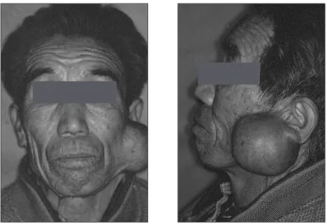
We report a case of the huge pleomorphic adenoma of the left parotid gland in a 67-year-old man who complained the large mass, measured about 10×7×5 cm-sized, in front of the left ear and on the mandibular ascending ramus. The diagnosis was confirmed by the clinical examination, computed tomo- graphic scan, fine needle aspiration, and incisional biopsy. Superficial parotidectomy including the tumor and preservation of the facial nerve using the modified Blair approach was performed. And satisfactory results have been obtained cosmetically and functionally.
Key words : Parotid gland, Huge pleomorphic adenoma, Superficial parotidectomy, Preservation of facial nerve
Abstract
적으로 만족할만한 결과를 얻었기에 이를 문헌고찰과 함께 보고하는 바이다.
Ⅱ. 증례보고
67세 남자 환자가 좌측 전이부, 하악지 및 하악각 부위에 생긴 거대한 종물을 주소로 2001년 12월 24일 전남대학교 병원 구강악안면외과에 내원하였다. 환자는 약 5년 전에 좌 측 전이부 및 하악지 부위의 종물을 처음 인지하였으며, 동 통이나 압박감 등의 자각증상 없이 종물의 크기가 서서히 증가했다고 하였다. 의학적 기왕력, 가족력 및 사회력에서 는 특기할 사항이 없었다.
임상검사 소견에서 좌측 전이부 및 하악지 부위에서 주먹
있었고, 관류(perfusion)는 우측 이하선과 동일하였다(Fig.
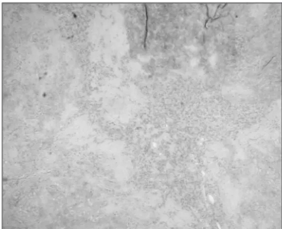
3). 동년 동월 동일 절개생검을 시행한 결과 간질조직 (stroma)과 도관상피 및 근상피세포로 구성된 다형성 선종 으로 진단되었다(Fig. 4). 술전 혈액검사, 뇨검사, 흉부 방 사선검사, 심전도검사 등을 포함한 이화학적 검사 결과 특 기할 만한 소견은 없었다.
2002년 1월 3일 비기관 삽관을 이용한 전신마취 하에 안 면신경을 보존하면서 종물을 포함하여 이하선 천층엽절제 술을 시행하였다. 먼저 변형 Blair 절개법
10)에 따라 피부 및 피부하층까지의 절개를 시행하였다(Fig. 5). 즉, 전이부에 서는 이하선 근막(parotid fascia)의 직상방까지, 경부에서 는 심경근막의 천층(superficial layer of deep cervical fascia)의 직상방까지 절개한 후 그 깊이에서 이하선 전연
Fig. 1. Frontal and left lateral facial photographs at the initial examination showing
a large, hard and well circumscribed mass in the left preauricular area and the
left mandibular ramus and angle region.
을 향해 박리 및 거상하여 표층근건막계(superficial mus- culoaponeurotic system; SMAS) 층의 상방에서 피부피 판을 형성하고 이하선 천층엽과 종물을 노출시켰다(Fig.
6). 연골성 외이도의 전연 및 흉쇄유돌근과 이하선 피막 사 이를 박리하고 대이개신경(great auricular nerve)과 외경 정맥(external jugular vein)을 절단 및 결찰하였다. 이하 선을 전방으로 견인하여 유양돌기(mastoid process)의 첨
(main trunk)를 찾아낸 다음, 안면신경의 분지부에 이를 때까지 이하선 천층엽과 종물을 한 덩어리로 조심스럽게 박 리하였다. 이하선 천층엽과 종물을 하방에 놓여 있는 안면 신경의 주요 5 분지들로부터 분리하고 이하선 도관을 절단 및 결찰한 다음 종물을 포함하여 이하선 천층엽절제술을 시 행하였다(Fig. 7). 종물은 이하선의 천층엽에만 국한되어 있었으므로 이하선 심층엽은 절제하지 않았다. 피부피판을 Fig. 3. Tc-99m pertechnetate salivary scan showing the
diminished uptake of the left parotid gland and the mass area, and the same perfusion of the both parotid glands.
Fig. 4. Microphotograph of the preoperative incisional biopsy showing a well-encapsulated mass and a bipha- sic appearance resulting from the admixture of epitheli- um and stroma. The stromal component shows abun- dant myxoid appearance. Focally, branching appear- ance of neoplastic duct is noted (hematoxyline-eosin stain, ×30).
Fig. 2. CT scans representing a well enveloped and heterogeneously enhancing mass, measured about 10×7×
5 cm-sized, in the superficial layer of the left parotid gland.
별한 합병증은 관찰되지 않고 있다(Fig. 10).
Fig. 5. Intraoperative photograph showing the modified Blair incision marked. The preauricular and neck exten- sion incisions are connected by an incision hidden in the lobular crease of the ear.
Fig. 8. The surgical specimen, measured about 10×7×5 cm-sized mass, and the cut sur- face of the resected tumor showing the well encapsulated mass composed of the brown and white myxoid tissues.
Fig. 6. The huge mass at the superficial lobe of the left parotid gland and the SMAS layer were exposed after the elevation of the skin flap.
Fig. 7. After the superficial parotidectomy including the
mass, the trunk of the facial nerve and its branches
were preserved.
Ⅲ. 고 찰
다형성 선종은 모든 타액선 종양 중에서 가장 흔히 발생하 며, 간엽과 상피의 두 가지 성분으로 구성된 종양이라고 하여 양성 혼합종(benign mixed tumor)이라고 명명되었 다
1,2). 그리고 연골양 및 점액양 요소를 포함한 모든 간엽조 직 성분이 상피세포의 이형성에 의해 생긴 것이라 하여 다 형성 선종으로 명명되었다
2,11). 타액선 종양은 전체 인체 종 양의 1%, 두경부 종양의 약 3%를 차지한다
1). 종양의 발생
5~10%는 악하선에, 1% 미만이 설하선에 발생하며, 소타 액선에서 발생하는 경우는 10~15%라고 하였다. 호발연령 은 일반적으로 40~50대에 가장 많이 발생하고, 성별 차이 는 여자가 남자보다 1.2~2.2배 정도 많이 발생하며, 좌우 측 발생부위의 차이는 없는 것으로 보고되었다
11). 본 증례는 67세 남자 환자의 좌측 이하선에 생긴 다형성 선종으로, 5 년 정도 방치한 결과 거대한 종물로 커지자 본과에 내원하 였다.
이하선의 다형성 선종은 대부분 완만한 성장을 하며 동통 Fig. 10. Frontal and lateral facial photographs at 2 months after the operation. There was no recurrence of the tumor or weakness of the facial nerve. The postoperative scars are relatively invisible, there is excellent cosmesis and the preoperative shape of the auricle has not been altered.
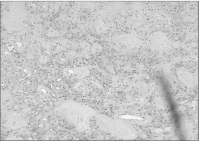
Fig. 9. Microphotograph of the permanent biopsy show-
ing many clusters of the epithelial cells and the myxoid
stroma, which diagnosed histopathologically as pleo-
morphic adenoma (Hematoxyline-Eosin stain, ×30).
×7×5 cm 크기의 잘 경계된 원형의 가동성 종물이 되었 다. 동통이나 압박감 등의 자각증상은 없었으며, 안면신경 마비 소견 또는 악하림프절 촉진시 비대 소견은 관찰되지 않았다. 그리고 내과적 기왕력, 가족력 및 사회력에서 특기 할 사항은 없었다.
조직학적 소견은 상피 및 간엽성분이 모두 있으며 도관세 포, 근상피세포, 중배엽세포들이 혼합되어 있는 것을 관찰 할 수 있고, 기질은 점액성, 섬유성, 연골성, 드물게는 골성 조직까지 단독 혹은 복합적으로 나타난다
2-4). 본 증례에서는 술전 절개생검 소견상 풍부한 점액성 기질조직(stroma)과 도관상피 및 근상피세포로 구성된 소견을 보여 다형성 선종 으로 진단하였다.
이하선 종양의 진단법으로는 세침흡인생검, 전산화단층촬 영, 절개생검, 방사선동위원소 타액선스캔, 자기공명영상촬 영 등이 있다
3-5). 세침흡인생검은 임상적으로 종양이 의심되 는 타액선 종양에서 진단적 및 경제적으로 유용하지만
12), 확 진율이 낮고 치료계획에 결정적인 영향을 줄 수 없으며 종 양세포 전파(seeding)의 위험성이 있으므로 주의해야 한 다
13). 전산화단층촬영은 종양의 존재 및 범위를 정확히 측정 할 수 있고, 주위 조직과의 관계와 침범 여부를 판단할 수 있는 장점이 있다. 종양의 경계가 명확하고 균질의 형태와 낮은 음영농도는 양성종양이나 저도의 악성종양을 의미하 며, 경계가 불분명하고 이질적인 형태와 높은 음영농도를 나타내면 고도의 악성종양이나 재발을 의미한다
14). Pertechnetate(
99mTcO
4-)를 이용한 방사선동위원소 타액선 스캔은 타액선 기능과 병리 양태를 평가하는 진단방법 중의 하나이다
15). 다형성 선종은 Pertechnetate(
99mTcO
4-)를 집 적하지 않기 때문에 섭취(uptake)는 감소하고 냉병소(cold spot)로 나타난다
15). Byrne 등
14)은 자기공명영상촬영은 종 양의 발견과 연조직 병소의 경계를 결정하는데 우수하다고 하였다. 본 증례에서는 전산화단층촬영 소견상 좌측 이하선 천엽에 약 10×7×5 cm 크기의 경계가 뚜렷한 균질의 조 영 증강을 나타내고, 주위조직으로 국소 침윤소견은 관찰되 지 않았으며, 방사선동위원소 타액선스캔 소견상 좌측 이하 선 종물 부위에서 Pertechnetate(
99mTcO
4-)가 감소된 섭취
경의 보존과 함께 전엽절제술을 시행한다
17). 방사선조사는 종양이 심층엽에 존재하거나 절제연이 충분하지 못할 경우 또는 치료후 재발된 종양과 같이 재발의 가능성이 높은 경 우에 시행할 수 있으며, 방사선조사량은 5,000 cGy 정도를 사용한다
18). 본 증례에서는 종물이 천층엽에 한정되고 주위 조직으로의 국소 침윤 소견이 관찰되지 않아, 안면신경을 보존하면서 종물을 포함하여 천층엽 일부를 제거하는 이하 선 천층엽절제술과 안면신경의 보존을 시행하였다.
본 증례에서 사용한 변형 Blair 절개법은 전이개 접근법 과 경부 연장 접근법을 귀의 귓불주름에 가리워지는 절개선 에 의해 서로 연결시킨 절개법으로, 이하선을 포함하는 수 술에 자주 사용되며 악관절 및 하악과두에 대한 수술에도 유용하다
10). 본 증례에서는 종물의 후방경계를 고려하여 통 상적인 후하악접근법보다 절개선을 더욱 후방에 위치시켰 지만, 전이개접근법을 변형시켜 측두부 방향으로 전상방으 로 약 2 cm 정도 연장시켰으므로 시야확보 및 수술부위로 의 접근에 어려움은 없었다.
이하선 종양에서 수술 후에 나타나는 합병증으로는 일시 적 또는 영구적 안면신경마비, Frey 증후군, 혈종, 재발, 타 액선 피부 누공(salivary fistula) 등이 있으며, 이중 가장 많이 발생하는 합병증은 일시적 안면신경마비이다
8,9). 본 증 례에서는 술후 일시적인 좌측 하순의 운동마비를 보인 것 외에는 별다른 합병증이 발생되지 않았다.
다형성 선종 절제술 후의 재발율은 과거에는 높은 편이었
지만, 최근에는 수술기법의 발전으로 인해 감소되고 있다
3-7).
Myssiorek 등
19)은 안면신경 보존과 함께 이하선 천층엽절
제술의 발전으로 인하여 재발율이 3.6% 이하로 줄어 들었
다고 하였다. 재발의 원인에는 여러 가지가 있지만, 가장 주
된 재발의 원인은 불완전한 절제와 술중 종양피막의 손상이
다
20). 그러므로 재발율을 낮추기 위해서는 종양피막의 손상
과 종양의 직접적인 접촉을 최소화하고, 종물과 함께 정상
이하선조직을 포함하여 절제해야 한다
18-20). 본 증례에서는
술후 약 4년 간의 추적관찰 결과 재발 또는 전이 소견은 관
찰되지 않고 있다.
Ⅳ. 요 약
우리는 좌측 전이부 및 하악지 부위에 생긴 10×7×5 cm 크기의 거대한 종물을 주소로 내원한 67세 남자 환자에서 임상검사, 방사선동위원소 타액선스캔, 전산화단층촬영 및 조직생검 등을 통해 좌측 이하선의 다형성 선종으로 진단하 고 변형 Blair 절개법을 통해 안면신경을 보존하면서 이하 선 천층엽절제술을 시행하여 기능적 및 심미적으로 만족스 러운 결과를 얻었다. 이하선 천층엽절제술과 안면신경의 분 리 및 보존을 시행할 경우 단순적출술에 비해 국소 재발율 이 낮고 안면신경이 보존되므로 이하선 다형성 선종의 수술 법으로 적절함을 알 수 있었다.
참고문헌