Copyright © 2021 Korean Stroke Society
This is an Open Access article distributed under the terms of the Creative Commons Attribution Non-Commercial License (http://creativecommons.org/licenses/by-nc/4.0/) which permits unrestricted non-commercial use, distribution, and reproduction in any medium, provided the original work is properly cited.
pISSN: 2287-6391 • eISSN: 2287-6405
Letter to the Editor
128 http://j-stroke.org
Cilostazol and Probucol for Cognitive Decline after Stroke: A Cognitive Outcome Substudy of the
PICASSO Trial
Jae-Sung Lim,
aSun U. Kwon,
bKyung-Ho Yu,
aSungwook Yu,
cJong-Ho Park,
dByung-Chul Lee,
aMi Sun Oh,
aYong-Jae Kim,
eJoung-Ho Rha,
fYang-Ha Hwang,
gJi Sung Lee,
hSung Hyuk Heo,
iSeong Hwan Ahn,
jWoo-Keun Seo,
kJong-Moo Park,
lJu-Hun Lee,
mJee-Hyun Kwon,
nSung-Il Sohn,
oJin-Man Jung,
pHahn Young Kim,
qEung-Gyu Kim,
rJae-Kwan Cha,
sMan-Seok Park,
tHyo Suk Nam,
uHee-Joon Bae,
vDong-Eog Kim,
wJaeseol Park,
aYeonwook Kang,
xJimi Choi,
yJuneyoung Lee
y, za Department of Neurology, Hallym University Sacred Heart Hospital, Hallym Neurological Institute, Hallym University College of Medicine, Anyang, Korea
bDepartment of Neurology, Asan Medical Center, University of Ulsan College of Medicine, Seoul, Korea
cDepartment of Neurology, Korea University Anam Hospital, Korea University College of Medicine, Seoul, Korea
dDepartment of Neurology, Myongji Hospital, Hanyang University College of Medicine, Goyang, Korea
eDepartment of Neurology, Eunpyeong St. Mary’s Hospital, College of Medicine, The Catholic University of Korea, Seoul, Korea
fDepartment of Neurology, Inha University Hospital, Incheon, Korea
gDepartment of Neurology, Kyungpook National University Hospital, School of Medicine, Kyungpook National University, Daegu, Korea
hClinical Research Center, Asan Institute for Life Sciences, Asan Medical Center, Seoul, Korea
iDepartment of Neurology, Kyung Hee University Medical Center, Seoul, Korea
jDepartment of Neurology, Chosun University Hospital, Gwangju, Korea
kDepartment of Neurology, Samsung Medical Center, Sungkyunkwan University School of Medicine, Seoul, Korea
lDepartment of Neurology, Nowon Eulji Medical Center, Eulji University, Seoul, Korea
mDepartment of Neurology, Hallym University Kangdong Sacred Heart Hospital, Hallym University College of Medicine, Seoul, Korea
nDepartment of Neurology, Ulsan University Hospital, University of Ulsan College of Medicine, Ulsan, Korea
oDepartment of Neurology, Keimyung University Dongsan Hospital, Keimyung University School of Medicine, Daegu, Korea
pDepartment of Neurology, Korea University Ansan Hospital, Ansan, Korea
qDepartment of Neurology, Konkuk University School of Medicine, Seoul, Korea
rDepartment of Neurology, Inje University Busan Paik Hospital, Inje University College of Medicine, Busan, Korea
sDepartment of Neurology, Dong-A University Hospital, Busan, Korea
tDepartment of Neurology, Chonnam National University Hospital, Chonnam National University Medical School, Gwangju, Korea
uDepartment of Neurology, Severance Hospital, Yonsei University College of Medicine, Seoul, Korea
vDepartment of Neurology, Seoul National University Bundang Hospital, Seoul National University College of Medicine, Seongnam, Korea
wDepartment of Neurology, Dongguk University Ilsan Hospital, Goyang, Korea
xDepartment of Psychology, Hallym University, Chuncheon, Korea
yDepartment of Biostatistics, Korea University College of Medicine, Seoul, Korea
zBK21 FOUR R&E Center for Learning Health Systems, Korea University, Seoul, Korea
Journal of Stroke 2021;23(1):128-131 https://doi.org/10.5853/jos.2020.03650
Dear Sir:
Previous clinical trials to prevent post-stroke cognitive impair- ment, such as Prevention Regimen for Effectively Avoiding Second Strokes (PRoFESS) and Secondary Prevention of Small Subcortical Strokes (SPS3), failed to show clinically meaningful results.1,2 There is an evidence that cilostazol, a phosphodies-
terase-3 inhibitor, could suppress cognitive decline in patients with dementia,3 and decrease amyloid beta accumulation.4 Probucol, a cholesteryl ester transfer protein activator with lip- id-lowering and anti-oxidative effects, has a beneficial effect on cognition by inhibiting amyloid beta-induced hippocampal synaptic impairment.5 Thus, we aimed to determine the effica- cy of cilostazol and probucol for preventing poststroke cogni-
Vol. 23 / No. 1 / January 2021
tive decline in patients with multiple cerebral microbleeds (CMBs) or a history of prior intracerebral hemorrhage (ICH); a population that is expected to have a high risk for future cog- nitive decline.
PreventIon of CArdiovascular events in iSchemic Stroke pa- tients with high risk of cerebral hemOrrhage for reducing COG- nitive decline (PICASSO-COG) is a predetermined substudy of the PICASSO trial, which is a randomized double-blinded pla- cebo-controlled trial with a 2×2 factorial design: cilostazol versus aspirin, and probucol versus no probucol.6 The design and analysis plan have been previously reported.7 The key in- clusion criteria were non-cardioembolic ischemic stroke or transient ischemia attack and previous ICH or multiple CMBs on gradient echo imaging. Cognitive function was assessed us- ing the Mini-Mental State Examination (MMSE) and the Mon-
treal Cognitive Assessment (MoCA) at randomization and at 4, 13, 25, 37, and 49 months after randomization. The cognitive function at the second visit (4 months after randomization) in patients who were randomized within 3 months after stroke was set as the baseline function, while the cognitive function at the first visit (1 month after enrollment) was set as the baseline for those randomized beyond 90 days after stroke. The baseline cognitive assessment was therefore conducted be- tween 4 and 7 months after stroke onset in all participants eli- gible for the PICASSO-COG substudy. The primary outcome was a change in MMSE score, and a restricted maximum likelihood- based mixed effects model with repeated measurements was used. The efficacy of each treatment was analyzed separately because the interaction effect between the antiplatelets and lipid-lowering treatment was not significant. Detailed informa-
Changes from baseline to follow-up (MMSE)
Baseline 1st 2nd
Visits
3rd 4th
0.5
Cilostazol Aspirin 0
-0.5 -1 -1.5 -2
-2.5 Changes from baseline to follow-up (MoCA)
Baseline 1st 2nd
Visits
3rd 4th
1
Cilostazol Aspirin 0.5
0 -0.5 -1 -1.5 -2
Cilostazol Aspirin
451 441
449 439
301 292
181 180
66 72
Cilostazol Aspirin
447 430
444 427
297 285
178 176
64 68
Changes from baseline to follow-up (MMSE)
Baseline 1st 2nd
Visits
3rd 4th
0.5
Probucol No probucol 0
-0.5 -1 -1.5 -2 -2.5
Probucol No probucol
459 433
456 432
303 290
197 164
82 56
Changes from baseline to follow-up (MoCA)
Baseline 1st 2nd
Visits
3rd 4th
1
Probucol No probucol 0.5
0 -0.5 -1 -1.5 -2
Probucol No probucol
452 425
447 424
298 284
192 162
77 55
A
C
B
D
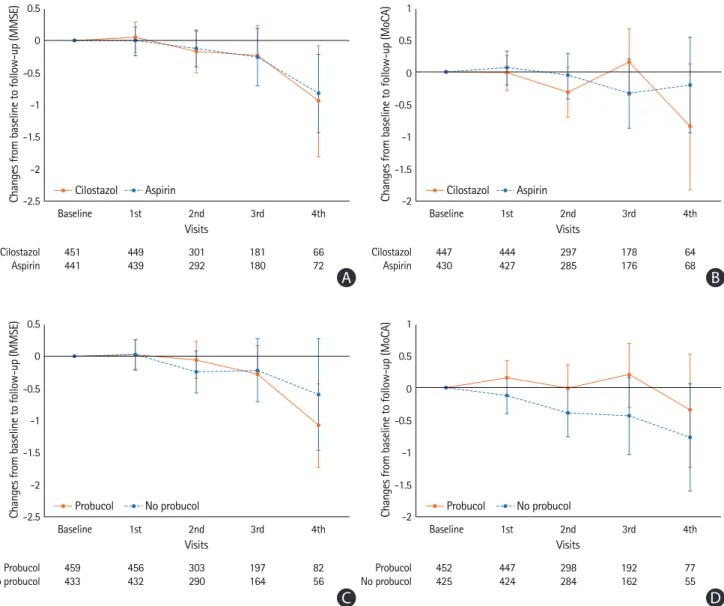
Figure 1. Mean changes in cognitive scores from baseline to each follow-up in (A, B) cilostazol vs. aspirin and (C, D) probucol vs. no probucol groups. (A, C) Mini-Mental State Examination (MMSE) and (B, D) Montreal Cognitive Assessment (MoCA).
Lim et al. Cilostazol and Probucol for Post-Stroke Cognition
https://doi.org/10.5853//jos.2020.03650
130 http://j-stroke.org
tion on the analyses, including sensitivity and subgroup analy- ses, are presented in the Supplementary methods.
As shown in Supplementary Figure 1, among 1,382 subjects, 1,240 completed cognitive evaluations at randomization and 892 subjects (877 for the MoCA) were finally included (Supple- mentary Table 1).7 The baseline characteristics were not signifi- cantly different between the treatment groups, except the pro- portion of those with baseline MMSE ≤24 (Supplementary Ta- ble 2).1 Cilostazol did not show any significant differences in preventing cognitive decline in comparison with aspirin (Figure 1 and Supplementary Table 3). In the subgroup analysis ac- cording to the baseline MMSE score, the decrease in the MMSE score in the aspirin group of those with baseline MMSE ≤24 was more pronounced than that in the cilostazol group al- though the treatment effect was not significant (Supplemen- tary Table 4). In the propensity score-matched subsets consid- ering the baseline differences in the proportions of those with baseline MMSE ≤24, the cilostazol group showed a favorable outcome in those with mild to moderate white matter hyperin- tensities (WMHs) (Supplementary Table 5). Otherwise, no sig- nificant results were found in the subgroups and sensitivity analysis. Probucol treatment did not show any beneficial effect in the primary outcome using the MMSE. When analyzed ac- cording to the MoCA scores, probucol showed a favorable ef- fect in preventing cognitive decline compared with the no pro- bucol group (Supplementary Table 3). This effect was also ob- served in the subgroups without diabetes mellitus, with con- comitant lipid-lowering agents, with baseline MMSE >24, and without severe WMH (Supplementary Table 4).
Longitudinal cognitive profiles of the study population might explain why this trial failed to prove the hypothesis. The demo- graphics of the study subjects were comparable to those of the SPS3 trial.2 However, 69.3% of the PICASSO-COG subjects had moderate or severe WMH, while half of the subjects in the SPS3 had none or mild WMH. In this distinctive population, the magnitude of observed cognitive change was smaller than what we had expected.7 There are several reasons to consider.
It has been reported that cognitive decline in patients with moderate to severe WMH was mainly observed in processing speed and executive function.8 In the SPS3 trial conducted in patients with lacunar infarction, verbal fluency was mainly im- paired in addition to episodic memory.9 Memory dysfunction has also been reported to be affected by actually mediating executive dysfunction.8 Therefore, the MMSE was not sensitive enough to capture these long-term cognitive changes. The MoCA has been reported to be more sensitive to the stroke population than the MMSE; however, the MoCA total score seems inadequate to quantify changes over a 2-year study pe-
riod. For subsequent clinical trials, neuropsychological tests that more sensitively assess changes over time in the target population, such as fluency, trail-making, and the Stroop test should be adopted. In another aspect, the active risk factor control in the trial setting might prevent the cognitive deterio- ration of study subjects, including the control group, and made it difficult to verify the effectiveness of the trial drug. This can be conceived from the findings from the previous trials for vas- cular cognitive impairment, which showed stable cognitive trajectories in placebo arms.10 Lastly, it is possible that the het- erogeneity of WMH might have been affected.11 The theoreti- cally hypothesized cognitive decline might not be actually ob- served in patients with WMH of causes other than ischemic origin. However, since the subjects of this trial had ischemic stroke based on the inclusion criteria and had preceding ICH/
multiple CMBs, the proportion of these patients is not expected to be high.
We predetermined the time window of baseline cognitive evaluation between 4 and 7 months after entry event to mini- mize the effects of acute stroke on cognitive function.12 The intervals between index-stroke and baseline evaluations were 1 month in the PRoFESS trial and 74 to 76 days in the SPS3 trial.1,2 If we were to include the spontaneous cognitive recov- ery after stroke in our analysis, the effects of the study medi- cation could be exaggerated or underestimated.
As a limitation, the current study population did not seem to fulfill the criteria of reliable cognitive decline, and the trial needed much longer follow-up to show a significant change in the MMSE score.7 In addition, a treatment effect could have occurred between the index-stroke and the baseline assess- ment. Since we limited our analysis to those who underwent baseline evaluations for 4 to 7 months after index-stroke, we could not address this possibility in our analysis.
To the best of our knowledge, this is the first clinical trial comparing the efficacy of aspirin, cilostazol, and probucol in preventing poststroke cognitive decline. Cilostazol and probu- col did not show any significant differences compared to aspi- rin and no probucol. However, when patients were assessed by the MoCA, probucol reduced cognitive decline after stroke.
Supplementary materials
Supplementary materials related to this article can be found online at https://doi.org/10.5853/jos.2020.03650.
References
1. Diener HC, Sacco RL, Yusuf S, Cotton D, Ounpuu S, Lawton
Vol. 23 / No. 1 / January 2021
WA, et al. Effects of aspirin plus extended-release dipyridam- ole versus clopidogrel and telmisartan on disability and cog- nitive function after recurrent stroke in patients with isch- aemic stroke in the Prevention Regimen for Effectively Avoiding Second Strokes (PRoFESS) trial: a double-blind, ac- tive and placebo-controlled study. Lancet Neurol 2008;7:
875-884.
2. Pearce LA, McClure LA, Anderson DC, Jacova C, Sharma M, Hart RG, et al. Effects of long-term blood pressure lowering and dual antiplatelet treatment on cognitive function in pa- tients with recent lacunar stroke: a secondary analysis from the SPS3 randomised trial. Lancet Neurol 2014;13:1177- 1185.
3. Ihara M, Nishino M, Taguchi A, Yamamoto Y, Hattori Y, Saito S, et al. Cilostazol add-on therapy in patients with mild de- mentia receiving donepezil: a retrospective study. PLoS One 2014;9:e89516.
4. Park SH, Kim JH, Bae SS, Hong KW, Lee DS, Leem JY, et al.
Protective effect of the phosphodiesterase III inhibitor cilo- stazol on amyloid β-induced cognitive deficits associated with decreased amyloid β accumulation. Biochem Biophys Res Commun 2011;408:602-608.
5. Santos DB, Peres KC, Ribeiro RP, Colle D, dos Santos AA, Moreira EL, et al. Probucol, a lipid-lowering drug, prevents cognitive and hippocampal synaptic impairments induced by amyloid β peptide in mice. Exp Neurol 2012;233:767-775.
6. Kim BJ, Lee EJ, Kwon SU, Park JH, Kim YJ, Hong KS, et al.
Prevention of cardiovascular events in Asian patients with ischaemic stroke at high risk of cerebral haemorrhage (PI- CASSO): a multicentre, randomised controlled trial. Lancet Neurol 2018;17:509-518.
7. Yu KH, Hong KS, Oh MS, Lee J, Lee JS, Kwon SU, et al. Design and rationale for a cognitive outcome substudy in ischemic stroke patients with high risk of cerebral hemorrhage. J Stroke Cerebrovasc Dis 2016;25:2061-2066.
8. Jokinen H, Kalska H, Mäntylä R, Ylikoski R, Hietanen M, Po- hjasvaara T, et al. White matter hyperintensities as a predic- tor of neuropsychological deficits post-stroke. J Neurol Neu-
rosurg Psychiatry 2005;76:1229-1233.
9. Jacova C, Pearce LA, Costello R, McClure LA, Holliday SL, Hart RG, et al. Cognitive impairment in lacunar strokes: the SPS3 trial. Ann Neurol 2012;72:351-362.
10. Black S, Román GC, Geldmacher DS, Salloway S, Hecker J, Burns A, et al. Efficacy and tolerability of donepezil in vascu- lar dementia: positive results of a 24-week, multicenter, in- ternational, randomized, placebo-controlled clinical trial.
Stroke 2003;34:2323-2330.
11. Pantoni L. Cerebral small vessel disease: from pathogenesis and clinical characteristics to therapeutic challenges. Lancet Neurol 2010;9:689-701.
12. Sachdev PS, Lo JW, Crawford JD, Mellon L, Hickey A, Wil- liams D, et al. STROKOG (stroke and cognition consortium):
an international consortium to examine the epidemiology, diagnosis, and treatment of neurocognitive disorders in rela- tion to cerebrovascular disease. Alzheimers Dement (Amst) 2016;7:11-23.
Correspondence: Kyung-Ho Yu
Department of Neurology, Hallym University Sacred Heart Hospital, Hallym Neurological Institute, Hallym University College of Medicine, 22 Gwanpyeong- ro 170beon-gil, Dongan-gu, Anyang 14068, Korea
Tel: +82-31-380-3740 Fax: +82-31-381-9474 E-mail: [email protected] https://orcid.org/0000-0002-8997-5626 Co-correspondence: Sun U. Kwon
Department of Neurology, Asan Medical Center, University of Ulsan College of Medicine, 88 Olympic-ro 43-gil, Songpa-gu, Seoul 05505, Korea
Tel: +82-2-3010-3960 Fax: +82-2474-4691 E-mail: [email protected] https://orcid.org/0000-0001-8302-331X Received: August 31, 2020
Revised: November 14, 2020 Accepted: November 18, 2020
This work was supported by Korea Otsuka Pharmaceutical Company.
Sun U. Kwon declares grants from Korea Otsuka Pharmaceutical Company. All other authors declare no competing interests.
Vol. 23 / No. 1 / January 2021
https://doi.org/10.5853//jos.2020.03650 http://j-stroke.org 1
Supplementary methods
The PreventIon of CArdiovascular events in iSchemic Stroke patients with high risk of cerebral hemOrrhage for reducing COGnitive decline (PICASSO-COG) substudy was conducted only in South Korea (59 centers) because the cognitive assess- ment tools had not been validated by cross-cultural studies in each language.
The primary outcome was the change in Mini-Mental State Examination (MMSE) score over time from baseline in an in- tention-to-treat population. A restricted maximum likelihood- based mixed effects model with repeated measurements (MMRM) was used to compare cognitive changes over time between groups. The model included the fixed categorical ef- fects of treatment group and sex as well as fixed continuous covariates of the patient’s age, duration of education, number of visits, baseline cognitive scores, and the National Institutes of Health Stroke Scale (NIHSS) score. The effect of the study sites was adjusted as a random factor in the model. An un- structured covariance structure, common to the treatments, was used to model the within-subject correlation. Although this study had a 2×2 factorial design, the efficacy of each treatment was analyzed separately because the interaction ef- fect between the antiplatelets and lipid-lowering treatment was not significant.
As cognitive impairment is an independent risk factor for at- trition in a longitudinal study, we performed sensitivity analysis to examine its influence on cognitive outcome. Sensitivity analyses included the following: (1) a restricted maximum like-
lihood-based MMRM analysis with further adjustment for the participant’s drop-out status during the trial period as well as treatment status at the previous visit, which is defined as the patient missing the cognitive evaluation in the scheduled visit before the current visit, in the model; (2) MMRM analyses of participants’ cognitive evaluation at baseline and follow-up visits at 13, 25, 37, and 49 months; (3) MMRM analyses of participants who completed all scheduled visits during the fol- lowing periods: baseline to 13 months, baseline to 25 months, baseline to 37 months, and baseline to 49 months; and (4) MMRM analyses of participants with ischemic stroke as an en- try event excluding transient ischemic attack.
The primary outcome between the comparative arms was compared for the following subgroups: diabetes versus non-di- abetes, mild to moderate (Fazekas grade 0–2) versus severe (Fazekas grade 3) white matter hyperintensities on magnetic resonance imaging, baseline MMSE ≤24 versus >24, and con- comitant use of statin versus non-use. The same analyses as performed in the sensitivity and subgroup analyses were re- conducted in the propensity score matched subsets, which were constructed using the variables of subject age, sex, edu- cational years, baseline cognitive score, and baseline NIHSS score to overcome the differences between the treatment groups arising from non-random missing in this substudy.
The longitudinal change in MoCA was also evaluated using the same statistical methods. A two-sided P-value of 0.05 was used to indicate statistical significance. All statistical analyses were performed using SAS version 9.3 (SAS Institute, Cary, NC, USA).
Lim et al. Cilostazol and Probucol for Post-Stroke Cognition
Supplementary Table 1. Baseline characteristics of the included and excluded subjects
Characteristic
PICASSO-COG study MMSE analysis MoCA analysis
Included
(n=1,240) Excluded
(n=142) P Included
(n=892) Excluded
(n=348) P Included
(n=877) Excluded
(n=363) P
Age (yr) 65.8±10.8 68.3±10.0 0.01 64.9±10.8 68.2±10.5 <0.01 64.8±10.8 68.3±10.4 <0.01
Female sex 480 (38.7) 53 (37.3) 0.75 327 (36.7) 153 (44.0) 0.02 318 (36.3) 162 (44.6) 0.01
Education (yr) 9 (6–12) 9 (6–12) 0.41 9 (6–12) 6 (5–12) <0.01 9 (6–12) 6 (5–12) <0.01
Hypertension 1,091 (88.0) 133 (93.7) 0.04 796 (89.2) 295 (84.8) 0.03 783 (89.3) 308 (84.9) 0.03
Diabetes 389 (31.4) 53 (37.3) 0.15 276 (30.9) 113 (32.5) 0.60 271 (30.9) 118 (32.5) 0.58
Hyperlipidemia 511 (41.2) 54 (38.0) 0.47 373 (41.8) 138 (40.0) 0.49 369 (42.1) 142 (39.1) 0.34
Use of lipid-lowering agent* 969 (78.2) 85 (59.9) <0.01 695 (77.9) 274 (78.7) 0.75 686 (78.2) 283 (78.0) 0.92
Coronary artery disease 59 (4.8) 8 (5.6) 0.65 37 (4.2) 22 (6.3) 0.11 37 (4.2) 22 (6.1) 0.17
Smoking 546 (44.0) 65 (45.8) 0.69 407 (45.6) 139 (39.9) 0.07 404 (46.1) 142 (39.1) 0.02
Index event 0.01 0.18 0.05
Ischemic stroke 1,175 (94.8) 142 (100.0) 850 (95.3) 325 (93.4) 838 (95.6) 337 (92.8)
Transient ischemic attack 65 (5.2) 0 (0.0) 42 (4.7) 23 (6.6) 39 (4.5) 26 (7.2)
Baseline NIHSS 1 (0–3) 3 (1-5) <0.01 1 (0–3) 2 (1–4) <0.01 1 (0–3) 2 (1–4) <0.01
Baseline MMSE 26 (21–28) - - 26 (23–29) 24 (17–27) <0.01 26 (23–29) 24 (17–27) <0.01
24 or less 492 (39.7) - 313 (35.1) 179 (51.4) <0.01 303 (34.6) 189 (52.1) <0.01
>24 748 (60.3) - 579 (64.9) 169 (48.6) 574 (65.4) 174 (47.9)
Baseline MoCA 20 (14–24) - - 20 (16–24) 17 (10–22) <0.01 20 (16–24) 17 (10–22) <0.01
Treatment
Cilostazol 618 (49.8) 71 (50.0) 0.97 451 (50.6) 167 (48.0) 0.42 447 (51.0) 171 (47.1) 0.22
Probucol 622 (50.2) 69 (48.6) 0.72 459 (51.5) 163 (46.8) 0.14 452 (51.5) 170 (46.8) 0.13
SBP (mm Hg) 135.4±18.4 133.7±19.1 0.30 135.4±18.6 135.5±17.8 0.96 135.3±18.7 135.6±17.6 0.79
DBP (mm Hg) 80.1±11.8 80.9±12.2 0.43 80.1±11.8 80.2±11.8 0.88 80.1±11.9 80.2±11.6 0.85
BP readings 7 (4–13) 10 (3–18) 0.01 9 (6–13) 2 (1–4) <0.01 9 (6–13) 2 (1–4) <0.01
Follow-up periods (yr) 1.9 (1.0–3.0) 2.7 (0.6–4.5) <0.01 2.1 (1.3–3.0) 0.5 (0.1–1.1) <0.01 2.1 (1.3–3.0) 0.6 (0.1–1.1) <0.01 Severe WMH 324 (27.1) 30 (22.1) 0.21 210 (24.5) 114 (33.5) <0.01 206 (24.4) 118 (33.2) <0.01 Outcome events
Recurrent stroke† 102 (8.2) 9 (6.3) 0.43 44 (4.9) 58 (16.7) <0.01 41 (4.7) 61 (16.8) <0.01
Ischemic 80 (6.5) 8 (5.6) 0.71 31 (3.5) 49 (14.1) <0.01 30 (3.4) 50 (13.8) <0.01
Hemorrhagic 23 (1.9) 1 (0.7) 0.50 14 (1.6) 9 (2.6) 0.23 12 (1.4) 11 (3.0) 0.048
Myocardial infarction 8 (0.7) 1 (0.7) 0.99 3 (0.3) 5 (1.4) 0.04 3 (0.3) 5 (1.4) 0.04
Death 39 (3.2) 9 (6.3) 0.08 17 (1.9) 22 (6.3) <0.01 17 (1.9) 22 (6.1) <0.01
Values are presented as mean±standard deviation, number (%), or median (interquartile range). Severe white matter hyperintensities were defined as Fazekas grade 3.
PICASSO-COG, PreventIon of CArdiovascular events in iSchemic Stroke patients with high risk of cerebral hemOrrhage for reducing COGnitive decline; MMSE, Mini-Mental State Examination; MoCA, Montreal Cognitive Assessment; NIHSS, National Institutes of Health Stroke Scale; SBP, systolic blood pressure; DBP, diastolic blood pressure; BP, blood pressure; WMH, white matter hyperintensity.
*Prior to randomization; †One subject had both ischemic and hemorrhagic stroke and was counted as a duplicate.
Vol. 23 / No. 1 / January 2021
https://doi.org/10.5853//jos.2020.03650 http://j-stroke.org 3
Supplementary Table 2. Baseline characteristics of the study subjects
Characteristic Antiplatelet treatment Lipid-lowering treatment
Cilostazol (n=451) Aspirin (n=441) Probucol (n=459) No probucol (n=433)
Age (yr) 65.0±10.8 64.8±10.8 64.7±10.8 65.2±10.8
Male sex 283 (62.7) 282 (63.9) 290 (63.2) 275 (63.5)
Education (yr) 9 (6–12) 9 (6–12) 9 (6–12) 9 (6–12)
Entry event
Ischemic stroke 432 (95.8) 418 (94.8) 441 (96.1) 409 (94.5)
Transient ischemic attack 19 (4.2) 23 (5.2) 18 (3.9) 24 (5.5)
Index of high risk of ICH
Prior history of ICH 69 (15.3) 76 (17.2) 72 (15.7) 73 (16.9)
Imaging findings of ICH without clinical history 83 (18.4) 77 (17.5) 87 (18.9) 73 (16.9)
Multiple microbleeds (≥2) 299 (66.3) 288 (65.3) 300 (65.4) 287 (66.3)
Time-to-randomization since entry event (day) 18 (9–41) 18 (9–42) 18 (9–43) 18 (8–41)
≤10 130 (28.8) 132 (29.9) 128 (27.9) 134 (30.9)
11–30 177 (39.2) 173 (39.2) 180 (39.2) 170 (39.3)
31–90 93 (20.6) 93 (21.1) 95 (20.7) 91 (21.0)
>90 51 (11.3) 43 (9.8) 56 (12.2) 38 (8.8)
Baseline NIHSS 1 (0–3) 1 (0–3) 1 (0–3) 1 (0–3)
Baseline MMSE 26 (22–28) 27 (23–29) 26 (22–28) 26 (23–29)
Baseline MMSE ≤24 173 (38.4) 140 (31.7) 160 (34.9) 153 (35.3)
Baseline MoCA 20 (15–24) 21 (16–24) 20 (15.5–24) 21 (16–24)
Time-to-baseline MMSE since entry event (day) 136 (125–148) 135 (127–151) 136 (126–151) 135 (127–148.5) Risk factors
Hypertension 402 (89.1) 394 (89.3) 411 (89.5) 385 (88.9)
Diabetes mellitus 134 (29.7) 142 (32.2) 136 (29.6) 140 (32.3)
Dyslipidemia 183 (40.6) 190 (43.1) 206 (44.9) 167 (38.6)
Current smoking 93 (20.6) 102 (23.1) 103 (22.4) 92 (21.2)
Coronary artery disease 15 (3.3) 22 (5.0) 22 (4.8) 15 (3.5)
Lipids (mg/dL)
Total cholesterol 165.7±39.2 169.0±41.1 170.5±40.8 164.0±39.3
LDL-C 101.0±36.0 102.7±35.4 104.7±36.4 98.7±34.7
HDL-C 45.2±11.7 45.9±12.1 45.5±12.1 45.5±11.7
Fazekas score for WMH
0 0 (0.0) 0 (0.0) 0 (0.0) 0 (0.0)
1 122 (27.1) 141 (32.0) 130 (29.1) 133 (32.4)
2 194 (43.0) 191 (43.3) 213 (47.6) 172 (41.8)
3 112 (24.8) 98 (22.2) 104 (23.3) 106 (25.8)
Concomitant therapy
Aspirin (after randomization) 218 (47.5) 223 (51.5)
Cilostazol (after randomization) 241 (52.5) 210 (48.5)
Probucol 241 (53.4) 218 (49.4)
Other lipid-lowering agents 355 (79.1) 353 (80.2) 360 (78.8) 348 (80.6)
Values are presented as mean±standard deviation, number (%), or median (interquartile range).
ICH, intracerebral hemorrhage; NIHSS, National Institutes of Health Stroke Scale; MMSE, Mini-Mental State Examination; MoCA, Montreal Cognitive Assess- ment; LDL-C, low-density lipoprotein cholesterol; HDL-C, high-density lipoprotein cholesterol; WMH, white matter hyperintensity.
Lim et al. Cilostazol and Probucol for Post-Stroke Cognition
Supplementary Table 3. MMRM analysis in cilostazol vs. aspirin and probucol vs. no probucol groups
Time point
Cilostazol vs. aspirin Probucol vs. no probucol
MMSE MoCA MMSE MoCA
Cilostazol
(n=451) Aspirin
(n=441) P Cilostazol
(n=447) Aspirin
(n=430) P Probucol
(n=459) No probucol
(n=433) P Probucol
(n=452) No probucol
(n=425) P
Baseline 24.76±4.48 25.07±4.89 0.81* 18.98±6.16 19.67±6.31 0.31* 24.93±4.73 24.90±4.65 0.57* 19.23±6.15 19.42±6.33 0.01*
1st Follow-up 24.85±4.65 25.10±4.95 <0.01† 19.02±6.37 19.78±6.62 0.045†25.00±5.03 24.94±4.55 <0.01† 19.44±6.43 19.34±6.58 0.03† 2nd Follow-up 24.61±5.12 25.13±4.92 18.75±6.66 19.91±6.57 24.91±5.03 24.83±5.03 19.31±6.81 19.33±6.46 3rd Follow-up 24.56±5.29 24.82±5.35 19.01±6.80 19.43±6.96 24.81±5.17 24.54±5.48 19.67±6.72 18.69±7.04 4th Follow-up 23.97±5.51 25.15±5.41 18.69±7.34 20.62±6.98 24.41±5.69 24.84±5.17 19.91±7.48 19.36±6.82 Values are presented as mean±standard deviation. P-values for treatment-by-time interaction were not significant in any analysis.
MMRM, maximum likelihood-based mixed effects model with repeated measurements; MMSE, Mini-Mental State Examination; MoCA, Montreal Cognitive Assessment.
*P-value for MMRM for treatment effect; †P-value for MMRM for the time effect.
Vol. 23 / No. 1 / January 2021
https://doi.org/10.5853//jos.2020.03650 http://j-stroke.org 5
Supplementary Table 4. Subgroup analysis of the cilosatzol/aspirin groups and probucol/no probucol groups
Variable Baseline 1st Follow-up 2nd Follow-up 3rd Follow-up 4th Follow-up
Cilosatzol/aspirin MMSE scores Baseline MMSE ≤24 (n=313)
Cilostazol (n=173) 20.10±3.56 20.70±4.52 19.84±5.03 19.94±5.26 19.40±5.21
Aspirin (n=140) 19.23±4.41 19.73±5.10 20.01±5.44 19.67±5.90 16.54±6.15
P 0.83* 0.01†
Baseline MMSE >24 (n=579)
Cilostazol (n=278) 27.66±1.65 27.41±2.30 27.28±2.62 27.40±2.64 26.76±3.46
Aspirin (n=301) 27.79±1.65 27.56±2.14 27.34±2.39 27.26±2.67 27.26±2.16
P 0.82* 0.01†
Probucol/no probucol MoCA scores Diabetes mellitus (n=271)
Probucol (n=135) 18.70±6.29 18.68±6.61 18.17±6.80 18.16±6.85 18.59±8.46
No probucol (n=136) 18.82±6.06 18.71±6.14 18.52±6.00 17.72±6.28 18.05±6.76
P 0.16* 0.18†
No diabetes mellitus (n=606)
Probucol (n=317) 19.45±6.09 19.77±6.34 19.77±6.78 20.19±6.61 20.28±7.22
No probucol (n=289) 19.70±6.45 19.63±6.77 19.72±6.65 19.21±7.40 20.24±6.82
P 0.02* 0.01†
Concomitant lipid-lowering agents (n=699)
Probucol (n=355) 19.46±6.08 19.73±6.28 19.81±6.60 20.06±6.51 20.69±7.19
No probucol (n=344) 19.47±6.35 19.44±6.62 19.59±6.38 19.23±6.70 19.45±6.73
P 0.08* 0.38†
No concomitant lipid-lowering agents (n=175)
Probucol (n=95) 18.35±6.41 18.44±6.94 17.71±7.30 18.58±7.28 17.06±8.23
No probucol (n=80) 19.21±6.34 18.84±6.46 18.30±6.75 16.62±7.95 19.13±7.30
P 0.03* 0.03†
Baseline MMSE ≤24 (n=303)
Probucol (n=155) 12.94±4.90 12.90±5.11 12.60±5.79 13.21±5.82 11.92±6.41
No probucol (n=148) 12.86±5.10 12.78±5.13 12.17±4.89 11.61±5.64 8.55±5.43
P 0.18* <0.01†
Baseline MMSE >24 (n=574)
Probucol (n=297) 22.51±3.69 22.78±4.03 22.75±4.26 23.14±4.10 23.75±4.22
No probucol (n=277) 22.92±3.52 22.82±4.17 22.55±4.05 22.42±4.30 22.07±3.77
P 0.01* 0.88†
Mild to moderate white matter hyperintensities (n=637)
Probucol (n=338) 20.37±5.51 20.75±5.86 20.70±6.10 20.97±5.95 21.42±6.42
No probucol (n=299) 20.48±5.91 20.43±6.15 20.47±5.76 20.16±6.10 20.85±5.28
P <0.01* 0.40†
Severe white matter hyperintensities (n=206)
Probucol (n=102) 15.49±6.58 15.18±6.44 14.69±7.06 15.35±7.41 14.20±8.85
No probucol (n=104) 16.35±6.61 16.03±6.82 15.99±7.24 14.05±7.87 15.30±9.29
P 0.79* 0.01†
Values are presented as mean±standard deviation. Mild to moderate white matter hyperintensities were defined as Fazekas grade 1 or 2, and severe white matter hyperintensities as Fazekas grade 3. P-values for treatment by time interactions were not significant for any analysis.
MMSE, Mini-Mental State Examination; MoCA, Montreal Cognitive Assessment.
*P-value by MMRM for the treatment effect; †P-value by MMRM for the time effect.
Lim et al. Cilostazol and Probucol for Post-Stroke Cognition
Supplementary Table 5. Comparisons of MMSE scores between cilostazol and aspirin group according to severity of white matter changes in propensity score matched subsets
MMSE scores Mild to moderate white matter hyperintensities (n=574) Severe white matter hyperintensities (n=148)
Cilostazol (n=287) Aspirin (n=287) P Cilostazol (n=74) Aspirin (n=74) P
Baseline 25.85±3.79 25.85±4.25 0.02* 22.66±4.69 22.27±6.01 0.12*
1st Follow-up 26.14±3.59 25.99±4.36 0.26† 21.95±5.36 22.01±5.72 <0.01†
2nd Follow-up 26.08±3.99 25.83±4.05 21.79±5.85 21.74±6.81
3rd Follow-up 26.43±3.30 25.47±4.63 20.03±6.53 21.47±7.16
4th Follow-up 26.30±3.06 25.75±5.18 16.90±6.87 20.73±7.04
Values are presented as mean±standard deviation. The propensity score was calculated using variables, including the participant’s age, sex, duration of educa- tion, baseline National Institutes of Health Stroke Scale (NIHSS) score, baseline MMSE score, baseline Montreal Cognitive Assessment (MoCA) score, coronary artery disease (yes/no), hypertension (yes/no), systolic blood pressure, and pattern of measurement within each white matter hyperintensity. Mild to moderate white matter hyperintensities were defined as Fazekas grade 1 or 2, and severe white matter hyperintensities as Fazekas grade 3. P-values for treatment by time interactions were not significant for any analysis.
MMSE, Mini-Mental State Examination.
*P-value by MMRM for the treatment effect; †P-value by MMRM for the time effect.
Vol. 23 / No. 1 / January 2021
https://doi.org/10.5853//jos.2020.03650 http://j-stroke.org 7
Supplementary Figure 1. Flow diagram of subject enrollment. PICASSO-COG, PreventIon of CArdiovascular events in iSchemic Stroke patients with high risk of cerebral hemOrrhage for reducing COGnitive decline; MMSE, Mini-Mental State Examination.
1,240 PICASSO-COG participants
316 Assigned to cilostazol plus probucol
241 Analyzed 218 Analyzed 210 Analyzed 223 Analyzed
306 Assigned to aspirin plus probucol
46 Beyond predefined
baseline MMSE window 51 Beyond predefined
baseline MMSE window 54 Beyond predefined
baseline MMSE window 64 Beyond predefined baseline MMSE window 4 Withdrew consent
24 Without follow-up MMSE 1 Improper cognitive tests
3 Withdrew consent 32 Without follow-up MMSE
2 Improper cognitive tests
0 Withdrew consent 34 Without follow-up MMSE
4 Improper cognitive tests
4 Withdrew consent 23 Without follow-up MMSE
2 Improper cognitive tests 316 Assigned to
aspirin 302 Assigned to
cilostazol