Effects of dental acid etchants in oral epithelial cells
7
0
0
전체 글
(2) Dental acid etchants on oral epithelial cells. problems, including chemical burning, irritation and in-. cells were washed with phosphate-buffered saline and. flammation, intra and extra-orally.. then fixed in 95% ethanol for an hour. After hydration and. P.A. (37%) can lead to necrosis in the oral mucosa and. dehydration, the cells were stained with H&E. After H&E. ulcerative lesions of the periodontal tissue [5,6]. These le-. staining, the nucleus appears deep purple, and the cyto-. sions can spread from the superficial to the deeper layers. plasm appears red or pink by light microscopy.. and can be accompanied by difficulty of swallowing and pronunciation and burning depending on the location.. Cell damage analysis. Furthermore, iatrogenic chemical burn caused by P.A. can cause itching, burning sensations, as well as vesicular and corrosive lesions on facial skin [7].. To quantify cell damaged by the dental acid-etchants, the damaged cells were counted in images of 5 random micro-. Some studies have been conducted on the hazards of. scopic fields (×400 magnification) after H&E staining. Cell. dental acid-etchants (37% P.A.) to the skin [7] and oral. damage contains both irreversible cell injuries, including. mucosa [5,6]. However, most of them were clinical case. karyorrhexis, pyknosis, karyolysis and membrane destruc-. studies and there are few basic studies on oral epithelial. tion, and reversible cell injuries, including vacuole and cell. cells. Therefore, our study aimed to investigate the effect of. swelling (enlargement). The percentage of damaged cells. dental acid-etchants on oral epithelial cells by comparing. was calculated in the total number of cells.. the degree of cell damage and cell viability in oral epithelial cells with application time and concentration of dental. Cell viability and cytotoxicity assay. acid-etchants. To identify the cytotoxic effect of dental acid-etchants. MATERIALS AND METHODS. on epithelial cell, a 3-(4, 5-dime-thylthiazol-2-yl)-2, 5-di-. Cell cultures. In brief, the cells (1×10⁵) were seeded in to 24 well plates. phenyltetrazolium bromide (MTT) assay was performed. and different concentration (non-treated, undiluted, dilu-. Immortalized human oral keratinocytes (IHOK) trans-. tion ratios of 1:2, 1:5, and 1:10) of dental acid-etchants. fected with human papilloma virus 16 E6/E7 were used. were applied for different times (0 second, 10 seconds, 30. [8]. The cells were grown in F medium, which is consisting. seconds, 1 minute, and 5 minutes). After cell stabilization. of Dulbecco’s Modified Eagles Medium (Gibco BRL, Grand. for 24 hours, MTT solution (Duchefa Biochemie, Haarlem,. Island, NY, USA) and Ham’s Nutrient Mixture-F12 (Gibco. Netherlands) was added to each well and incubated for 4. BRL) at a ratio of 3:1, supplemented with 10% fetal bo-. hours at 37°C. After removing the MTT solution, dimethyl. vine serum and 1% penicillin/streptomycin. The cells were. sulfoxide (Duchefa Biochemie) was added to dissolve the. maintained in an incubator at 37°C, with an atmosphere. formazan dye crystals. The optical density was measured. of 5% CO2. The cell culture medium was changed every 3. at a wavelength of 540 nm by microplate reader (Bio-Rad,. days.. Hercules, CA, USA). The percentage of damaged cells the experimental group was normalized to each control.. Hematoxylin-eosin staining Statistical analysis To observe the morphological changes caused by dentalacid etchants, hematoxylin-eosin (H&E) staining was per4. All statistical analyses were performed by SPSS ver. 20.0. formed. IHOK cells (5×10 ) were seeded in chamber slides. (IBM Corp., Armonk, NY, USA). Mann– Whitney U-tests. (Lab-Tek Chamber slide; Nalge Nunc, Roskilde, Denmark). were used to compare between control and experimental. and were treated with dental acid-etchant (eDent, Seoul,. groups. Each experiment was performed at least in trip-. Korea) for the indicated time or concentration. Briefly, the. licate. The results were reported as the mean±standard. 300. www.chosunobr.org.
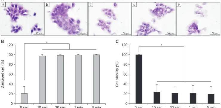
(3) Do-kyeong Kim, et al.. deviation. A value of p <0.05 was considered statistically. including vacuoles in some cells, pyknosis, and karyolysis.. significant.. In 30 seconds of etchant application, almost cells showed cell injury in the nucleus. In one minute of application, the. RESULTS. cells were enlarged with nuclear injury and subsequent. Effect of dental acid-etchant application time on cell damage and viability of oral epithelial cells. cell damage significantly was increased from 10 seconds of. membrane destruction. Consistently, the percentage of etchant application (Fig. 1B). These results demonstrated that cell damage was induced by dental acid-etchant, even. To examine the effect of application time on cell damage. for a short time.. caused by dental acid-etchants, we first observed morpho-. To further investigate cell viability by different applica-. logical changes in the oral epithelial cells. After treatment. tion times of dental acid-etchants, MTT assays were per-. with dental acid-etchants for the indicated time (0 second,. formed (Fig. 1C). After treatment with dental acid-etchants. 10 seconds, 30 seconds, 1 minute, 5 minutes), H&E stain-. for the indicated times (0 second, 10 seconds, 30 seconds, 1. ing was performed (Fig. 1A). The concentration of dental. minute, and 5 minutes), it was removed and the cells were. acid-etchants is fixed in 37% P.A., which commonly used. regrown in fresh culture medium for 24 hours. Compared. in clinical practice. In the first 10 seconds of application,. to control (0 second), cell viability was decreased 4.29-. the dental acid-etchants showed remarkable cell damage,. fold (23.30%±16.16%) in cells treated with etchants for 10. A a. c. b. 50 m. B. 50 m. 50 m. C. *. 120. 50 m. 120. 50 m. *. 100. Cell viability (%). 100. Damaged cell (%). e. d. 80 60 40 20. 80 60 40 20. 0. 0 0 sec. 10 sec. 30 sec. 1 min. 5 min. 0 sec. 10 sec. 30 sec. 1 min. 5 min. Fig. 1. Effect of dental acid-etchant application time on cell damage and viability in oral epithelial cells. (A) Morphology of oral epithelial cells by etchant gel application time. After application of dental acid-etchants (37% phosphoric acid) for the indicated times ([a] 0 second, [b] 10 seconds, [c] 30 seconds, [d] 1 minute and [e] 5 minutes), the cells were stained with hematoxylin-eosin. Representative images are shown (scale bar 50 µm, magnification ×400). (B) Cell damage by etchant gel application time. Cell damage includes both nuclear and membrane injuries. All damaged cells were counted and then bar graph shows the relative percentage of cells damaged by etchant gel application time. (C) Cell viability by application time of dental acid-etchant in oral epithelial cells. Immortalized human oral keratinocytes cells were seeded into 24-well plates and then treated with 37% phosphoric acid (dental acid-etchant) for the indicated times. The cells were re-grown in fresh medium (10% fetal bovine serum+1% penicillin/streptomycin in P medium) for 24 hours and then 3-(4, 5-dime-thylthiazol-2-yl)-2, 5-diphenyltetrazolium bromide assays were performed. The bar graph shows the cell viability percentage relative to the control (0 second). *p <0.05 by Mann–Whitney U-test.. 301.
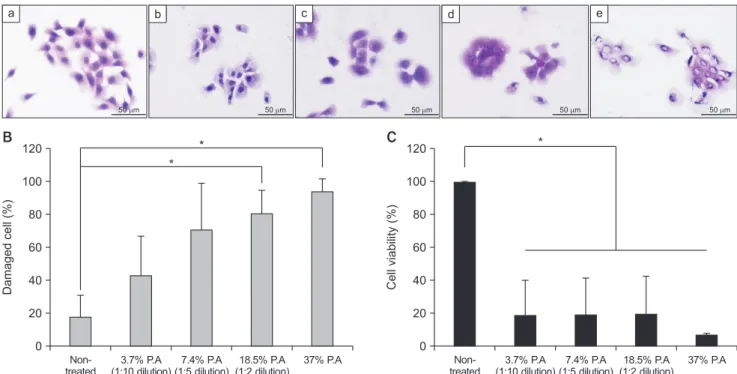
(4) Dental acid etchants on oral epithelial cells. seconds. As the application time increased, the cell viability. Morphological changes were observed by H&E stain-. decreased 4.55-fold (21.96%±13.45%), 4.69-fold (21.32%. ing in oral epithelial cells (Fig. 2A). To clearly observe the. ±15.54%) and 5.15-fold (19.44%±15.00%) at 30 seconds,. cell morphological damage and cell viability, dental acid-. 1 minute, and 5 minutes, respectively (p =0.014). As the. etchants were applied for 1 minute. Dental acid-etchants. etchant-applied time increased, cell viability was signifi-. were diluted with distilled water at ratios of 1:2 (18.5%. cantly reduced.. P.A.) to 1:10 (3.7% P.A.). When the cells were treated with. Taken together, dental acid-etchants (37% P.A.) induced. etchants diluted 1:10 ratio (3.7% P.A.), there were no vis-. irreversible cell injury, including karyolysis and pyknosis, in. ible morphological changes compared to control cells. time as short as 10 seconds.. (non-treated). After the application of etchants diluted 1:5 (7.4% P.A.), the cells were enlarger than the control cells,. Effects of dental acid-etchant concentration on cell damage and viability of oral epithelial cells. and some cells showed nuclear injury, including pyknosis. When treated with etchants diluted 1:2 (18.5% P.A.), karyorrhexis and vacuoles were shown in the cells. Con-. Next, we examined the effects of different dental acidetchants concentrations on cell damage.. a. sistently, the percentage of damaged cells was significantly increased from the application of etchants diluted 1:2. c. b. 50 m. 50 m. 50 m. *. 120. e. d. 50 m. 50 m. *. 120. * 100. Cell viability (%). Damaged cell (%). 100 80 60 40 20. 80 60 40 20. 0. 0 Nontreated. 3.7% P.A 7.4% P.A 18.5% P.A (1:10 dilution) (1:5 dilution) (1:2 dilution). 37% P.A. Nontreated. 3.7% P.A 7.4% P.A 18.5% P.A (1:10 dilution) (1:5 dilution) (1:2 dilution). 37% P.A. Fig. 2. Effect of dental acid-etchant concentration on cell damage and viability in oral epithelial cells. (A) Morphology of oral epithelial cells by etchant gel concentration. After application with each concentration ([a] non-treated, [b] 3.7% phosphoric acid [P.A.; 1:10 dilution], [c] 7.4% P.A. [1:5 dilution], [d] 18.5% P.A. [1:2 dilution], and [e] 37% P.A. [undiluted dental-etchant]) for 1 minute, the cells were fixed and stained with hematoxylin-eosin. Representative images are shown (scale bar 50 µm, magnification ×400). (B) Cell damage by etchant gel application time. Cell damage includes both nuclear and membrane injuries. All damaged cells were counted, and the bar graph shows the percentage of damage cell by etchant gel application time relative to the control cells. (C) Cell viability by concentration of dental acid-etchant in oral epithelial cells. Immortalized human oral keratinocytes cells were seeded into 24-well plates and then treated with the indicated concentration (dilution ratio) of dentaletchant. The cells were re-grown in fresh medium (10% fetal bovine serum+1% penicillin/streptomycin in P medium) for 24 hours and 3-(4, 5-dime-thylthiazol-2-yl)-2, 5-diphenyltetrazolium bromide assays were performed. The bar graph shows the percentage of cell viability relative to the control cells (non-treated). P.A., phosphoric acid. *p <0.05 by Mann–Whitney U-test.. 302. www.chosunobr.org.
(5) Do-kyeong Kim, et al.. (18.5% P.A.) (p <0.05) (Fig. 2B). Compared to nontreated. etchants on oral epithelial cells. First, we identified the. cells, cell injury was increased 2.44-fold (42.79%±23.49%). effects by dental acid-etchant’s application time on oral. in cells treated with 3.7% P.A. (1:10 diluted etchants) and. epithelial cells (Fig. 1). The concentration of dental-acid-. 4.01-fold (70.45%±27.86%) in cells treated with 7.4% P.A.. etchants was fixed to 37% P.A., which commonly used. (1:5 diluted etchants), although it is not significant. When. clinically. In the etch technique, dental acid-etchants must. the cells were treated with more 18.5% P.A. (1:2 diluted. be rinsed with water immediately after 15-30 seconds,. etchants), the percentage of damaged cells increased 4.58-. which is application time in manufacturer’s instruction.. fold (80.37%±13.89%) (p <0.05). Collectively, as the con-. At this time, inadequate rinse can remain the dental acid-. centration of P.A. increased, the number of damaged cells. etchants with various diluted concentration of P.A., and. increased. In particular, oral epithelial cells showed that in-. then we hypothesized that it can irritate on oral soft tissues.. creased cell injury in the nucleus and cell membrane when. Seconds, we examined the effects by dental acid-etchant’. they were treated with dental acid-etchants containing. s various diluted concentrations on oral epithelial cells (Fig.. more than 18.5% P.A.. 2). When dental acid-etchants were applied to oral epi-. Following treatment with each concentration of dental. thelial cells for 10 seconds or the etchant contained at least. acid-etchant, we observed cell viability. After treatment. 18.5% P.A., vacuoles and nuclear damage were observed in. with dental acid-etchants, the cells were stabilized for. cells, including karyolysis and karyorrhexis. Furthermore,. 24 hours and then MTT assays were performed (Fig. 2C).. cell injuries were remarkedly shown with increasing ap-. When the cells were treated with diluted etchants at a ratio. plication time and concentration. Among the cell mor-. of 1:10, cell viability decreased 5.33-fold (18.75%±20.98%). phological changes, vacuole formation is an early stage of. compared to control (non-treated). Moreover, cell viability. cell damage and can occur in different types of cell death,. decreased 5.17-fold (19.35%±21.93%) in cells treated with. such as autophagy, apoptosis, and necrosis [9,10]. Also,. 1:5 diluted etchants (7.4% P.A.) and 5.07-fold (19.72%±. nuclear damage, such as pyknosis and karyolysis, is caused. 23.01%) with 1:2 diluted-etchant (18.5% P.A.) (p =0.014).. by cell necrosis [11]. Consistently, the cell survival rate was. Thus, when oral epithelial cells were treated with dental. significantly reduced by dental acid-etchant application,. acid-etchants containing more than 3.7% P.A., the cell sur-. even though the etchants were applied for as short as 10. vival rate was significantly reduced.. seconds or a P.A. concentration as low as 3.7%. The dentin adhesive system is one of the fastest de-. DISCUSSION. veloping dental materials and can classify to the etch & rinse system and self-etching system [3,12]. Self-etching. Dental acid-etchants commonly used clinically are highly. adhesives have advantages, such as reducing the damage. acidic, containing 37% P.A., and contact with human tis-. to oral soft tissues and contamination to the surrounding,. sues can cause tissue necrosis or chemical burns [6,7]. In-. and short chair time, because there is no acid corrosion. tra- and extra-oral tissues are at greater risk of exposure to. and rinse. However, self-etching adhesives can induce cell. dental acid-etchants. In severe cases, the gingiva recessed. shrinkage, irregular cell morphology, cytotoxicity, and ne-. and necrotized by etchants was treated with subepithelial. crosis [13-15]. Consistent with our results, cell viability was. connective tissue graft [5]. The chemical burns of facial. decreased, even though the self-etching adhesives were. skin and tongue that occurred during composite restoration. applied at low concentrations.. and orthodontic treatment [6,7]. However, most reports. Acid-mediated corrosion can maximize the ability for. were clinical research and few studies have examined the. adhesion to dentin [4,16]. Both dental acid-etchants and. effects of oral epithelial cells by direct contact with dental. self-etching adhesives can cause irreversible injury to the. acid-etchants.. cells when they come in direct contact with the cells at low. We investigated morphological changes, cell damage,. concentrations and for short application times. To maxi-. and cell viability to identify the effects by dental acid-. mize the function of dental acid-etchants and minimize the. 303.
(6) Dental acid etchants on oral epithelial cells. adverse effects on soft tissue, it is important to prevent the inflow of dental acid-etchants into soft tissues by exposing only the treatment area. First, the dentist should thoroughly isolate the treatment area from soft tissue, by using a rubber dam or cotton roll and use of gel-type dental acidetchants. For dental hygienists, it is important to use strong suction or a large size suction tip to eliminate completely acid-etchants so that residual acid-etchants do not irritate the oral mucosa. This study examined that effects of dental acid-etchants on oral epithelial cells as a basic experimental study in vitro and specifically showed the risk of dental acid-etchants. Therefore, dentists and dental hygienists should pay strict attention to the handling of acid-etchants during restoration and orthodontic treatment. Furthermore, we expect that this study will provide as the basis for the development of biocompatible acid-etchants and dentin adhesive systems in the future.. CONFLICTS OF INTEREST The authors declare that they have no competing interests.. ORCID Do-kyeong Kim https://orcid.org/0000-0002-5154-9613 Jae-won Kwak https://orcid.org/0000-0002-8055-4542 Ryeong-mi Jo https://orcid.org/0000-0002-7157-0460 Da-som Jung https://orcid.org/0000-0001-7442-1084 Da-young Youn https://orcid.org/0000-0002-2737-6580 Na-yeon Oh https://orcid.org/0000-0003-1809-4925 Ji-hye Jang https://orcid.org/0000-0001-5917-2374. 304. www.chosunobr.org. REFERENCES 1. Jeong WG, Gang YM, Gung HS, Gim DH, Gim RY, Gim AJ, Gim EJ, Gim HE, Mun HJ, Bak SS, Bak JH, Bae SS, Seong GY, Song JJ, Sim SS, An SH, Yang SY, U SH, Yu JS, Yun MS, Yun HY, Yun HS, Lee MO, Lee SY, Lee SS, Lee Y, Lee H, Lee HK, Im GO, Im SY, Jeong MH, Jeong UJ, Jo MS, Jo HE, Ji MG, Choe MH, Hong SM, Hwang SH. Principles and practice of conservative dentistry. 4th ed. Seoul: Daehan Narae Publishing, Inc.; 2016. 2. Buonocore MG. A simple method of increasing the adhesion of acrylic filling materials to enamel surfaces. J Dent Res 1955;34:849-853. doi: 10.1177/00220345550340060801. 3. Sofan E, Sofan A, Palaia G, Tenore G, Romeo U, Migliau G. Classification review of dental adhesive systems: from the IV generation to the universal type. Ann Stomatol (Roma) 2017;8:1-17. doi: 10.11138/ads/2017.8.1.001. 4. Lopes GC, Thys DG, Klaus P, Oliveira GM, Widmer N. Enamel acid etching: a review. Compend Contin Educ Dent 2007;28:18-24; quiz 25, 42. 5. Akman AC, Demiralp B, Güncü GN, Kiremitçi A, Sengün D. Necrosis of gingiva and alveolar bone caused by acid etching and its treatment with subepithelial connective tissue graft. J Can Dent Assoc 2005;71:477-479. 6. Rao R. Report of etchant induced oral chemical burna rare incidence. Int J Adv Res 2018;6:257-260. doi: 10.21474/IJAR01/6212. 7. Park JH, Shin HJ, Park SH, Kim JW, Cho KM. Iatrogenic chemical burn on facial skin by 37% phosphoric acid etchant. J Korean Acad Conserv Dent 2009;34:38-41. doi: 10.5395/JKACD.2009.34.1.038. 8. Lee HJ, Guo HY, Lee SK, Jeon BH, Jun CD, Lee SK, Park MH, Kim EC. Effects of nicotine on proliferation, cell cycle, and differentiation in immortalized and malignant oral keratinocytes. J Oral Pathol Med 2005;34:436-443. 9. Schauer A, Knauer H, Ruckenstuhl C, Fussi H, Durchschlag M, Potocnik U, Fröhlich KU. Vacuolar functions determine the mode of cell death. Biochim Biophys Acta 2009;1793:540-545. doi: 10.1016/j.bbamcr.2008.11.006. 10. Nakajima A, Kurihara H, Yagita H, Okumura K, Nakano H. Mitochondrial extrusion through the cytoplasmic vacuoles during cell death. J Biol Chem 2008;283:24128-24135. doi: 10.1074/jbc.M802996200. 11. Kumar V, Abbas AK, Aster JC, Robbins SL. Robbins basic pathology. 9th ed. Philadelphia: Elsevier/Saunders; 2013;xii, 910. 12. Lawson NC, Robles A, Fu CC, Lin CP, Sawlani K, Burgess JO. Two-year clinical trial of a universal adhesive in total-etch and self-etch mode in non-carious cervical lesions. J Dent 2015;43:1229-1234. doi: 10.1016/ j.jdent.2015.07.009. 13. Rhee CH, Kim IR, Kim GC, Kim SS, Son WS. In vitro.
(7) Do-kyeong Kim, et al. cytotoxicity of self-etching primers. Korean J Orthod 2006;36:422-433. 14. Pupo YM, Bernardo CFF, de Souza FFFA, Michél MD, Ribeiro CNM, Germano S, Maluf DF. Cytotoxicity of etchand-rinse, self-etch, and universal dental adhesive systems in fibroblast cell line 3T3. Scanning 2017;2017:9650420. doi: 10.1155/2017/9650420. 15. Kusdemir M, Gunal S, Ozer F, Imazato S, Izutani N, Ebisu S,. Blatz MB. Evaluation of cytotoxic effects of six self-etching adhesives with direct and indirect contact tests. Dent Mater J 2011;30:799-805. doi: 10.4012/dmj.2011-046. 16. Lee AR, Kim HS, Ku HW, Hwang HK, Jo HH, Min JB. Evaluation of etched enamel using quantitative light-induced fluorescence. Oral Biol Res 2018;42:10-15. doi: 10.21851/ obr.42.01.201803.10.. 305.
(8)
수치
관련 문서
Abstract – In this study, we investigated whether adenosine, adenine, uridine and homogentisic acid derived from Pinellia ternata affect the secretion, production and gene expression
Schematic diagram of anti- oral microbial activity and anti-in- flammatory effects of rosmarinic acid in MC3T3-E1 osteoblastic cells on a titanium (Ti)
