저작자표시-비영리-변경금지 2.0 대한민국 이용자는 아래의 조건을 따르는 경우에 한하여 자유롭게 l 이 저작물을 복제, 배포, 전송, 전시, 공연 및 방송할 수 있습니다. 다음과 같은 조건을 따라야 합니다: l 귀하는, 이 저작물의 재이용이나 배포의 경우, 이 저작물에 적용된 이용허락조건 을 명확하게 나타내어야 합니다. l 저작권자로부터 별도의 허가를 받으면 이러한 조건들은 적용되지 않습니다. 저작권법에 따른 이용자의 권리는 위의 내용에 의하여 영향을 받지 않습니다. 이것은 이용허락규약(Legal Code)을 이해하기 쉽게 요약한 것입니다. Disclaimer 저작자표시. 귀하는 원저작자를 표시하여야 합니다. 비영리. 귀하는 이 저작물을 영리 목적으로 이용할 수 없습니다. 변경금지. 귀하는 이 저작물을 개작, 변형 또는 가공할 수 없습니다.
MASTER’S THESIS
Anti-cancer effects of Lapathoside A from
Fagopyrum esculentum
in human pancreatic
cancer cells and neuroprotective properties of
Ecklonia cava
ferment extract against
glutamate-induced oxidative stress in HT22
hippocampal cell
Mi Sook Kang
(Supervised by professor Jae Hoon Kim)
Department of Biotechnology
GRADUATE SCHOOL
JEJU NATIONAL UNIVERSITY
PART Ⅰ
Lapathoside A isolated from Fagopyrum esculentum induces apoptosis
in human pancreatic cancer cells. CONTENTS
CONTENTS ··· ⅰ
LIST OF FIGURES ··· ⅲ
LIST OF TABLES ··· ⅳ
ABSTRACT ··· 1
1. INTRODUCTION ··· 2
2. MATERIALS AND METHODS ··· 4
2.1. Extraction and isolation of lapathoside A from buckwheat roots ··· 4
2.2. Identification of lapathoside A ··· 4
2.3. Cell culture ··· 5
2.4. Cell viability assay ··· 5
2.5. Flow cytometric analysis for cell apoptosis detection ··· 6
2.6. Flow cytometric analysis for Reactive Oxygen Species (ROS) detection ··· 6
2.7. Western blot analysis ··· 7
2.8. Statistical analysis ··· 7
3. RESULTS ··· 9
3.1. Isolation and identification of lapathoside A ··· 9
3.2. Lapathoside A inhibits pancreatic cancer cell growth ··· 14
3.3. Lapathoside A induces the apoptosis of PANC-1 and SNU-213 cells ··· 16
3.4. Lapathoside A induces ROS generation in human pancreatic cancer cells ··· 20
3.5. Effects of lapathoside A on the phosphorylation of FAK, Akt proteins and MAPKs ··· 22
4. DISCUSSION ··· 25
5. REFERENCES ··· 27
LIST OF FIGURES
Figure 1. Extraction and isolation of lapathoside A. ··· 10 Figure 2. NMR spectrum of lapathoside A. ··· 11 Figure 3. Chemical structure of lapathoside A. ··· 13 Figure 4. Cytotoxic effects of buckwheat root extract and lapathoside A in PANC-1 and SNU-213 cells. ··· 15 Figure 5. Flow cytometric analysis of human pancreatic cancer cells after treatment of lapathoside A for 24 hours. ··· 17 Figure 6. Flow cytometric analysis of human pancreati cancer cells after treatment of lapathoside A for 48 hours. ··· 18 Figure 7. Lapathoside A induced apoptosis in pancreatic cancer cells. ·· 19 Figure 8. Lapathoside A induces ROS generation in pancreatic cancer cells. ··· 21 Figure 9. Expression levels of cellular signaling pathway proteins treated with lapathoside A in 293T and PANC-1 cells. ··· 23 Figure 10. Expression levels of MAPK proteins treated with lapathoside A in 293T, PANC-1, and SNU-213 cells. ··· 24
LIST OF TABLES
ABSTRACT
Lapathoside A, a phenylpropanoid ester compound, was isolated from buckwheat roots. Ethanol (70%) extract of buckwheat roots was separated into n-hexane, methylene chloride, ethyl acetate, n-butanol, and water fractions by solvent partitioning. Seven fractions were obtained from the ethyl acetate fraction by liquid chromatography. Of the seven fractions, fraction No. 6 contained lapathoside A. The structure of lapathoside A was determined by LC-MS, LC-MS/MS, and NMR analysis. Next, we investigated the anticancer activity of lapathoside A in human pancreatic cancer cell lines (PANC-1 and
SNU-213). After treatment with lapathoside A at 25 M, viabilities of PANC-1 μ
and SNU-213 cells decreased 40% and 27%, respectively. Treatment with lapathoside A also increased the number of apoptotic cells and regulated expression levels of apoptotic proteins. Results using flow cytometric analysis indicated that treatment with lapathoside A increased the number of apoptotic cells in pancreatic cancer cell lines. These results suggest that lapathoside A might have potential application of the treatment of pancreatic cancer.
1. INTRODUCTION
Pancreatic cancer is one of the deadliest cancers. Its 5-year relative survival rate is about 9% [1, 2]. According to a report of Statistics Korea in 2017, deaths from pancreatic cancer have increased steadily in Korea over the last decade [3]. In addition, pancreatic cancer is the fifth leading cause of cancer death in Korea [3]. Despite advances in surgery, chemotherapy, and radiotherapy for other cancers over the past several years, pancreatic cancer still has a very poor prognosis. When pancreatic cancer is diagnosed, the cancer spreads to other organs, making it difficult for most patients to undergo surgery. This is because pancreatic cancer does not show symptoms until the disease has progressed [4,5]. In addition, pancreatic cancer is resistant to chemotherapy and radiation therapy, leading to limited effectiveness of the treatment [6-8]. Therefore, it is very meaningful to find new drug candidates from natural products for pancreatic cancer treatment [9].
Buckwheat is a common pseudo-cereal plant of the genus Fagopyrum in the
family Polygonaceae. The most commonly cultivated species are common
buckwheat (Fagopyrum esculentum) and tartary buckwheat (Fagopyrum
tataricum). They are grown in Asia, Middle East, Europe, and North America [10].
According to previous studies, buckwheat seeds contain not only complex carbohydrates, but also many health-conscious ingredients such as minerals, dietary fiber, flavones, flavonoids, phytosterols, and pagopyrins [11-13]. Although compositions of these compounds vary depending on the species, growth environment, these compounds are known to have antioxidant, anti-inflammatory, and anticancer effects [14-16]. However, little is known about the natural compounds present in buckwheat roots. In the present study, lapathoside A was isolated from buckwheat roots as a bioactive compound. Cell viability assay, flow cytometric analysis, and western blot analysis were
performed to investigate the anticancer effect of lapathoside A on human pancreatic cancer cell lines.
2. MATERIALS AND METHODS
2.1 Extraction and isolation of lapathoside A from buckwheat roots
Buckwheat was cultivated in Aewol, Jeju Island, South Korea. Its roots were collected in June, 2017. Collected buckwheat roots were washed, dried in a cold wind, and ground. The dried powder of buckwheat roots was extracted with 70% ethanol for 24 hours at room temperature. The crude extract of buckwheat roots was filtered, concentrated under reduced pressure, and lyophilized. Sequential solvent fractionation was carried out for 10 g of the buckwheat root extract using n-Hexane (n-Hex), methylene chloride (MC), ethyl acetate (EtOAc), and butanol (BuOH). Solvent fractions of buckwheat root extract were subjected to vacuum liquid chromatography (VLC) using n-Hex (100%), MC (100-50%), and EtOAc (0-50%) as mobile phases.
2.2 Identification of lapathoside A
High-performance liquid chromatography (HPLC) analysis was performed for buckwheat root extract, solvent fraction, and VLC fraction. A high-performance liquid chromatography system e2695 (Waters Corp., Milford, MA, USA) equipped with a photodiode array detector 2998 (Waters Corp., USA) and a
Cadenza CD-C18 column 59 (3 m, 150 mm × 4.5 mm) was used for HPLC μ
analysis. The column temperature was set at 40 . Extracts and fractions were ℃
dissolved in 70% ethanol. Lapathoside A was dissolved in methanol and then
filtered with a 0.5 m syringe filter. 0.5% acetic acid in Hμ 2O (A) and
acetonitrile (B) were used as mobile phases. The composition was changed from A 75% to A 60% for 40 min. The flow rate was set at 1 mL/min.
LCQ-Fleet Ion Trap Mass Spectrometer (Thermo Fisher Scientific, Waltman, MA, USA) was used for LC-MS/MS analysis to identify the structure of a
single isolated compound. The ionization method of LC-MS/MS was ESI
negative using a Hypersil GOLD (50 x 2.1 mm, 1.9 m) column at 275μ ℃ of
capillary temperature. As mobile phases, 0.1% formic acid in H2O (A) and
acetonitrile (B) were used. The flow started with composition of A 95% and maintained for 1 min. The composition was then changed to A 0% for 13 min.
The flow rate was 200 L/min. Finally, the structure of the isolated compound μ
was characterized using 1H and 13C NMR (CD3OD, 500MHz) with an Avance
III NMR spectrometer (Bruker BioSpin, Rheinstetten, Germany). 2.3 Cell culture
Human pancreatic cancer cell lines PANC-1 and SNU-213 were purchased from Korean Cell Line Bank (KCLB, Seoul, Korea). 293T kidney epithelial cells were obtained from KCLB as a noncancerous cell line. PANC-1 and 293T cells were maintained in DMEM (Gibco-BRL, Gaithersburg, MD, USA) containing 10% fetal bovine serum (FBS, Gibco-BRL, USA) and 1% Penicillin-Streptomycin (Pen-Strep, Gibco-BRL, USA). SNU-213 was cultured in RPMI 1640 medium (Gibco-BRL, USA) supplemented with 10% FBS and 1%
Pen-Strep. Cells were incubated at 37℃ with 5% CO2 and maintained.
2.4 Cell viability assay
Cell viability was measured by WST-1 analysis using an EZ-Cytox cell viability assay kit (Daeil Lab Service, Seoul, Korea). PANC-1, SNU-213, and 293T
cells were seeded into 24-well plates at a density of 1.25×10⁴ cells/well and
treated with lapathoside A for 72 hours. After treatment, cells were incubated with 10% of WST-1 reagent for cell viability analysis. After incubation at 37℃
in a 5% CO2 incubator for approximately 30 minutes, the absorbance was
(Thermo Fisher Scientific, USA).
2.5 Flow cytometric analysis for cell apoptosis detection
Apoptotic cell death was detected with a FITC Annexin V Apoptosis Detection Kit from BD Pharmingen (San Diego, CA, USA). PANC-1, SNU-213, and 293T
cells were seeded into 35 mm dishes at a density of 1×10⁵ cells/dish and
incubated for 24 hours before treatment with lapathoside A for 72 hours. After treatment, cells were trypsinized, harvested, and washed with cold PBS. Cells
were then resuspended in 500 L of binding buffer containing Annexin V-FITC μ
(5 L) and propidium iodide (5 L). Cells were then incubated at 37μ μ ℃ in the
dark for 15 minutes. Apoptotic cells were detected with a LSRFortessa flow cytometer (BD Biosciences, CA, USA). Cells that were negative for both Annexin V and PI were considered as viable cells. Cells that were positive for Annexin V but negative for PI were considered as early-stage apoptotic cells. Cells that were positive for both Annexin V and PI were considered as late-stage apoptotic cells or necrotic cells. The percentage of total apoptotic cells was calculated by combining percentages of early-stage apoptotic cells and late-stage apoptotic cells.
2.6 Flow cytometric analysis for Reactive Oxygen Species (ROS) detection
ROS generation was detected using oxygen-sensitive 2 ,7′ ′
-dichloro-dihydro-fluoroscein diacetate (H2DCFDA). PANC-1, SNU-213, and
293T cells were seeded at a density of 1×10⁵ cells/dish onto 35 mm dishes
and incubated for 24 hours prior to treatment with lapathoside A for 72 hours.
and incubated for 24 hours prior to treatment with the extracts alone or extracts in combination with 4 mM glutamate. After treatment, cells were
incubated in Fresh culture medium containing 10 M Hμ 2DCFDA for 15 minutes.
Then cells were harvested and washed with cold PBS and resuspended in
500 L of DPBS. Finally, ROS generation was measured by flow cytometric μ
analysis.
2.7 Western blot analysis
The M-PER Mammalian Protein Extraction Reagent as cell lysis buffer was purchased from Thermo Fisher Scientific (USA). Primary antibodies (AKT, FAK, GAPDH, etc.) were purchased from Cell Signaling Technology (Beverly, MA, USA). Secondary antibodies were obtained from Merck (Darmstadt, Germany). Cells were treated with lapathoside A at different concentrations after 24 hours of seeding on 6-well plates. After treatment, cells were lysed with cell lysis buffer (M-PER Mammalian Protein Extraction Reagent containing 2 mM sodium vanadate, 30 mM sodium pyrophosphate, 100 mM sodium fluoride, 0.1 M PMSF, and protein inhibitors). Proteins were separated by SDS-PAGE on 12% SDS polyacrylamide gel and transferred to nitrocellulose membranes. These membranes were blocked with 5% skim milk in TBST for 8 hours and incubated with primary antibodies (FAK, phospho-FAK, AKT, phospho-AKT,
GAPDH) at 4℃ overnight. Protein bands were detected using HIGH X-DOL
(Poohung, Gyeonggi-do, Korea) and HIGH X-FIX (Poohung, Korea) followed by exposure to X-ray films.
All data results are represented as mean ± standard deviation (SD) or standard error (SE). Student’s t-test was used for comparison between non-treated control group and treated data groups. (*p < 0.05, **p < 0.01, ***p < 0.001)
3. RESULTS 3.1 Isolation and identification of lapathoside A
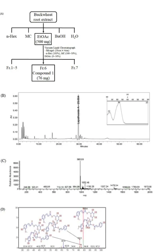
Extraction of buckwheat root and separation of lapathoside A were performed according to the scheme described in Figure 1A. The dried powder of buckwheat root was extracted with 70% ethanol for 24 hours at room temperature to obtain a crude extract. Analysis of the buckwheat root extract by HPLC showed the highest peak at retention time of 29.664 minutes (Figure 1B). A total of 10 g of the buckwheat root extract was subjected to sequential solvent fractionation. About 500 mg of powder was obtained from the EtOAc fraction. Seven fractions were obtained after subjecting the EtOAc fraction to vacuum liquid chromatography. A total of 76 mg of a compound was obtained from No. 6 fraction. The compound had the same retention time as the single peak identified in the crude extract. The isolated compound was a light brown solid powder. It was subjected to further characterization. LC-MS analysis confirmed the m/z 985.09 [M-H]- molecular weight peak (Figure 1C). A m/z 838.96 [M134 coumaroyl]-daughter-ion was identified in LC-MS/MS fragmentation ions (Figure 1D). In the 1H NMR, a multiplet signal was
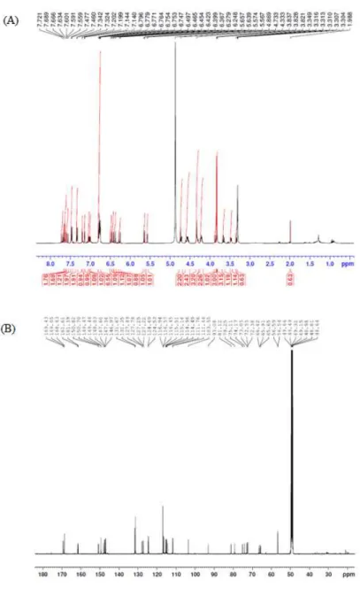
detected near δ 3 to 4 and an anomeric proton was detected at 5.57 (d, J =
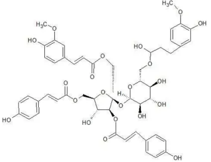
3.5) (Figure 2A), indicating the presence of one sugar or more. The 13C NMR spectrum (Figure 2B) was compared with the DEPT135 data, confirming the presence of four carbonyl groups. Typical O-Me signals were detected at δ 3.87 and 3.82 peaks. Results confirmed that two p-coumaric acids and two ferrous acids were acylated in sucrose (Table 1). Compared to a previous report [17], the isolated compound was identified as lapathoside A (Figure 3).
Figure 1. Extraction and isolation of lapathoside A. (A) The scheme of isolation process from Buckwheat root extracts. (B) HPLC-PDA Chromatogram of buckwheat root extract. (C) LC-MS spectrum of compound 1 in Figure 1A. (D) LC-MS/MS fragmentation ions of compound 1 in Figure 1A.
Figure 2. NMR spectrum of lapathoside A. The spectrum data was obtained
using Avance III NMR spectrometer (500MHz) in methanol-d4. (A) 1H NMR
No. 1H 13C fructose 1 4.33(2H, m) 66.4 2 103.5 3 5.65(d, 9.0) 79.2 4 4.75(m) 74.1 5 4.24(m) 81.1 6 4.58(m), 4.56(m) 65.6 glucose 1’ 5.57(d, 3.5) 93.0 2’ 3.48(dd, 9.5, 4.0) 73.0 3’ 3.65(dd, 9.5, 9.0) 75.1 4’ 3.30(m) 72.3 5’ 3.30(m) 72.5 6’ 4.73(m), 4.21(m) 65.9 phenylpropanoids (glu-6’) feruloyl 9’‘ 169.4 8’‘ 6.49(d, 16.0) 115.1 7’‘ 7.63(d, 16.5) 147.3 1’‘ 127.8 2’‘ 7.20(d, 1.5) 111.7 3’‘ 149.4 4’‘ 150.7 5’‘ 6.77(d, 8.5) 116.5 6’‘ 7.01(dd, 8.5, 2.0) 124.6 O-Me 3.87(3H, s) 56.5 (fruc-1) feruloyl 9’‘’ 168.6 8’‘’ 6.39(d, 16.0) 115.1 7’‘’ 7.66(d, 16.0) 147.6 1’‘’ 127.6 2’‘’ 7.14(d, 2.0) 111.6 3’‘’ 149.4 4’‘’ 150.7 5’‘’ 6.76(d, 8.5) 116.4 6’‘’ 7.04(dd, 8.5, 2.0) 124.5 O-Me 3.82(3H, s) 56.5 (fruc-3) p-coumaroyl 9’‘’‘ 168.6 8‘’‘’ 6.45(d, 15.5) 114.4 7’‘’‘ 7.72(d, 16.0) 148.0 1’‘’‘ 127.2 2’‘’‘, 6’‘’‘ 7.47(d, 8.5) 131.6 3’‘’‘, 5’‘’‘ 6.79(d, 8.5) 116.9 4’‘’‘ 161.6 (fruc-6) p-coumaroyl 9’‘’‘’ 169.0 8’‘’‘’ 6.27(d, 15.5) 114.9 7’‘’‘’ 7.59(d, 16.0) 146.9 1’‘’‘’ 127.2 2’‘’‘’, 6’‘’‘’ 7.34(d, 9.0) 131.3 3’‘’‘’, 5’‘’‘’ 6.77(d, 9.0) 116.9 4’‘’‘’ 161.3
3.2 Lapathoside A inhibits pancreatic cancer cell growth
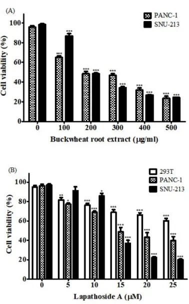
To investigate the cytotoxic effect of the buckwheat root extract 143 on pancreatic cancer cells, PANC-1 and SNU-213 cells were treated with
buckwheat root extract at concentrations of 0 - 500 g/ml for 72 hours. Cell μ
viability was then measured using the WST-1 assay. After cells were treated
with 200 g/ml of buckwheat root extract, viabilities of PANC-1 and SNU-213 μ
cells were about 48.41 and 48.47%, respectively (Figure 4A). Buckwheat root extract decreased viabilities of both PANC-1 and SNU-213 cells in a concentration dependent manner. This suggests that the buckwheat root extract might be a source of novel natural antitumor agents. Next, cell viability assay was performed for cells treated with lapathoside A isolated from buckwheat root extract. As shown in Figure 4B, lapathoside A decreased the proliferation of both PANC-1 and SNU-213 cells in a concentration dependent manner. The
cell viability of PANC-1 cells after treatment with 25 M of lapathoside A was μ
39.73%. Viabilities of SNU-213 cells after treatment with lapathoside A at 15 μ
M and 25 M were decreased to 36.90% and 20.07%, respectively (Figure μ
4B). Lapathoside A showed less effect on viability of control 293T cells than on pancreatic cancer cell lines.
Figure 4. Cytotoxic effects of buckwheat root extract and lapathoside A in PANC-1 and SNU-213 cells. (A) Cell viability of PANC-1 and SNU-213 cells after treatment with buckwheat root extract for 72 hours. (B) Cell viability of PANC-1 and SNU-213 cells after lapathoside A treatment for 72 hours. (Data represent the percentage ± SD and are representative of three individual experiments, *p < 0.05, **p < 0.01, ***p < 0.001.)
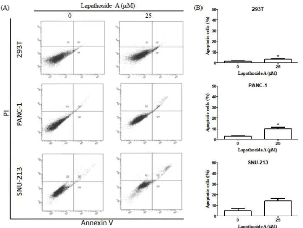
3.3 Lapathoside A induces the apoptosis of PANC-1 and SNU-213 cells To determine the apoptotic effect of lapathoside A, flow cytometric analysis was performed after staining lapathoside A-treated PANC-1 and SNU-213 cells with Annexin V/PI. Twenty four hours of Lapathoside A treatment had no significant effect for 293T, PANC-1, and SNU-213 cells (Figure 5). Treatment of lapathoside A for 48 hours increased the percentage of apoptotic cells slightly in 293T and PANC-1 cells (Figure 6). Percentages of early and late apoptotic cells were increased after 72 hours of lapathoside A treatment (Figure 7). As shown in Figures 7, lapathoside A treatment had no significant effect on 293T cells. However, the percentage of apoptotic PANC-1 cells increased from 5.47% to 21.83% and that of apoptotic SNU-213 cells increased from 11.33% to 38.77% after treatment with lapathoside A. These results suggest that lapathoside A can induce apoptosis of both PANC-1 and SNU-213 cells.
Figure 5. Flow cytometric analysis of human pancreatic cancer cells after treatment of lapathoside A for 24 hours. (A) Flow cytometric analysis were
performed in PANC-1 and SNU-213 cells after treatment with 25 M of μ
lapathoside A for 24 hours. (B) The percentage of total apoptotic cells was calculated from Figure 7A (*P < 0.05).
Figure 6. Flow cytometric analysis of human pancreati cancer cells after treatment of lapathoside A for 48 hours. (A) Flow cytometric analysis were
performed in PANC-1 and SNU-213 cells after treatment with 25 M of μ
lapathoside A for 48 hours. (B) The percentage of total apoptotic cells was calculated from Figure 7A (*P < 0.05).
Figure 7. Lapathoside A induced apoptosis in pancreatic cancer cells. (A) Flow cytometric analysis were performed in PANC-1 and SNU-213 cells after
treatment with 25 M of lapathoside A for 72 hours. (B) The percentage of μ
total apoptotic cells was calculated from Figure 7A (*P < 0.05). (C) Protein levels of cleaved-PARP were determined by Western blot analysis.
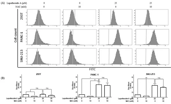
3.4 Lapathoside A induces ROS generation in human pancreatic cancer cells
ROS, as significant signaling molecules, are involved in signal transduction and sustaining cellular redox homeostasis in aerobic organisms. ROS are not only supporters of tumor cells, but are also efficient therapeutic tools in treating cancer. In the present study, intracellular ROS production was detected using
a H2DCF DA probe in human pancreatic cancer cells. Following treatment with ‑
lapathoside A for 12 hours, the flow cytometric analysis revealed that 25 M μ
of lapathoside A significantly increased ROS generation (Figure 8). To further analyze the role of ROS, the ROS scavenger NAC was used to inhibit the accumulation of intracellular ROS.
Figure 8. Lapathoside A induces ROS generation in pancreatic cancer cells.
(A) Cells were pretreated with NAC (10mM) and treated with 25 M of μ
lapathoside A for 12 hours. Then cells were labeled with H2DCF-DA. ROS
production were measured by flow cytometric analysis. (B) Fold change of geometric mean was calculated as a ratio of sample group to control group.
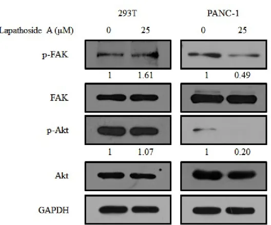
3.5 Effects of lapathoside A on the phosphorylation of FAK, Akt proteins and MAPKs
It has been reported that Akt is overexpressed in most human cancers, including pancreatic cancer. Local adhesion kinase (FAK) is upstream of Akt. To investigate the cellular signaling pathway regulated by lapathoside A, we examined the expression and phosphorylation of FAK and Akt. As shown in Figure 9, Fak and Akt proteins were easily detected in PANC-1 cells. Phosphorylation levels of FAK (Tyr397) and Akt (Ser473) were decreased after treatment with lapathoside A. In contrast, lapathoside A did not show any significant effect on 293T control cells under the same conditions. These results demonstrate that lapathoside A specifically acts on pancreatic cancer cells.
Figure 9. Expression levels of cellular signaling pathway proteins treated with lapathoside A in 293T and PANC-1 cells. Cells were treated with lapathoside
A at 25 M for 24 hours. Total protein was collected and the expression of μ
FAK and Akt were analyzed by western blot assay. GAPDH was used as an loading control.
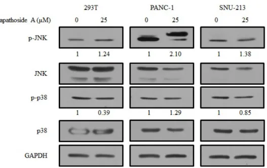
Figure 10. Expression levels of MAPK proteins treated with lapathoside A in 293T, PANC-1, and SNU-213 cells. Cells were treated with lapathoside A at
25 M for 24 hours. Total protein was collected and the expression of JNK μ
and p38 were analyzed by western blot assay. GAPDH was used as an loading control.
4. DISCUSSION
Buckwheat contains more organic compounds than other agriculture plants [14]. The root of buckwheat has been used as folk medicine in China traditionally [18]. We found that buckwheat root extract had anticancer activity against pancreatic cancer. We also identified lapathoside A as a major component in buckwheat root extract. Lapathoside A is one of the phenylpropanoid sucrose esters (PSEs). It has four Ph-CH=CH-CO- moieties through ester linkage with a sucrose. Various PSEs have been found in many plants. They have been reported to posses antioxidant, antivirus, antibacterial, anti-inflammatory, and anti-cancer effects [19, 20].
Lapathoside A isolated from Polygonum lapathifolium has been shown to be able to inhibit Epstein-Barr virus-early antigen (EBV-EA) induced by tetradecanoylphorbol-13-acetate (TPA) [17]. TPA, an agonist of protein kinase
C (PKC), is known to induce EBV reactivation through NF- B and AP-1. It κ
regulates the gene expression of EBV reactivation via PKC [20-22]. Besides, lapathoside A exhibits anti-tumor effects on mouse two-stage skin carcinogenesis induced by 7,12-dimethylbenz[a]anthracene (DMBA) and TPA [23].
To date, anticancer effects of lapathoside A in pancreatic cancer cell lines have not been reported yet. In our study, lapathoside A was able to induce apoptosis of pancreatic cancer cells. Gemcitabine was expected to be a potential agent for pancreatic cancer. However, it did not produce clinically good results in practice [24]. On the other hand, it had serious side effects such as anemia, extreme tiredness, diarrhea, and hair loss [25]. The development of anticancer compounds without showing side effects can significantly reduce the social cost of pancreatic cancer. Therefore, many studies have been conducted to develop more effective and less toxic
anticancer drugs from natural products as new treatments for pancreatic cancer [26, 27]. Since deregulation of apoptosis is a feature of all cancers, induction of apoptosis in cancer cells is an effective strategy of cancer treatment. Focal adhesion kinase (FAK) is overexpressed in tumor cells. Attenuation of FAK activity can lead to apoptosis [28]. The activity of Akt/protein kinase B suppresses apoptosis by preventing cytochrome c release from mitochondria [29]. Consistent with these reports, lapathoside A reduced FAK and Akt phosphorylation and induced apoptosis of pancreatic cancer cell lines.
In summary, we demonstrated that lapathoside A isolated from buckwheat roots could inhibit the proliferation of PANC-1 and SNU-213 cells in vitro. Considering its anti-proliferation activity and low toxicity, lapathoside A has the potential to be developed as an anticancer agent for the treatment of pancreatic cancer.
5. REFERENCES
1. Capella C, Albarello L, Capelli P, Sessa F, Zamboni G (2011) Carcinoma of the exocrine pancreas: The histology report. Dig Liver Disease 43:S282-92 2. Klöppel G, Hruban RH, Longnecker DS, et al. (2000) Ductal adenocarcinomas of the pancreas. In: Hamilton SR, Aaltonen LA, editors. WHO Classification of Tumours of the digestive system, IARC Press, Lyon, pp. 221 30–
3. ANNUAL REPORT ON THE CAUSES OF DEATH STATISTICS. (2017) Statistics Korea Retrieved from the original. See p. 15 for estimated deaths and trends in cancer death rates.
4. Vincent A, Herman J, Schulick R, Hruban RH, Goggins M. (2011) Pancreatic cancer. Lancet 378(9791):607-20
5. Liu Y, Bi T, Wang G, Dai W, Wu G, Qian L, Gao Q, Shen G. (2015) Lupeol inhibits proliferation and induces apoptosis of human pancreatic cancer PCNA-1 cells through AKT/ERK pathways. Naunyn Schmiedeberg’s Arch Pharmacol, 388(3):295-304
6. Stathis A, Moore MJ (2010) Advanced pancreatic carcinoma: current treatment and future challenges. Nat Rev Clin Oncol 7(3):163-72
7. Bu H, Luo J, Chen H, Zhang J, Li H, Guo H, Wang Z, Lin S (2012) Oridonin enhances antitumor activity of gemcitabine in pancreatic cancer through MAPK-p38 signaling pathway. Int J Oncol, 41(3):949-58
8. Gaianigo, Nicola et al. EMT and Treatment Resistance in Pancreatic Cancer. Cancers vol. 9,9 122. 12 Sep. 2017
9. Demain AL, Vaishnav P (2011) Natural products for cancer chemotherapy. Microb biotechnol 4(6):687-99
10. Li S, Zhang QH (2001) Advances in the development of functional foods
from buckwheat. Crit Rev Food Sci Nutr 41(6):451 64–
11. Dziadek K, Kopeć A, Pastucha E, Piątkowska E, Leszczyńska T, Pisulewska E, Witkowicz R, Francik R (2016) Basic chemical composition and bioactive compounds content in selected cultivars of buckwheat whole seeds, dehulled seeds and hulls. J Cereal Sci 69:1-8
12. Verardo V, Arráez-Román D, Segura-Carretero A, Marconi E, Fernández-Gutiérrez A, Caboni MF (2010) Identification of buckwheat phenolic compounds by reverse phase high performance liquid chromatography–
electrospray ionization-time of flight-mass spectrometry (RP-HPLC ESI-TOF-MS) –
J Cereal Sci 52(2):170-6
13. Gabr AM, Sytar O, Ghareeb H, Brestic M (2019) Accumulation of amino acids and flavonoids in hairy root cultures of common buckwheat (Fagopyrum esculentum). Physiol Mol Biol Plants 25(3):787-97
14. Liu C-L, Chen Y-S, Yang J-H, Chiang B-H (2007) Antioxidant Activity of Tartary ( Fagopyrum tataricum (L.) Gaertn.) and Common ( Fagopyrum esculentum Moench) Buckwheat Sprouts. J Agric Food Chem 56(1):173-8
15. Kiprovski B, Mikulic-Petkovsek M, Slatnar A, Veberic R, Stampar F, Malencic D, Latkovic D (2015) Comparison of phenolic profiles and antioxidant properties of European Fagopyrum esculentum cultivars. Food Chem 185:41-7 16. Vojtíšková P, Kmentová K, Kubáň V, Kracmar S (2012) Chemical composition of buckwheat plant (Fagopyrum esculentum) and selected buckwheat products. J Microbiol Biotechnol Food Sci 1:1011
17. Takasaki M, Kuroki S, Kozuka M, Konoshima T (2001) New Phenylpropanoid Esters of Sucrose from Polygonum lapathifolium. J Nat Prod 64(10):1305-8
18. Kim S-H, Cui C-B, Kang I-J, Kim SY, Ham S-S (2007) Cytotoxic Effect of Buckwheat (Fagopyrum esculentum Moench) Hull Against Cancer Cells. J Med Food 10(2):232-8
19. Panda P, Appalashetti M, Natarajan M, Chan-Park MB, Venkatraman SS, Judeh ZMA (2012) Synthesis and antitumor activity of lapathoside D and its analogs. Eur J Med Chem 53:1-12
20. Panda P, Appalashetti M, Judeh ZMA (2011) Phenylpropanoid Sucrose Esters: Plant-Derived Natural Products as Potential Leads for New Therapeutics. Curr Med Chem 18(21):3234-51
21. Gradoville L, Kwa D, El-Guindy A, Miller G (2002) Protein kinase C-independent activation of the Epstein-Barr virus lytic cycle. J Virol
76(11):5612 26–
22. Gao X, Ikuta K, Tajima M, Sairenji T (2001)
12-O-Tetradecanoylphorbol-13-acetate Induces Epstein Barr Virus Reactivation via NF- B and AP-1 as Regul– κ
ated by Protein Kinase C and Mitogen-Activated Protein Kinase. Virology, 286 (1):91-9
23. Takasaki M, Konoshima T, Kuroki S, Tokuda H, Nishino H (2001) Cancer chemopreventive activity of phenylpropanoid esters of sucrose, vanicoside B
and lapathoside A, from Polygonum lapathifolium. Cancer Lett. 173(2):133 8–
24. Koay EJ, Truty MJ, Cristini V, et al (2014) Transport properties of pancreatic cancer describe gemcitabine delivery and response. J Clin Invest
124(4):1525 36–
25. Chatelut E, Delord JP, Canal P (2003) Toxicity patterns of cytotoxic drugs.
Invest New Drugs 21:141 8–
26. Lee J, Lee J, Kim M, Kim JH (2017) Dietary approach to attenuate human pancreatic cancer growth and migration with innoxiousness. J Funct Food
30:303-12
27. Lee J, Kim JH (2016) Kaempferol inhibits pancreatic cancer cell growth and migration through the blockade of EGFR-related pathway in vitro. PLOS ONE 11(5):e0155264
28. Kurenova E, Xu L-H, Yang X, Baldwin AS, Craven RJ, Hanks SK, Liu Z-G, Cance WG (2004) Focal Adhesion Kinase Suppresses Apoptosis by Binding to the Death Domain of Receptor-Interacting Protein. Mol Cell Biol
24(10):4361 71–
29. Majewski N, Nogueira V, Robey RB, Hay N (2004) Akt Inhibits Apoptosis Downstream of BID Cleavage via a Glucose-Dependent Mechanism Involving Mitochondrial Hexokinases. Mol Cell Biol 24(2):730-40
PART Ⅱ
Ecklonia cava ferment extract attenuates glutamate-induced oxidative stress in HT22 hippocampal cell.
CONTENTS
CONTENTS ··· ⅰ
LIST OF FIGURES ··· ⅱ
ABSTRACT ··· 1
1. INTRODUCTION ··· 2
2. MATERIALS AND METHODS ··· 4
2.1. Fermentation and extraction ··· 4
2.2. Cell culture ··· 4
2.3. Cell viability assay ··· 4
2.4. Flow cytometric analysis for cell apoptosis detection ··· 4
2.5. Flow cytometric analysis for ROS detection ··· 5
2.6. Statistical analysis ··· 5
3. RESULTS ··· 6
3.1. EFE protects against glutamate-induced cytotoxicity ··· 6
3.2. EFE protects against glutamate-induced apoptotic cell death ··· 8
3.3. EFE prevents ROS production induced by glutamate ··· 10
4. DISCUSSION ··· 12
5. REFERENCES ··· 13
LIST OF FIGURES
Figure 1. Protective effect of EFE against glutamate-induced cytotoxicity in HT22 cells. ··· 7 Figure 2. Effects of EFE and EE on apoptotic cell death in glutamate-trea ted HT22 cells. ··· 9 Figure 3. Effects of EFE and EE on ROS production in glutamate-treated HT22 cells. ··· 11
ABSTRACT
Glutamate induces neuronal cell death and thought to be involved in advance of neurodegenerative diseases through Reactive Oxygen Species (ROS)
generation. ROS is known to cause neurodegenerative diseases. Ecklonia cava
is known to have biological activities such as antioxidant, anti-inflammatory,
anticancer, antidiabetic effects. In the present study, protective effects of E.
cava ethanol extract and E. cava ferment extract was investigated against
glutamate-induced neuronal cell death through oxidative stress. Cell viability was measured using WST-1 assay and confirmed that EFE had no toxicity on HT22. And EFE protected HT22 cells from glutamate-induced cell death. Also, apoptotic cell death induced by glutamate was attenuated by EFE. Flow cytometric analysis for detection of intracellular ROS generation showed that EFE reduced glutamate-induced ROS production. These results suggest that EFE have neuroprotective effect by attenuates glutamate-induced apoptosis and ROS generation in HT22 cells.
1. INTRODUCTION
Recently, many countries became aging society due to rising life expectancy and declining birthrates. Age-related neurodegenerative disease are rapidly rising in incidence, such as Alzheimer‘s disease (AD), Parkinson’s disease (PD), Huntington’s disease (HD), amyotrophic lateral sclerosis (ALS) [1]. Neurodegenerative diseases are incurable and debilitating conditions that result in progressive degeneration and death of nerve cells. Neuronal cell death is one of the major pathological characteristics of neurodegenerative diseases. Glutamate is an endogenous excitatory transmitter in central nervous system (CNS). At high concentratioins, it causes neurotoxicity and is involved several degenerative brain disease [3]. Also, Glutamate induces neuronal cell death through ROS generation [3]. Reactive Oxygen Species (ROS) is known to cause neurodegenerative diseases. An immortalized mouse hippocampal cell
line, HT22 has been widely used as a in vitro model to study
glutamate-induced oxidative neurotoxicity [4,5]. HT22 cell death is induced by non-receptor-mediated oxidative stress due to lack of ionotropic glutamate receptors [5].
Seaweed refers to some species of marine algae. It includes some type of Phaeophyta (brown), Rhodophyta (red), Chlorophyta (green). In the present
study, we used Ecklonia cava, a kind of brown alga of the genus Ecklonia in
the family Lessoniaceae. It is found in the ocean of Korea and Japan. E. cava
has been used as a traditional food and folk herb [6]. It contains many bioactive compounds such as polysaccharides, carotenoids, fucoidans,
phlorotannins [7]. Recently, E. cava is reported to have antioxidant [8],
anti-inflammatory [9], anticancer [10], antidiabetic [11] effects. In the present
study, E. cava was fermented using mushroom mycelia. Neuroprotective effects
investigated using WST-1 assay and flow cytometric analysis. ROS generation
2. MATERIALS AND METHODS 2.1. Fermentation and extraction
Fermentation using mushroom mycelia was conducted and the optimal fermentation conditions were established by Quegen Biothch Co.,Ltd. Mushroom mycelia was cultured in medium supplemented with E. cava.
2.2. Cell culture
HT22 Mouse hippocampal cell line was gifted by Jae-ran Lee from Korean Research Institute of Bioscience and Biotechnology (KRIBB). Cells were maintained in DMEM containing 10% FBS and 1% Pen-Strep. Cells were
incubated at 37℃ with 5% CO2 and maintained.
2.3. Cell viability assay
Cell viability was measured by WST-1 analysis using an EZ-Cytox cell viability
assay kit. Cells were seeded at a density of 3×10³ cells/well into 96-well
plates, and then cells were treated with different concentrations of extracts alone or extracts in combination with 4 mM glutamate. After treatment, cells were incubated with 10% of WST-1 reagent for cell viability analysis. After
incubation at 37℃ in a 5% CO2 incubator for approximately 30 minutes, the
absorbance was measured at wavelength of 450 nm using a Multiskan GO spectrophotometer.
Apoptotic cell death was detected with a FITC Annexin V Apoptosis Detection Kit from BD Pharmingen (San Diego, CA, USA). HT22 cells were seeded at a
density of 6×10⁴ cells/dish onto 35 mm dishes. Then, cells were incubated for
24 hours prior to treatment with the extracts alone or extracts in combination with 4 mM glutamate. After treatment, cells were trypsinized, harvested, and
washed with cold PBS. Cells were then resuspended in 500 L of binding μ
buffer containing Annexin V-FITC (5 L) and propidium iodide (5 L). Cells μ μ
were then incubated at 37℃ in the dark for 15 minutes. Apoptotic cells were
detected with a LSRFortessa flow cytometer.
2.5. Flow cytometric analysis for ROS detection
ROS generation was detected using oxygen-sensitive 2 ,7′ ′
-dichloro-dihydro-fluoroscein diacetate (H2DCFDA). HT22 cells were seeded into
35 mm dishes at a density of 6×10⁴ cells/dish and incubated for 24 hours
prior to treatment with the extracts alone or extracts in combination with 4 mM glutamate. After 12 hours of treatment, cells were incubated in Fresh culture
medium containing 10 M Hμ 2DCFDA for 15 minutes. Then cells were harvested
and washed with cold PBS and resuspended in 500 L of DPBS. Finally, μ
ROS generation was measured by flow cytometric analysis.
2.6. Statistical analysis
All data results are represented as mean ± standard deviation (SD) or standard error (SE). Student’s t-test was used for comparison between non-treated control group and treated data groups. (*p < 0.05, **p < 0.01, ***p < 0.001)
3. RESULTS
3.1. EFE protects against glutamate-induced cytotoxicity
Cell viability was measured using WST-1. HT22 cells were treated with varying
concentration of EFE and EE (10, 20, 30, 40, 50 g/ml). Treatment of EFE μ
did not cause significant toxicity at the highest concentration except that 40 μ g/ml of EFE decreased cell viability about 17% (Figure 1A). However,
Treatment with different EE concentrations ranging from 30 to 50 g/ml μ
induced HT22 cell death in a dose-dependent manner (Figure 1C). Exposure to glutamate caused a significantly decreased the cell viability, approximately 68%. Pretreatment with EFE reduced cytotoxicity over a concentration rage of
30-50 g/ml in a dose-dependent manner (Figure 1B). Pretreatment with EE μ
did not show any significant change. These results suggest that EFE have a neuroprotective effect against glutamate-induced cytotoxicity in HT22 hippocampal cells.
Figure 1. Protective effect of EFE against glutamate-induced cytotoxicity in HT22 cells. Toxicity of (A) EFE and (C) EE in HT22 cells was measured by WST-1 assay after treatment at different concentrations for 24 hours. Cells were treated with different concentration of (B) EFE and (D) EE prior to expose of 4 mM glutamate. (All data are shown as the mean ± SEM of at least three independent experiments, #P < 0.05 vs. control; *p < 0.05, **p < 0.01, ***p < 0.001 vs. glutamate alone.)
3.2. EFE protects against glutamate-induced apoptotic cell death
Flow cytometric analysis using Annexin V/PI staining was conducted to confirm apoptotic cell death in HT22 cells. Glutamate treatment induced apoptosis upto 72% compaired to control (5%), with majority of apoptotic cells in a late stage
of apoptosis (Figure 2A). However, pretreatment with 30 and 50 g/ml of EFE μ
markedly reduced apoptotic cells to 32 and 14%. However, EE pretreatment did not show significant reduction of apoptotic cell death. These results indicate that EFE have the neuroprotective effect through inhibition of apoptotic cells death induced by glutamate.
Figure 2. Effects of EFE and EE on apoptotic cell death in glutamate-treated HT22 cells. HT22 cells were treated with or without 4 mM of glutamate in
combination with 30, 50 μg/ml of EFE and EE for 12 hours. (A)
Representative scatter plots of control and treated cells were measured using Annexin V/PI staining. (B) Percentages of apoptotic cells were calculated by combining early stage of apoptosis and late stage of apoptosis. (All data are shown as the mean ± SEM of at least three independent experiments, #P < 0.05 vs. control; *p < 0.05, **p < 0.01, ***p < 0.001 vs. glutamate alone.)
3.3. EFE prevents ROS production induced by glutamate
Effects of EFE on glutamate-induced ROS generation were detected using flow
cytometric analysis of H2DCFDA. Cells were treated with 4 mM glutamate with
or without 50 g/ml EFE and EE for 12 hours and then stained with μ
H2DCFDA. Glutamate treatment increased intracellular ROS generation in HT22
cells about 3.4 folds compaired to control. Both of EFE and EE suppressed glutamate-induced intracellular ROS levels in HT22 cells. ROS levels were significantly reduced by treatment with NAC, ROS scavenger. Considering these results, treatment EFE and EE combined with glutamate significantly reduced ROS production in HT22 cells.
Figure 3. Effects of EFE and EE on ROS production in glutamate-treated
HT22 cells. (A) After staining with H2DCFDA, ROS levels were detected using
flow cytometric analysis. (B) Quantitative analysis is shown by the geometric means of the histograms expressing the percentage of ROS generation in HT22 cells. (All data are shown as the mean ± SEM of at least three independent experiments, #P < 0.05 vs. control; *p < 0.05, **p < 0.01, ***p < 0.001 vs. glutamate alone.)
4. DISCUSSION
Glutamate is an excitatory transmitter in CNS, and it causes neurotoxicity and is involved several neurodegenerative disease at high concentration [3]. Also, Glutamate induces neuronal cell death, necrotic cell death in early stage and apoptotic cell death in late stage, through ROS generation [3].
In the present study, E. cava was extracted with ethanol and fermented using
mushroom mycelia. And effects of EFE and EE on glutamate-induced neuronal cell death was measured by WST-1 assay and flow cytometric analysis. As a results, EFE had no significant cytotoxicity on HT22 cells and protected cells from neurotoxicity induced by glutamate. Especially, EFE attenuated glutamate-induced apoptotic cell death. The ROS generation measured using
H2DCFDA was confirmed that EFE reduced intracellular ROS production
induced by glutamate. Considering these results, we demonstrated that EFE could protect HT22 cell against glutamate-induced cell death through decrease of intracelluar ROS generation.
5. REFERENCES
1. Aaron D. Gitler, Paraminder Dhillon, James Shorter (2017) Neurodegenerative disease: models, mechanisms, and a new hope. Disease Models & Mechanisms. 10: 499-502
2. Sohn, E., & Sohn, E. (2011). A Study on the R&D Trend and Patent Analysis of Treatments for Degenerative Brain Diseases.
3. Lo EH, Dalkara T, Moskowitz MA. Mechanisms, challenges and opportunities in stroke. Nat Rev Neurosci. 2003;4(5):399-415.
4. Jin ML, Park SY, Kim YH, Oh JI, Lee SJ, Park G. The neuroprotective effects of cordycepin inhibit glutamate-induced oxidative and ER stress-associated apoptosis in hippocampal HT22 cells. Neurotoxicology. 2014;41:102-111.
5. Tan S, Wood M, Maher P. Oxidative stress induces a form of programmed cell death with characteristics of both apoptosis and necrosis in neuronal cells. J Neurochem. 1998;71(1):95-105.
6. Wijesinghe WA, Jeon YJ. Exploiting biological activities of brown seaweed Ecklonia cava for potential industrial applications: a review. Int J Food Sci Nutr. 2012;63(2):225-235.
7. Heo SJ, Ko SC, Cha SH, et al. Effect of phlorotannins isolated from Ecklonia cava on melanogenesis and their protective effect against photo-oxidative stress induced by UV-B radiation. Toxicol In Vitro. 2009;23(6):1123-1130.
8. Li Y, Qian ZJ, Ryu B, Lee SH, Kim MM, Kim SK. Chemical components and its antioxidant properties in vitro: an edible marine brown alga, Ecklonia cava. Bioorg Med Chem. 2009;17(5):1963-1973.
9. Lee SH, Ko CI, Jee Y, et al. Anti-inflammatory effect of fucoidan extracted from Ecklonia cava in zebrafish model. Carbohydr Polym. 2013;92(1):84-89.
10. LI, Y., QIAN, Z. J., KIM, M. M. and KIM, S. K. (2011), CYTOTOXIC ‐ ‐ ‐
ACTIVITIES OF PHLORETHOL AND FUCOPHLORETHOL DERIVATIVES ISOLATED FROM LAMINARIACEAE ECKLONIA CAVA. Journal of Food Biochemistry, 35: 357-369.
11. Kang C, Jin YB, Lee H, et al. Brown alga Ecklonia cava attenuates type 1 diabetes by activating AMPK and Akt signaling pathways. Food Chem Toxicol. 2010;48(2):509-516.
ACKNOWLEDGEMENT 년 반 동안의 석사과정을 마치고 학위 논문을 제출하는 과정에서 많은 것을 2 배울 수 있었습니다 그동안 많은 분들의 도움이 있었기 때문에 가능했습니다. . 미흡하지만 짧은 지면으로나마 감사의 마음을 전합니다. 우선 가장 큰 도움을 주시고 부족한 저를 가르쳐주신 김재훈 교수님께 감사드, 립니다 학위 과정 동안 다양한 실험을 배울 수 있도록 하시고 꼼꼼히 논문 지도. 를 해 주신 덕분에 무사히 마칠 수 있었습니다 바쁘신 중에도 제 학위 논문발표 . 심사위원을 맡아 주신 운노 타쯔야 교수님 김창숙 교수님께도 감사의 말씀을 드, 립니다. 친환경 농업연구소 217호에서 실험하시는 박사님들께도 크고 작은 도움을 받 았음에 감사드립니다 실험실 생활을 시작하고 여기까지 오는 데 많은 조언과 도. 움을 주신 한송이 박사님께 감사를 전합니다 또한 단백질공학 실험실의 이중회 . , 박사님과 선후배님 모두 감사드립니다. 마지막으로 늦게나마 석사과정을 하고 싶다는 저의 의견을 존중하고 지지해주 신 부모님과 학생인 언니 누나를 많이 챙겨준 동생들에게 감사합니다, . 미처 언급하지 못했지만 그동안 저에게 많은 도움을 주신 분들께 다시 한번 , 감사의 말씀을 드립니다.