submit.radiology.or.kr J Korean Soc Radiol 2011;65(1):23-26
23 INTRODUCTION
Myoepitheliomas are rare benign tumors composed entire- ly of myoepithelial cells with no ductal differentiation (1) and account for 1.5% of all tumors in the major and minor sali- vary glands of which, 40% of these arise in the parotid glands (1, 2). Only a few reports of myoepitheliomas occurring only in the soft palate exist in the literature. Here, we report the ap- pearance of a myoepithelioma in the soft palate on CT and MRI.
CASE REPORT
A 53-year-old woman visited the department of otorhinolar- yngology in our hospital with a palpable mass on the soft palate that had existed for several years. A round, movable, pinkish mass originating from the right posterior portion of the soft palate was found during physical examination. The attending
physician ordered an imaging evaluation to determine the tu- mor diagnosis and location. On the non-contrast enhanced CT scan, the mass had a smooth and partial lobulating con- tour, and was approximately 3.5 cm in size. The tumor was lo- cated in the right soft palate and had slightly compressed the right parapharyngeal space.
Contrast-enhanced CT was performed; after administering 90 mL of contrast material into an antecubital vein at a rate of 3 mL/sec with a power injector, enhanced images were ob- tained with a scanning delay of 35 sec. The contrast enhanced CT images showed faint contrast enhancement inside the mass while the periphery of the mass was moderately en- hanced (Fig. 1). We assumed that moderate peripheral en- hancement was due to the capsular structure of the mass.
Magnetic resonance imaging was performed for further eval- uation. On the T1-weighted images, the mass had slightly heterogeneous, iso-signal intensity compared to the pharyn- geal muscle. The mass was clearly visible on the T2-weighted
Case Report
pISSN 1738-2637
J Korean Soc Radiol 2011;65(1):23-26
Received February 7, 2011; Accepted June 20, 2011 Corresponding author: In-Kyu Yu, MD
Department of Radiology, Eulji University Hospital, 1306 Dunsan 2-dong, Seo-gu, Daejeon 302-799, Korea.
Tel. 82-42-611-3562 Fax. 82-42-611-3590 E-mail: [email protected]
Copyrights © 2011 The Korean Society of Radiology
We report the appearance of myoepithelioma arising from minor salivary glands in the soft palate observed on computed tomography (CT) and magnetic resonance imaging (MRI). CT, the tumor was round with a smooth and partial lobulating con- tour, and slightly marginal contrast enhancement. On T1-weighted images, the mass had heterogeneous iso-signal intensity compared to the pharyngeal muscle.
Additionally, the tumor had heterogeneously high T2 signal intensity with heteroge- neously strong enhancement on the Gd-enhanced T1-weighted image. Radiologists should consider myoepithelioma in the radiological differential diagnosis of soft palate tumors.
Index terms Myoepithelioma
Tomography, Spiral Computed Magnetic Resonance Imaging
Computed Tomography and Magnetic Resonance Imaging of Myoepitheliloma in the Soft Palate: A Case Report
1연구개에 발생한 근상피종의 CT와 MRI 영상 소견: 증례 보고
1Hun-Cheol Lim, MD
1, In-Kyu Yu, MD
1, Mi-Ja Park, MD
2, Dong-Sik Jang, MD
3Departments of 1Radiology, 2Pathology, 3Otorhinolaryngology, Eulji University Hospital, Daejeon, Korea
Computed Tomography and Magnetic Resonance Imaging of Myoepitheliloma in the Soft Palate
submit.radiology.or.kr
J Korean Soc Radiol 2011;65(1):23-26
24
images, and was heterogeneous with high signal intensity along with a smooth and partial lobulating contour. The tu- mor was heterogeneously enhanced on the Gd-enhanced T1- weighted images and had a partial non-enhancing portion (Fig. 2).
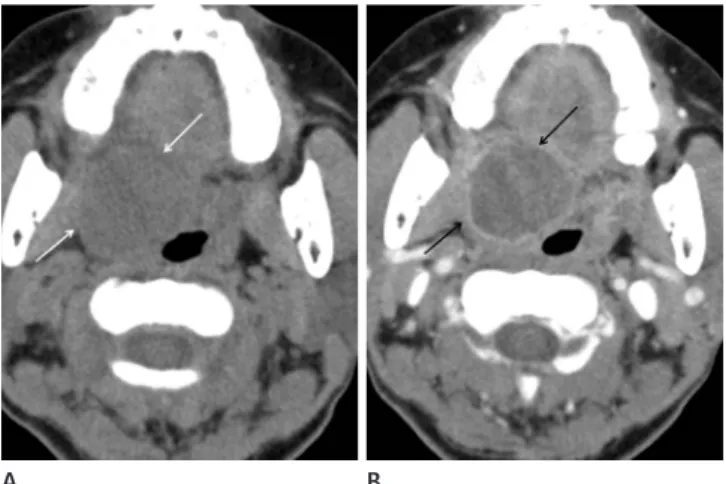
The patient underwent tumor resection under general an- esthesia and the specimen was a grayish brown soft tissue mass measuring 3.5 × 2.5 × 2.0 cm. Once sectioned, the tu- mor was found to be mainly solid with some necrotic chang- es. We believe that the non-enhancing portion of the tumor on the Gd-enhanced T1-weighted images was consistent with the intratumoral necrotic portion. Haematoxylin and Eosin- stained sections of the resected mass showed marked plasma- cytoid cell proliferation, polygonal cells with eccentric nuclei, and abundant hyaline eosinophilic cytoplasm in the myxoid Fig. 1. A 53-year-old woman with a palpable mass on the soft palate.
A. Pre-enhanced CT images show a mass on the right soft palate (white arrows).
B. On the contrast-enhanced CT images, the mass is faintly enhanced, and shows peripheral wall enhancement (black arrows), suggesting a capsular structure.
Fig. 2. Axial (A) and coronal (B) T1-weighted images show a slightly heterogeneous mass with iso-signal intensity on the right soft palate (arrows).
Axial (C) and coronal (D) T2-weighted images show the heterogeneous hyperintense mass on the right soft palate (arrows).
Axial (E) and coronal (F) Gd-enhanced T1-weighted images show a heterogeneously enhancing tumor with a poorly enhancing intratumoral area (arrow), suggesting the presence of a focal necrotic component. A poorly enhancing portion on Gd-enhanced T1-weighted images is nearly consistent with a subtle low attenuated portion on the contrast-enhanced CT images (Fig. 1B).
A B
A B C
D E F
Hun-Cheol Lim, et al
submit.radiology.or.kr J Korean Soc Radiol 2011;65(1):23-26
25
components. We believe that faint enhancement inside the mass on contrast-enhanced CT images corresponded to the plasmacytoid cells and surrounding myxoid stroma occupy- ing the greater part of the tumor (1). Furthermore, intratu- moral necrosis showed poor enhancement on the contrast- enhanced CT and Gd-enhanced T1-weighted images. The intratumoral necrotic portion was suspected of representing malignant changes of myoepithelioma. There are several patho- logic findings associated with malignancy including infiltra- tive growth, necrosis, cytologic atypia, high mitotic rates, and cellular pleomorphism (7, 8). Thus, the lesion in our patient had uncertain malignancy potential; because, although there was focal necrosis, there were no other pathologic findings that suggested malignancy.
Palatal masses may be associated with many diseases in- cluding minor salivary gland tumors, neurinoma, haemangi- oma, malignant tumors originating from the oral mucosa, and inflammatory disease (6). However, it is easy to distin- guish inflammatory from neoplastic diseases based on clinical findings such as fever or tenderness. Minor salivary glands are located everywhere in the oral mucosa. Thus, minor sali- vary gland tumors are primarily located in the palate (50%), lips (15%), cheek mucosa (12%), tongue (5%), and floor of the mouth (5%). Minor salivary gland tumors are a heteroge- neous group of neoplasms with a broad range of histological types, occurring in the minor salivary gland. The soft palate is the second most common site of minor salivary gland tu- stroma background (Fig. 3). S-100 protein and cytokeratin
immunostaining was strongly positive in the tumor cells, whereas smooth muscle actin staining was focally positive in the tumor cells. These histologic and immunostaining obser- vations were consistent with plasmacytoid myoepithelioma.
DISCUSSION
Myoepithelioma is a benign tumor composed of sheets and islands of various proportions of spindle, plasmacytoid, epi- thelioid, and clear cells that exhibit myoepithelial but not duc- tal differentiation (3). Myoepithelioma was initially consid- ered to be a subtype of pleomorphic adenoma, but was later distinguished from other types of pleomorphic adenoma.
This type of tumor has been classified as a distinct clinico- pathological entity by the World Health Organization since 1991 (4). Myoepithelioma may originate from the salivary gland, lacrimal gland, breast, bronchus, or skin. The most common location of myoepitheliomas is the salivary gland, particularly the parotid gland. Myoepitheliomas account for less than 1% of salivary gland neoplasms (4). The ages of pa- tients with myoepitheliomas range from 9 to 85 years, with an average of 44 years; this disease occurs most commonly among patients in their 30s (2).
A few reports have described the appearance of myoepithe- lioma in the soft palate on CT and MRI. Monzen et al. (5) re- ported CT and magnetic resonance imaging features of myo- epithelioma arising from the soft palate. In this report, benign myoepiethelioma was found to have a smooth surface and faint contrast enhancement inside the mass on contrast-en- hanced CT images. This lesion appears as a well-defined ovoid mass with iso-signal intensity compared to muscle on T1-weighted images. On T2-weighted images, the mass ap- pears as an area with high signal intensity while the well-en- hanced periphery of the mass shows low T2-signal intensity, suggesting a capsular structure. Slightly heterogeneous en- hancement of the mass is evident on contrast-enhanced T1- weighted images. Such findings are similar to our case and we can assume that the imaging features of our patient were con- sistent with myoepithelioma.
There are many biologic factors that influence the enhance- ment patterns of tumors such as vascularity and histological
Fig. 3. Haematoxylin and Eosin-stained sections (400 × magnification) of the resected mass show marked plasmacytoid cell proliferation, po- lygonal cells with eccentric nuclei, and abundant hyaline eosinophilic cytoplasm with some myxoid stroma background. No ductal differenti- ation is observed. Histologic findings suggest myoepithelioma.
Computed Tomography and Magnetic Resonance Imaging of Myoepitheliloma in the Soft Palate
submit.radiology.or.kr
J Korean Soc Radiol 2011;65(1):23-26
26
lioma of the soft palate. Acta Cytol 1997;41:1856-1858 4. Seifert G, Brocheriou C, Cardesa A, Eveson JW. WHO Inter-
national Histological Classification of Tumours. Tentative Histological Classification of Salivary Gland Tumours.
Pathol Res Pract 1990;186:555-581
5. Monzen Y, Fukushima N, Fukuhara T. Myoepithelioma and malignant myoepithelioma arising from the salivary gland:
computed tomography and magnetic resonance findings.
Australas Radiol 2007;51 Suppl:B169-B172
6. Hiwatashi A, Matsumoto S, Kamoi I, Yamashita H, Nakashi- ma A. Imaging features of myoepithelioma arising from the hard palate. A case report. Acta Radiol 2000;41:417-419 7. Suba Z, Németh Z, Gyulai-Gaál S, Ujpál M, Szende B, Sz-
abó G. Malignant myoepithelioma. Clinicopathological and immunohistochemical characteristics. Int J Oral Max- illofac Surg 2003;32:339-341
8. Savera AT, Sloman A, Huvos AG, Klimstra DS. Myoepithelial carcinoma of the salivary glands: a clinicopathologic study of 25 patients. Am J Surg Pathol 2000;24:761-774
9. Pons Vicente O, Almendros Marqués N, Berini Aytés L, Gay Escoda C. Minor salivary gland tumors: a clinicopathologi- cal study of 18 cases. Med Oral Patol Oral Cir Bucal 2008;
13:E582-E588 mors. The most common histological type of minor salivary
gland tumors is pleomorphic adenoma. Myoepithelioma is also a rare histologic type of minor salivary gland tumor (9).
It is difficult to differentiate myoepithelioma from other tu- mors based on radiological findings. Histopathologic studies area needed to differentially diagnose myoepitheliomas. Some imaging studies have suggested that myoepitheliomas contain a relatively large cystic component compared to pleomorphic ad- enomas. Additionally, pleomorphic adenomas show relatively delayed enhancement patterns on contrast-enhanced CT imag- es (1, 2). We suggest that when there is a submucosal soft palate mass, radiologists should consider myoepitheliomas in the ra- diological differential diagnosis of soft palate tumors.
REFERENCES
1. Kim HS, Lee WM, Choi SM. Myoepitheliomas of the soft palate: helical CT findings in two patients. Korean J Radiol 2007;8:552-555
2. Wang S, Shi H, Wang L, Yu Q. Myoepithelioma of the pa- rotid gland: CT imaging findings. AJNR Am J Neuroradiol 2008;29:1372-1375
3. Katsuyama E, Kaneoka A, Higuchi K, Takasu K. Myoepithe-
연구개에 발생한 근상피종의 CT와 MRI 영상 소견: 증례 보고
1임훈철1
·
유인규1·
박미자2·
장동식3우리는 연구개에 있는 소타액선에서 발생한 근상피종의 CT와 MRI 영상 소견을 증례보고하고자 한다. CT상에서 종양 은 둥근 모양이고 부드러운, 일부 엽상 형태를 가지며 조영제 주입 후에 변연부에 약간의 조영 증강을 보인다. T1 강조 영상 에서 종양은 주변 인두 근육과 비교하여 불균질한 동신호 강도를 보였다. 그리고 종양은 T2 강조 영상에서는 불균질한 고 신호 강도를 보이며 강한 비균질한 조영 증강을 보였다. 영상의학과 의사들은 연구개에서 발생하는 종양들 중에서 감별 진 단으로 근상피종을 반드시 고려해야 한다.
을지대학병원 1영상의학과, 2병리과, 3이비인후과