354
Print ISSN 1738-5520 / On-line ISSN 1738-5555 Copyright © 2010 The Korean Society of Cardiology IMAGES IN CARDIOVASCULAR MEDICINE
DOI 10.4070 / kcj.2010.40.7.354
Open Access
External Mass Compressing the Left Atrium on Transthoracic Echocardiography
Eun-Joo Im, MD, Su-Sung Oh, MD, Kyung-Mi Kang, MD, Ji-Hoon Kim, MD, Keon-Woong Moon, MD, Ki-Dong Yoo, MD and Chul-Min Kim, MD
Division of Cardiology, Department of Internal Medicine, St. Vincent’s Hospital, The Catholic University of Korea, Suwon, Korea
Received: December 5, 2009 / Accepted: December 29, 2009
Correspondence: Chul-Min Kim, MD, Division of Cardiology, Department of Internal Medicine, St. Vincent’s Hospital, The Catholic University of Korea, 93-6 Ji-dong, Paldal-gu, Suwon 442-723, Korea
Tel: 82-31-247-7139, Fax: 82-31-249-7139, E-mail: [email protected]
cc This is an Open Access article distributed under the terms of the Creative Commons Attribution Non-Commercial License (http://creativecommons.org/licens- es/by-nc/3.0) which permits unrestricted non-commercial use, distribution, and reproduction in any medium, provided the original work is properly cited.
A 74-year-old man who had suffered from dysphagia for about 2 months was referred to our hospital for treatment of an esophageal mass. He had diabetes mellitus and hyperten- sion for the previous 3 years. On physical examination, his blood pressure measured 110/70 mmHg at a heart rate of
85/min. No murmurs were heard on auscultation. An electro- cardiogram was a normal sinus rhythm and chest radiography revealed no active lung lesions. Esophagogastroduodenos- copy showed an ulcero-infiltrative mass in the mid-to-lower esophagus (Fig. 1). Chest computed tomography confirmed
Fig. 1. Esophagogastroduodenoscopy shows a 3.0×2.4×8.0 cm esophageal mass.
Fig. 2. Chest CT reveals an esophageal mass (*) which compress- es the left atrium (†).
*
†
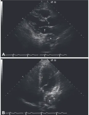
Fig. 3. Two-dimensional echocardiographic parasternal long axis view (A) and apical four-chamber view (B). A round and well-de- marcated esophageal mass (*) compresses the left atrium (†), which resembles the image on chest CT.
A
B
*
*
†
†
Eun-Joo Im, et al. 355
an obstructive, 3.0×2.4×8.0 cm intraluminal esophageal mass arising from the right anterior side (Fig. 2); there was no evi- dence of direct invasion into the major organs of the medias- tinum. The final biopsy result was squamous cell carcinoma.
Transthoracic echocardiography demonstrated normal sys- tolic and diastolic function. As noted on the parasternal long axis view and the apical 4-chamber view, the left atrium was compressed by the esophageal mass (Fig. 3). He underwent an esophagectomy, esophagogastrostomy, and feeding jeju- nostomy for a malignancy.
Extrinsic compression of the left atrium is an uncommon clinical event. Several case reports exist in patients with esoph- ageal carcinomas,1) and esophageal hematomas,2) dissecting an- eurysms of the ascending aorta,3) and mediastinal masses.4) The transthoracic echocardiography of the patient presented
herein showed a typical esophageal mass that compressed the left atrium.
REFERENCES
1) Shah A, Tunick PA, Greaney E, Pfeffer RD, Kronzon I. Diagnosis of esophageal carcinoma because of findings on transesophageal echo- cardiography. J Am Soc Echocardiogr 2001;14:1134-6.
2) Im E, Shim CY, Hwang HJ, et al. Transthoracic echocardiographic detection, differential diagnosis, and follow-up of esophageal hemato- ma. Korean Circ J 2007;37:666-70.
3) Walpot J, Amsel B, Pasteuning WH, Olree M. Left atrial compres- sion by dissecting aneurysm of the ascending aorta. J Am Soc Echo- cardiogr 2007;20:1220.e4-6.
4) Pehlivan Y, Sevinc A, Ozer O, Sari I, Davutoglu V. Mediastinal tes- ticular tumor compressing the left atrium in a young male presenting initially with symptoms of left heart failure. Intern Med 2009;48:169-71.