155 Introduction
Forced separation of the left atrial (LA) wall layers by blood, is known as left atrial dissection (LatD). LatD is a rare and severe complication of cardiac surgery1) occurring in up to 0.84% of mitral valve replacements (MVR).2)
Other rarer causes include mitral valve (MV) repair, aortic valve (AV) surgeries, myocardial infarction, percutaneous cor- onary intervention, cardiac mass excision, left ventricular (LV) aneurysm repair, coronary artery bypass graft (CABG), blunt cardiac trauma, pulmonary vein cannulation, infective endo- carditis (IE) and spontaneous occurrence.1)
It is most frequently associated with atrioventricular junc- tion injuries. However, there are other less common cases in- cluding those remote from the atrioventricular junction.1)
We report a case of infected LatD after CABG, MVR, AV and ascending aortic root replacement which was presented with disseminated intravascular coagulation (DIC) and septi- cemia.
To the best of our knowledge, this is the first report of LA dissecting flap associated with infective vegetations which was identified by transesophageal echocardiography (TEE).
Case
A 69-year-old man was admitted in our hospital due to fe- ver, chill, orthopnea and paroxysmal nocturnal dyspnea. In
physical examination, respiratory sounds were reduced in both his lower half of lung fields.
He had undergone a major cardiac surgery including CABG, MVR, aortic valve replacement (AVR) and ascending aorta replacement due to severe coronary artery disease, signif- icant valvular heart disease (rheumatic) and aneurysmally di- lated ascending aorta, 75 days prior to his recent admission.
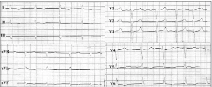
An electrocardiogram revealed atrial fibrillation rhythm, normal axis and non-specific ST-T changes in inferior and lat- eral leads (Fig. 1). The posteroanterior view of the chest radi- ography showed massive bilateral pleural effusion. Transtho- racic echocardiography followed by TEE showed normal LV size with preserved systolic function, bioprosthetic MV with acceptable hemodynamic and mobility and no paravalvular re-
pISSN 1975-4612/ eISSN 2005-9655 Copyright © 2014 Korean Society of Echocardiography www.kse-jcu.org http://dx.doi.org/10.4250/jcu.2014.22.3.155
CASE REPORT J Cardiovasc Ultrasound 2014;22(3):155-157
Infective Left Atrial Dissecting Flap after Cardiac Surgery
Maryam Nabati, MD, Sasan Tabiban, MD, Ali Ghaemian, MD, Babak Bagheri, MD, and Mojtaba Shokri, MSc
Department of Cardiology, Faculty of Medicine, Mazandaran University of Medical Sciences, Sari, Iran
Left atrial dissection (LatD), defined as the forced separation of the left atrial (LA) wall layers by blood, is a rare and severe compli- cation of cardiac surgery. It is most frequently associated with atrioventricular junction injuries. We report a case of infected LatD after coronary artery bypass graft, mitral valve replacement, aortic valve replacement and ascending aortic root replacement. The patient was presented with septicemia and disseminated intravascular coagulation. To the best of our knowledge, this is the first case report of LA dissecting flap concomitant with attached infective vegetations identified by transesophageal echocardiography.
KEY WORDS: Atrial dissection · Transesophageal echocardiography · Mitral valve · Prosthesis · Infective endocarditis.
• Received: April 8, 2014 • Revised: May 18, 2014 • Accepted: August 20, 2014
• Address for Correspondence: Maryam Nabati, Department of Cardiology, Fatemeh Zahra Teaching Hospital, Faculty of Medicine, Mazandaran University of Medical Sciences, Artesh Boulevard, Sari 48188-13771, Iran Tel: +98-11-3332-4002, Fax: +98-11-3332-4002, E-mail: [email protected]
• This is an Open Access article distributed under the terms of the Creative Commons Attribution Non-Commercial License (http://creativecommons.org/licenses/by-nc/3.0) which permits unrestricted non-commercial use, distribution, and reproduction in any medium, provided the original work is properly cited.
Fig. 1. An electrocardiogram revealed atrial fibrillation rhythm, normal axis and non-specific ST-T changes in inferior and lateral leads.
Journal of Cardiovascular Ultrasound 22 | September 2014
156
gurgitation. Bioprosthetic AV had acceptable hemodynamic and mobility without paravalvular regurgitation. There was normal functioning ascending aorta Dacron tube graft with- out leakage from proximal and distal part of prosthesis.
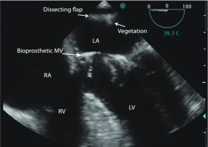
The LA was enlarged and dissection was found on the pos- terior wall. Two homogenous particles (1.18 × 0.4 cm and 0.9
× 0.58 cm) were attached to the dissecting flaps which were highly suggestive for vegetations and IE (Fig. 2, 3, and 4, Supplementary movie 1, 2, and 3).
Laboratory tests showed severe coagulopathy, high levels of fibrin degradation products, leukocytosis (white blood cells count: 26700/cm3, predominantly poly morphonucleated), se- vere anemia, thrombocytopenia, elevated liver enzymes and serum creatinine levels. These data were suggestive for DIC.
The blood culture revealed enterococci septicemia.
His general condition deteriorated rapidly. He suffered from tonic-clonic seizures, diminished consciousness, widespread mucosal bleeding and oliguria. Bilateral chest tubes were in- serted for massive pleural effusion. The patient was treated with intravenous vancomycin, cefepim, gentamycin and oral rifampin. He received infusion of fresh frozen plasma, packed cells, valproic acid, diazepam and phenytoin.
Based on his poor general condition, cardiac surgery was not performed and finally he expired from multi organ failure.
Autopsy findings were consistent with clinical, laboratory and echocardiographic findings.
Discussion
LatD is defined as a gap from the mitral annular area to the LA wall, developing a new cavity with or without connection into the true LA. The false chamber presents as an echolucent area and may cause partial elimination of atrial cavity.3) Similar to what happens in aortic dissection, the false chamber may be compressed during ventricular systole as LA is being filled.4)
The etiology of LatD after MVR is unknown. Suggested causes include: inappropriate suturing of the annulus to the prosthesis, inadvertent trauma to the posterior part of mitral annulus, intense debridement of the annulus, intense tension on posterior annulus sutures and inadvertent injury to the LA endocardium during atrial thrombectomy.5)
Suggested etiologies for LatD after AVR are division of the non-coronary aortic annulus and entrance into the LA wall due to removal of calcification or inadvertent incision of LV outflow tract.2)
LA wall is formed by two muscular layers that are longitu- dinally and circumferentially aligned. Disruption in this site may be a predisposing factor for LatD.2)
In our case, dissecting flap was remote from the atrioven- tricular junction. Therefore, flap was probably induced by un- intended LA endocardial injury. Time to presentation of LatD after MVR ranged from 1 day to 9 years.3) In our patient, it was diagnosed after seventy five days.
Reported clinical presentations include: acute low output
Fig. 2. Transesophageal echocardiography in mid esophageal four chamber view (0 degree) demonstrated a linear dissecting flap and its attached (irregular shaped) particles originated from left atrial wall. LA:
left atrium, LV: left ventricle, MV: mitral valve, RA: right atrium, RV: right ventricle.
Fig. 3. Transesophageal echocardiography in mid esophageal two chamber view (106 degree) demonstrated a linear dissection flap originated from posterior side of left atrial wall. LA: left atrium, LV: left ventricle, MV: mitral valve.
Fig. 4. Transesophageal echocardiography in mid esophageal two chamber view (97 degree) demonstrated a linear dissection flap originated from posterior side of left atrial wall. LA: left atrium, LV: left ventricle, MV: mitral valve.
Infective Left Atrial Dissecting Flap | Maryam Nabati, et al.
157 syndrome and congestive heart failure in the immediate post-
operative period, symptoms of moderate cardiac failure, per- sistent fever or it may be an incidental finding during routine echocardiography assessment.3)
Our patient presented as persistent fever and progressive dyspnea. Dyspnea might be caused by bilateral severe pneu- monia with parapneumonic effusion, septicemia and DIC.
TEE clearly improved the diagnostic capacity of echocardiog- raphy as a technique of choice in the semi-invasive diagnosis of LatD.3) LatD usually requires surgical treatment to stop blood flow to the false chamber.3)
Rupture of atrial wall with fistula formation into the LA or rupture into the pericardial space with bleeding or tamponade may occur. Also, pulmonary vein obstruction with reduced blood return and pulmonary edema have been reported.3) In patients with LatD and no or minimal symptoms, close fol- low-up is recommended.5)
In conclusion, previous cardiac surgery seems to be the main risk factor for developing LatD. Our study showed that a complicated infective flap in LA wall can be a source for pro- ducing septic systemic emboli and DIC. To the best of our knowledge, this form of LA dissecting flap has not been re-
ported in the literature previously.
Supplementary movie legends Movie 1. For Fig. 2.
Movie 2. For Fig. 3.
Movie 3. For Fig. 4.
References
1. Fukuhara S, Dimitrova KR, Geller CM, Hoffman DM, Ko W, Tranbaugh RF. Left atrial dissection: etiology and treatment. Ann Thorac Surg 2013;95:1557-62.
2. Leissner KB, Srinivasa V, Beutler S, Matyal R, Badr R, Haime M, Mahmood FU. Left atrial dissection and intramural hematoma after aor- tic valve replacement. J Cardiothorac Vasc Anesth 2011;25:309-10.
3. Gallego P, Oliver JM, González A, Domínguez FJ, Sanchez-Recalde A, Mesa JM. Left atrial dissection: pathogenesis, clinical course, and trans- esophageal echocardiographic recognition. J Am Soc Echocardiogr 2001;
14:813-20.
4. Tolpin DA, Collard CD, Thomas Z, Pan W. Left atrial dissection associ- ated with pulmonary vein cannulation. Anesth Analg 2009;109:1409-12.
5. Martinez-Sellés M, García-Fernandez MA, Moreno M, Bermejo J, Delcán JL. Echocardiographic features of left atrial dissection. Eur J Echo- cardiogr 2000;1:147-50.