Introduction
Echocardiography is a useful diagnostic technique for detect- ing cardiac masses. Cardiac masses have various echocardio- graphic manifestations following etiology. Commonly observed intracardiac masses are tumors such as myxoma, metastatic tumor, or thrombi due to atrial fibrillation or systolic dysfunc- tion, and vegetation due to infective endocarditis. Extrinsic le- sion is mediastinal tumor, coronary and aortic aneurysm, and so on.
Diaphragmatic hernias include Morgagni hernia and hiatal hernia that are causes of cardiac mass lesion. However, they are relatively rare on routine echocardiographic procedures. Thus we can easily overlook or misdiagnose them. We present three cases of diaphragmatic hernia resembling cardiac mass on transthoracic echocardiography, which are subsequently iden- tified hiatal hernia and Morgagni hernia.
Case Case 1
A 75-year-old female with hypertension was presented to our
pISSN 1975-4612/ eISSN 2005-9655 Copyright © 2015 Korean Society of Echocardiography www.kse-jcu.org http://dx.doi.org/10.4250/jcu.2015.23.2.107
department because of dyspnea on exertion. On physical exam- ination, blood pressure was 156/74 mm Hg. Crackle or mur- mur was not audible on auscultation. A 12-lead electrocardio- gram showed normal sinus rhythm. Chest X-ray showed cardiomegaly and air-fluid level that suggest large hiatal her- nia. A two-dimensional transthoracic echocardiogram showed huge extrinsic mass compress left atrium and filled with spon- taneous echo contrast that has internal swirling flow (Fig. 1).
Case 2
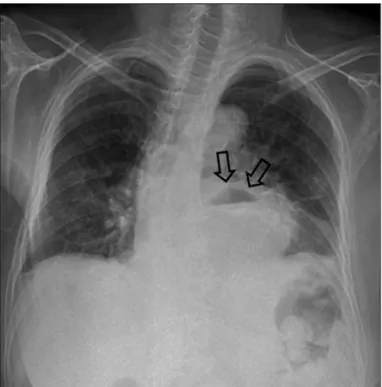
A 71-year-old female with prior history of hypertension and asthma presented neurosurgery department complaining of recently aggravated lower motor weakness and back pain. On physical examination, vital sign was stable. Crackle and mur- mur was not audible on auscultation. A 12-lead electrocardio- gram showed normal sinus rhythm and left anterior fascicular block. Chest X-ray showed mild cardiomegaly, air-fluid level adjacent left cardiac boarder suggesting hiatal hernia, and blunt- ing of left costophrenic angle suggesting left pleural effusion (Fig. 2). She underwent a two-dimensional transthoracic echo- cardiography for the preoperative evaluation.
CASE REPORT J Cardiovasc Ultrasound 2015;23(2):107-112
• Received: November 4, 2014 • Revised: March 9, 2015 • Accepted: May 19, 2015
• Address for Correspondence: Wook-Jin Chung, Department of Cardiovascular Medicine, Gachon University Gil Medical Center, 21 Namdong-daero 774beon-gil, Namdong-gu, Incheon 405-760, Korea Tel: +82-32-460-3054, Fax: +82-32-469-1906, E-mail: [email protected]
• This is an Open Access article distributed under the terms of the Creative Commons Attribution Non-Commercial License (http://creativecommons.org/licenses/by-nc/3.0) which permits unrestricted non-commercial use, distribution, and reproduction in any medium, provided the original work is properly cited.
Unusual Diaphragmatic Hernias Mimicking Cardiac Masses
Si Hun Kim, MD1,2, Myoung Gun Kim, MD1,2, Su Ji Kim, MD1,2, Jeonggeun Moon, MD1,2, Woong Chol Kang, MD, PhD1,2, Mi-Seung Shin, MD, PhD1,2, and Wook-Jin Chung, MD, PhD1,2
1Department of Cardiovascular Medicine, Gachon University Gil Medical Center, Incheon, Korea
2Gachon Cardiovascular Research Institute, Gachon University School of Medicine, Incheon, Korea
Hiatal hernia and Morgagni hernia are sorts of diaphragmatic hernias that are rarely detected on transthoracic echocardiography.
Although echocardiographic findings have an important role for differential diagnosis of cardiac masses, we often might overlook diaphragmatic hernia. We report three cases of diaphragmatic hernias having specific features. The first case is huge hiatal hernia that encroaches left atrium with internal swirling flow on transthoracic echocardiography. The second case is a hiatal hernia that encroaches on both atria, incidentally detected on preoperative echocardiography. The third case is Morgagni hernia which encroaches on the right atrium only. So, we need to consider possibility of diaphragmatic hernia when we find a cardiac mass with specific echocardiographic features.
KEY WORDS: Cardiac mass · Echocardiography · Diaphragmatic hernia · Hiatal hernia · Morgagni hernia.
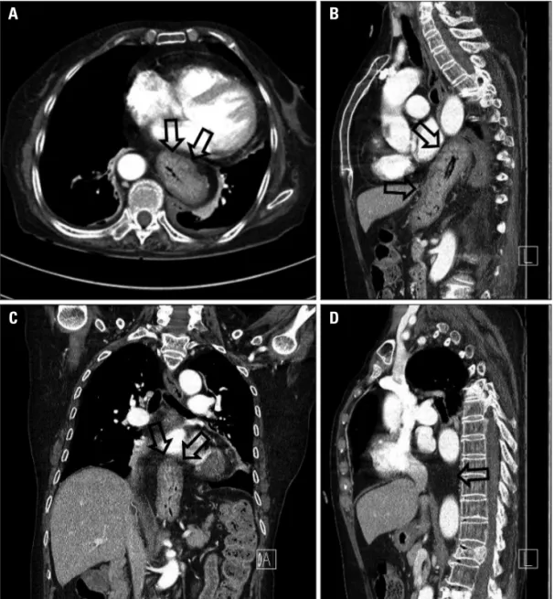
A two-dimensional transthoracic echocardiography showed huge extracardiac mass (3.8 × 3.5 cm) adjacent to the left heart compressing left atrium and mobile echogenic mass (3.4 × 2.8 cm) in right atrium (Fig. 3). The patient subsequently under- went chest computed tomography for differential diagnosis of mass lesion. Chest computed tomography showed large hiatal hernia in the posterior mediastinum (Fig. 4). After consulting a surgeon, the patient underwent an open hiatal hernia repair.
Seven weeks later, a two-dimensional transthoracic echocar- diography showed disappearance of preexisting mass adjacent to the left atrium and decreased echogenic mass in right atrium.
Case 3
A 28-year-old female without past medical history was pre-
sented to our department because of recently identified right pericardial mass like lesion on chest X-ray. But, she did not have any symptom. On physical examination, blood pressure was 125/75 mm Hg. Crackle and murmur was not audible on aus- cultation. A 12-lead electrocardiogram showed normal sinus rhythm. Chest X-ray showed opacity in right pericardial area suspected pericardial fat tissue. We performed a two-dimen- sional transthoracic echocardiogram and chest computed to- mography to identify right pericardial opacity.
A two-dimensional transthoracic echocardiogram showed hypoechoic mass encroaching posterior aspect of both atria on parasternal short axis view (Fig. 5). Chest computed tomogra- phy showed prominent fat tissue in right cardiophrenic area with suspicious internally radiating vasculature that was sus-
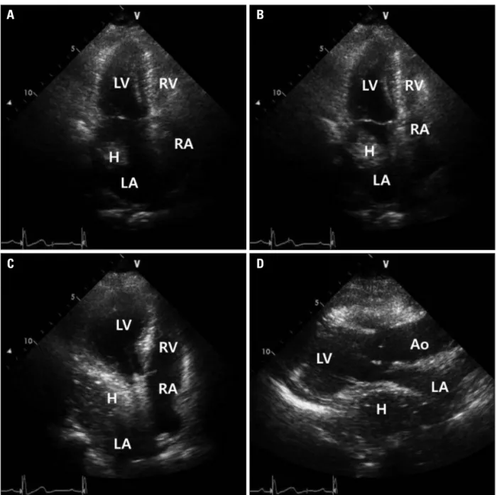
Fig. 1. Hiatal hernia with internal swirling flow. A: Apical 4-chamber view of transthoracic echocardiography shows intracardiac mass in left atrium. B:
Posterior plane of (A) shows larger intracardiac mass in left atrium. C: More posterior plane of (B) shows huge extrinsic mass that compress left atrium with internal swirling flow. D: Parasternal long axis view of transthoracic echocardiography shows huge extrinsic mass that compress left atrium with internal swirling flow. Ao: aorta, H: hiatal hernia, LA: left atrium, LV: left ventricle, RA: right atrium, RV: right ventricle.
A
C
B
D
Unusual Diaphragmatic Hernias | Si Hun Kim, et al.
pected to be Morgagni’s hernia (Fig. 6).
Discussion
Diaphragmatic hernia such as hiatal and Morgagni hernia can be mimic cardiac masses on transthoracic echocardiography.
Previously, there was a report of hiatal hernia and Morgagni hernia that mimic pericardial lipoma.1) They only focused on differential diagnosis of hiatal hernia and echocardiographic findings wasn’t concerned. For the rarity of these cases, it could be easily overlooked or misdiagnosed. So, we would like to re- port and discuss about that echocardiographic findings could give us useful information for the differential diagnosis of car- diac masses including hiatal hernia.
Hiatal hernia is characterized by the displacement of the gastroesophageal junction and part of the stomach into the me- diastinum. Often asymptomatic, it may present wide spectrum of clinical manifestation from epigastric pain due to concomi- tant gastroesophageal reflux to postprandial syncope.2) It can be easily diagnosed with upper gastrointestinal barium exami- nation or endoscopy. However, it can mimic a cardiac mass on transthoracic echocardiography. Echocardiographic feature that
Fig. 2. Chest X-ray showing air-fluid level that suggest large hiatal hernia (arrows).
Fig. 3. Echocardiographic findings of hiatal hernia showing cardiac mass involving both atria. A: Parasternal long axis view of transthoracic echocardiography shows huge extracardiac mass (3.8 × 3.5 cm) adjacent to the left heart compressing left atrium. B: Parasternal long axis RV inflow view of transthoracic echocardiography shows mobile echogenic mass (3.4 × 2.8 cm) in right atrium. Apical 4-chamber view (C) and apical 2-chamber view (D) of transthoracic echocardiography shows mobile echogenic mass that compressing left atrium. Ao: aorta, H: hiatal hernia, LA:
left atrium, LV: left ventricle, RA: right atrium, RV: right ventricle.
A
C
B
D
may suggest a hiatal hernia was first described by Nishimura et al.3) They reported that left atrial mass like lesion was in a maximal size when the left atrium was seen in a posterior plane, but smaller or absent as the left atrium was seen in more anteri- or planes. D’Cruz and Hancock4) reported additional findings.
One is changes in the degree of encroachment of the hiatal her- nia on the left atrium depending on respiratory cycle. Another is loss of the normal sharply defined sonolucency of the descend- ing thoracic aorta due to superimposition of the hiatal hernia.
The other is the visualization of swirling echodensities follow- ing the oral ingestion of carbonated beverage.4)
In case 1, transthoracic echocardiography showed spontane- ous echo contrasts that had internal swirling flow without in- gestion of carbonated beverage and this finding is helpful for diagnosing hiatal hernia. But we didn’t undergo additional in- gestion of carbonated beverage in case 2 and 3, because chest X- ray and computed tomography showed definite hiatal hernia in case 2 and computed tomography showed Morgagni hernia
A
C
B
D
Fig. 4. Chest computed tomography of hiatal hernia showing cardiac mass involving both atria. Chest computed tomography shows large hiatal hernia compressing left atrium (A and B) and right atrium (C and D) (arrows).
Fig. 5. Echocardiographic findings of Morgagni’s hernia. Parasternal short axis view of transthoracic echocardiography shows large hypoechoic mass encroaching posterior aspect of right atrium. Ao:
aorta, LA: left atrium, MH: Morgagni hernia, RA: right atrium, RV: right ventricle.
Unusual Diaphragmatic Hernias | Si Hun Kim, et al.
consisting of omental fat without stomach or bowel in case 3.
The first case showed typical echocardiographic findings hia- tal hernia. At first, extrinsic mass that compressing left atrium is maximal size which was seen posterior plane. And it gradually decreased when it was seen anterior plane. The next, it showed internal swirling flow on parasternal ling axis view and apical 4-chamber view (Fig. 1). These findings were helpful for dif- ferential diagnosis of hiatal hernia without other imaging mo- dalities.
Interestingly, although hiatal hernia is usually known to en- croach on the left atrium, transthoracic echocardiography in the second case showed both atrial mass-like lesion separately be- cause hiatal hernia was large enough to encroach both atria. So, we need to consider hiatal hernia as a differential diagnosis of extrinsic cardiac mass involving both atria.
Morgagni hernia is rarely reported diaphragmatic hernia due to defect of anterior diaphragm and first described by Giovan- ni Morgagni in 1769.5) Foramina of Morgagni is located imme- diately adjacent to the xiphoid process of the sternum and her- niation occurred through it mostly right side of body.6) Mostly hernia sac contains transverse colon, omentum, liver, and, less frequently, small bowel or stomach. Morgagni hernia gets di- agnosed late because it can be asymptomatic or present only ambiguous gastrointestinal or respiratory symptoms.7) In as- ymptomatic patient like this case, smooth supradiaphragmatic shadow of cardiac margin can be founded on routine chest X- ray. Nonetheless echocardiography has been shown to be use- ful, computed tomography is the most sensitive because it can give anatomical feature of the hernia and its complications such as strangulation.7) In our case, patient was asymptomatic and only had supradiaphragmatic shadow of right cardiophrenic angle on chest X-ray. Parasternal short axis view of transthorac- ic echocardiography showed encroachment of extrinsic mass le- sion on the posterior aspect of both atria. Chest computed to-
mography showed prominent fat tissue in right cardiophrenic area and it contained radiating vasculature. These findings can be seen in Morgagni hernia that consists of omental fat and li- poma. The most important finding for differential diagnosis is that the presence of radiating fine linear or curvilinear densities indicates omental blood vessels and radiating vasculature indi- cates Morgagni hernia.8) Additionally, magnetic resonance im- aging can be helpful. Since it readily provides images in the cor- onal and sagittal planes, it is better suited to recognize this diaphragmatic defect which lies predominantly in the axial plane.9)
In summary, diaphragmatic hernia such as hiatal and Mor- gagni hernia can be mimic cardiac masses on transthoracic echocardiography. As those were rare, we could easily overlook several specific echocardiographic findings which would help distinguish diaphragmatic hernia from other cardiac mass. So, we need to consider possibility of diaphragmatic hernia when we would find cardiac mass with specific echocardiographic features.
References
1. Kim J, Han D, Sohn C, Kim JS, Park YH. Catheter ablation of ven- tricular arrhythmias via the radial artery in a patient with prior myocardial infarction and peripheral vascular disease. Korean Circ J 2012;42:632-7.
2. Oishi Y, Ishimoto T, Nagase N, Mori K, Fujimoto S, Hayashi S, Ochi Y, Kobayashi K, Tabata T, Oki T. Syncope upon swallowing caused by an esophageal hiatal hernia compressing the left atrium: a case re- port. Echocardiography 2004;21:61-4.
3. Nishimura RA, Tajik AJ, Schattenberg TT, Seward JB. Diaphrag- matic hernia mimicking an atrial mass: a two-dimensional echocardiographic pitfall. J Am Coll Cardiol 1985;5:992-5.
4. D’Cruz IA, Hancock HL. Echocardiographic characteristics of diaphrag- matic hiatus hernia. Am J Cardiol 1995;75:308-10.
5. Khouzam RN, Akhtar A, Minderman D, Kaiser J, D’Cruz IA. Echo- cardiographic aspects of hiatal hernia: a review. J Clin Ultrasound 2007;35:
196-203.
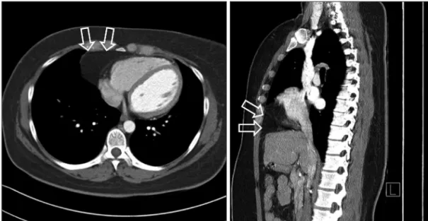
Fig. 6. Chest computed tomography of Morgagni’s hernia. Chest computed tomography shows prominent fat tissue in right cardiophrenic area with suspicious internal vasculature that suggested Morgagni’s hernia (arrows).
6. Torfs CP, Curry CJ, Bateson TF, Honoré LH. A population-based study of congenital diaphragmatic hernia. Teratology 1992;46:555-65.
7. Lin ST, Moss DM, Henderson SO. A case of Morgagni hernia presenting as pneumonia. J Emerg Med 1997;15:297-301.
8. Sutro WH, King SJ. Computed tomography of Morgagni hernia. N Y
State J Med 1987;87:520-1.
9. Yeager BA, Guglielmi GE, Schiebler ML, Gefter WB, Kressel HY.
Magnetic resonance imaging of Morgagni hernia. Gastrointest Radiol 1987;
12:296-8.