904 www.eymj.org
INTRODUCTION
Inspissated bile syndrome (IBS) is defined as the obstruction of the extrahepatic duct by a bile plug, sludge without bile duct malformation, congenital chemical defects of the bile, or he- patocellular lesions.1 This inspissation of bile and mucus with- in the bile ducts is usually caused by blood transfusion, pro- longed parenteral nutrition, or diuretics.2,3 Management options include medication, surgery, chemotherapy, radiotherapy, and invasive surgical interventions, such as a percutaneous cholecystostomy, endoscopic retrograde cholangiopancrea- tography, internal biliary drainage, and percutaneous transhe- patic biliary drainage (PTBD).4-7 However, there are limited surgical and procedural options for newborns, because these treatments are invasive, difficult, and frequently accompanied
by postoperative complications. Consequently, very few re- ports of such cases are present in the literature. We herein de- scribe our experience with a two-month-old infant presenting with IBS who successfully underwent PTBD.
CASE REPORT
A female newborn weighing 2790 g at 36+6 weeks of gestation was born at a local hospital through vaginal delivery with me- conium staining. The Apgar score was 5 at both 1 and 5 min.
The baby displayed no movement or respiratory drive. Thus, positive pressure ventilation was performed, and she was then transferred to the newborn intensive care unit.
The newborn had features of meconium aspiration, hypoxic ischemic encephalopathy, disseminated intravascular coagu- lation, and retinal hemorrhage. Gastrointestinal motility was decreased, and there was intermittent gastrointestinal bleed- ing for 20 days. Initial feeding was started on postnatal day one; however, continuation of feeding was difficult due to re- current ileus and bleeding. An initial abdominal ultrasonog- raphy conducted on the second day after birth showed normal findings, but at 40 days of age, abdominal ultrasonography de- tected sludge in the gall bladder. Cycles of improvement and deterioration of the sludge in the gall bladder continued there- after. Total parenteral nutrition (TPN) was started on the first
Percutaneous Transhepatic Biliary Drainage in a Two-Month-Old Infant
with Inspissated Bile Syndrome
Sung Hui Chang1, Seung-Moon Joo2, Choon-Sik Yoon2, Kwang-Hun Lee2, and Soon Min Lee1
Departments of 1Pediatrics and 2Radiology, Yonsei University College of Medicine, Gangnam Severance Hospital, Seoul, Korea.
Inspissated bile syndrome (IBS) is a relatively rare condition. Many treatment options are available, including medication, sur- gery, and surgical interventions, such as insertion of cholecystostomy drain, endoscopic retrograde cholangiopancreatography, internal biliary drainage, and percutaneous transhepatic biliary drainage (PTBD). We herein report the first case of IBS that was successfully treated with PTBD in a two-month-old infant in Korea. PTBD was initiated on postnatal day 72. On postnatal day 105, we confirmed complete improvement and successfully removed the catheters. This report suggests that PTBD is a viable and safe treatment option for obstructive jaundice in very young infants.
Key Words: Infants, percutaneous transhepatic biliary drainage, inspissated bile syndrome, obstructive jaundice
Case Report
pISSN: 0513-5796 · eISSN: 1976-2437
Received: May 21, 2018 Revised: July 6 2018 Accepted: July 15, 2018
Corresponding author: Soon Min Lee, MD, PhD, Department of Pediatrics, Yonsei University College of Medicine, Gangnam Severance Hospital, 211 Eonju-ro, Gang- nam-gu, Seoul 06273, Korea.
Tel: 82-2-2019-3350, Fax: 82-2-2019-4601, E-mail: [email protected]
•The authors have no financial conflicts of interest.
© Copyright: Yonsei University College of Medicine 2018
This is an Open Access article distributed under the terms of the Creative Com- mons Attribution Non-Commercial License (https://creativecommons.org/licenses/
by-nc/4.0) which permits unrestricted non-commercial use, distribution, and repro- duction in any medium, provided the original work is properly cited.
Yonsei Med J 2018 Sep;59(7):904-907 https://doi.org/10.3349/ymj.2018.59.7.904
905
Sung Hui Chang, et al.
https://doi.org/10.3349/ymj.2018.59.7.904
day, and 100 mL/kg of enteral feeding was achieved on the 57 days of age. TPN was discontinued at 66 days of age.
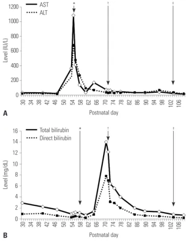
Fifty-five days after birth, liver function tests showed abnor- mal findings with elevation of aspartate aminotransferase (AST) 1086 IU/L, alanine aminotransferase (ALT) 679 IU/L,
and γ-glutamyl transpeptidase 399 IU/L (Fig. 1). Coagulation times were in the normal range. Congenital TORCH infection was excluded. Ursodeoxycholic acid (UDCA) was started for treatment of hepatitis and was continued for 18 days. Multivi- tamins and phenobarbital were administered together. At 71 days of age, acholic stools were noted, and the infant was con- firmed as having cholestatic jaundice (total bilirubin 14.0 mg/
dL, RR <1.2 mg/dL; direct bilirubin 7.8 mg/dL, RR <0.2 mg/
dL). Ultrasonography revealed the presence of newly devel- oped common bile duct sludge (length approximately 2.0 cm) and extra- and intra-hepatic bile duct dilatation (Fig. 2A).
PTBD was performed under ultrasound guidance in our in- terventional radiology suite at 72 days postnatally. The patient was intubated, and vital signs were monitored during the pro- cedure. Pain was managed with fentanyl citrate. A 21-G nee- dle was advanced under ultrasonographic guidance, using color Doppler to avoid vessels with a sub-xiphoid approach.
After puncturing the intrahepatic duct branch B2, contrast material was injected, and correct positioning was confirmed by opacification of the intrahepatic duct. A 0.018-guidewire was passed into the common bile duct, and a 5-Fr Pigtail catheter (Cook Endoscopy Inc., Limerick, Ireland) was placed there (Fig. 3).
After PTBD insertion, natural drainage of approximately 15–
114 mL of greenish bile was done daily. Patient follow-up was done via clinical assessment, liver function tests, bilirubin lev- el tests, and abdominal ultrasonography (Fig. 2B). Four weeks after PTBD initiation, a follow-up cholangiogram was per- formed to confirm resolution of dilatation of the biliary tree and complete removal of the bile plug. Subsequently, the pig- tail catheter was removed without complications.
DISCUSSION
IBS, first described in 1935, is diagnosed based on medical history and typical ultrasound features.8 The incidence in Fig. 1. Results from liver function and serum bilirubin tests. (A) The liver
function test during the patient’s clinical course showed gradual improve- ment after PTBD. (B) Serum bilirubin level during the infant’s clinical course showed marked improvement after PTBD. *Medication start,
†PTBD insertion, ‡PTBD removal. AST, aminotransferase; ALT, alanine aminotransferase; PTBD, percutaneous transhepatic biliary drainage.
A B
Fig. 2. Abdominal ultrasound imaging before and after PTBD. (A) On the day before PTBD, a 2.0 cm sized common bile duct (CBD) sludge and multiple bile sludges at not-tensile gall bladder, as well as extrahepatic/intrahepatic bile duct dilatation, were observed. (B) Three days after PTBD, extrahe- patic/intrahepatic bile duct dilatation had improved. PTBD, percutaneous transhepatic biliary drainage.
30 34 38 42 46 50 54 58 62 66 70 74 78 82 86 90 94 98 102 106
16 14 12 10 8 6 4 2 0
* † ‡
Total bilirubin Direct bilirubin
B Postnatal day
30 34 38 42 46 50 54 58 62 66 70 74 78 82 86 90 94 98 102 106
1200 1000 800 600 400 200 0
* † ‡
AST ALT
A Postnatal day
Level (IU/L)Level (mg/dL)
906
Percutaneous Transhepatic Biliary Drainage in Infant
https://doi.org/10.3349/ymj.2018.59.7.904 England is 1 in 175000 live births, accounting for about 8% of
all cases of surgical jaundice during infancy.2
Infants with biliary obstruction develop jaundice, and pass pale stool and dark urine.9 Blood tests reveal elevated AST, ALT, and direct bilirubin levels. The pathological findings are nonspecific.1 In present case, elevated liver enzymes and se- rum bilirubin levels, as well as clinical symptoms, were consis- tent with the findings of previous studies. The causes of biliary obstruction without structural abnormality include long-term fasting, long-term TPN, hemolytic anemia, hepatitis, transfu- sion, and antibiotics, such as ceftriaxone or diflucan.8-12 In our case, long-term fasting, transfusion, and TPN could have caused IBS. Treatment of IBS includes various challenges. In some cases, biliary sludge reportedly resolved spontaneously with- out treatment within 1 week.13,14 In one case, omega-3 PUFAs were used as an alternative to surgery.15 In some medical tri- als, use of UDCA alone to dissolve the bile sludge failed to im- prove biochemical test results, and ultimately, surgery was re- quired.7,16 Surgical therapy is normally only needed when the extrahepatic bile ducts are dilated to more than 3 mm. Among the various surgical interventions, percutaneous transhepatic cholangiography is difficult in infants because of the small size of the intrahepatic gall ducts.6 Failure of this treatment will en- tail a laparotomy for drainage of the inspissated bile.
The feasibility of PTBD in adults and children has been dem- onstrated, such as for the palliative treatment of inoperable cases, decompression before surgery, recurrent obstructive jaundice after surgery, purulent cholangitis and hepatic ab- scess, and biliary interventions for gallstones and bile duct bi- opsy. In contrast to adult cases, temporary biliary drainage
could be used in children without congenital anomalies.17 How- ever, published data concerning its usage in infants are very limited. A study on PTBD for obstructive jaundice in a three- month-old child with a brain tumor was reported recently.18 Therefore, in newborns or infants without congenital anoma- lies, PTBD should be considered if recovery does not occur af- ter active feeding and medical treatment in order to prevent invasive surgery and progression to hepatic failure.
To our knowledge, this is the first report from Korea to de- scribes a case of obstructive jaundice treated by PTBD in a two-month-old infant. PTBD appears to be a safe and effec- tive treatment for obstructive jaundice in very young infants.
ORCID
Soon Min Lee https://orcid.org/0000-0003-0174-1065
REFERENCES
1. Bernstein J, Braylan R, Brough AJ. Bile-plug syndrome: a correct- able cause of obstructive jaundice in infants. Pediatrics 1969;43:
273-6.
2. Davenport M, Betalli P, D'Antiga L, Cheeseman P, Mieli-Vergani G, Howard ER. The spectrum of surgical jaundice in infancy. J Pedi- atr Surg 2003;38:1471-9.
3. Lightwood R, Bodian M. Biliary obstruction associated with icter- us gravis neonatorum. Arch Dis Child 1946;21:209-17.
4. Bollu BK, Dawrant MJ, Thacker K, Thomas G, Chenapragadda M, Gaskin K, et al. Inspissated bile syndrome; safe and effective min- imally invasive treatment with percutaneous cholecystostomy in neonates and infants. J Pediatr Surg 2016;51:2119-22.
5. Helin R, Bhat R, Rao B. Ultrasound-guided percutaneous chole- Fig. 3. Radiographic findings of percutaneous transhepatic biliary drainage procedures. The intrahepatic duct branch was punctured with a 21-G nee- dle (A-C), and the guidewire was passed into the common bile duct (D-F). The needle was replaced with a 5-Fr pigtail catheter and percutaneous tran- shepatic biliary drainage was achieved (G and H). There is a plug in the distal common bile duct, visible as a filling defect.
A
E
B
F
C
G
D
H
907
Sung Hui Chang, et al.
https://doi.org/10.3349/ymj.2018.59.7.904
cystostomy for acute neonatal biliary obstruction. Neonatology 2007;91:266-70.
6. Duman L, Büyükyavuz BI, Akcam M, Koroglu M, Tepeli H. Percu- taneous management of bile-plug syndrome: a case report. J Pe- diatr Surg 2011;46:e37-41.
7. Gunnarsdóttir A, Holmqvist P, Arnbjörnsson E, Kullendorff CM.
Laparoscopic aided cholecystostomy as a treatment of inspissat- ed bile syndrome. J Pediatr Surg 2008;43:e33-5.
8. Brownschidle S, Sullivan J, Sartorelli K, Potenta S, Zenali M. Neo- natal cholestasis due to biliary sludge- review and report of a case associated with use of diflucan. Ann Clin Path 2014;2:1018.
9. Fawaz R, Baumann U, Ekong U, Fischler B, Hadzic N, Mack CL, et al. Guideline for the evaluation of cholestatic jaundice in infants:
joint recommendations of the North American Society for Pediat- ric Gastroenterology, Hepatology, and Nutrition and the European Society for Pediatric Gastroenterology, Hepatology, and Nutrition.
J Pediatr Gastroenterol Nutr 2017;64:154-68.
10. Suchy FJ. Neonatal cholestasis. Pediatr Rev 2004;25:388-96.
11. Doty JE, Pitt HA, Porter-Fink V, DenBesten L. The effect of intrave- nous fat and total parenteral nutrition on biliary physiology. JPEN J Parenter Enteral Nutr 1984;8:263-8.
12. Soysal A, Eras¸ov K, Akpinar I, Bakir M. Biliary precipitation dur-
ing ceftriaxone therapy: frequency and risk factors. Turk J Pediatr 2007;49:404-7.
13. Lang EV, Pinckney LE. Spontaneous resolution of bile-plug syn- drome. AJR Am J Roentgenol 1991;156:1225-6.
14. Jee KB, Song JY, You KY, Min KS, Kim DH, Lee KS. A case of spon- taneous resolution of bile plug syndrome in a 4-year-old girl. Ko- rean J Pediatr Gastroenterol Nutr 1999;2:262-6.
15. Jun WY, Cho MJ, Han HS, Bae SH. Use of omega-3 polyunsaturat- ed fatty acids to treat inspissated bile syndrome: a case report. Pe- diatr Gastroenterol Hepatol Nutr 2016;19:286-90.
16. Berger S, Schibli S, Stranzinger E, Cholewa D. One-stage laparo- scopic surgery for inspissated bile syndrome: case report and re- view of surgical techniques. Springerplus 2013;2:648.
17. Park JS, Baek JG, Yeom JS, Park ES, Seo JH, Lim JY, et al. A case of obstructive jaundice secondary to traumatic pancreatitis treated with percutaneous transhepatic biliary drainage. Korean J Pediatr Gastroenterol Nutr 2010;13:204-9.
18. Saettini F, Agazzi R, Giraldi E, Foglia C, Cavalleri L, Morali L, et al.
Percutaneous transhepatic biliary drainage in an infant with ob- structive jaundice caused by neuroblastoma. Pediatr Hematol On- col 2015;32:223-8.