Injection of Bupivacaine into Disc Space to Detect
Painful Nonunion after Anterior Lumbar Interbody Fusion (ALIF) Surgery in Patients with Discogenic Low Back Pain
Seiji Kimura, Seiji Ohtori, Sumihisa Orita, Gen Inoue, Yawara Eguchi, Masashi Takaso, Nobuyasu Ochiai, Kazuki Kuniyoshi, Yasuchika Aoki, Tetsuhiro Ishikawa, Masayuki Miyagi, Hiroto Kamoda, Miyako Suzuki, Yoshihiro Sakuma, Gou Kubota, Yasuhiro Oikawa, Kazuhide Inage,
Takeshi Sainoh, Kazuyo Yamauchi, Tomoaki Toyone, Junichi Nakamura, Shunji Kishida, Jun Sato, and Kazuhisa Takahashi
Department of Orthopaedic Surgery, Graduate School of Medicine, Chiba University, Chiba, Japan.
Received: May 9, 2013 Revised: June 27, 2013 Accepted: August 8, 2013
Corresponding author: Dr. Seiji Ohtori, Department of Orthopaedic Surgery, Graduate School of Medicine, Chiba University, 1-8-1 Inohana, Chuo-ku, Chiba 260-8670, Japan.
Tel: 81-43-226-2117, Fax: 81-43-226-2116 E-mail: [email protected]
∙ The authors have no financial conflicts of interest.
© Copyright:
Yonsei University College of Medicine 2014 This is an Open Access article distributed under the terms of the Creative Commons Attribution Non- Commercial License (http://creativecommons.org/
licenses/by-nc/3.0) which permits unrestricted non- commercial use, distribution, and reproduction in any medium, provided the original work is properly cited.
Purpose: Bupivacaine is commonly used for the treatment of back pain and the diagnosis of its origin. Nonunion is sometimes observed after spinal fusion sur- gery; however, whether the nonunion causes pain is controversial. In the current study, we aimed to detect painful nonunion by injecting bupivacaine into the disc space of patients with nonunion after anterior lumbar interbody fusion (ALIF) surgery for discogenic low back pain. Materials and Methods: From 52 patients with low back pain, we selected 42 who showed disc degeneration at only one level (L4-L5 or L5-S1) on magnetic resonance imaging and were diagnosed by pain provocation on discography and pain relief by discoblock (the injection of bupivacaine). They underwent ALIF surgery. If the patients showed low back pain and nonunion 2 years after surgery, we injected bupivacaine into the non- union disc space. Patients showing pain relief after injection of bupivacaine un- derwent additional posterior fixation using pedicle screws. These patients were followed up 2 years after the revision surgery. Results: Of the 42 patient subjects, 7 showed nonunion. Four of them did not show low back pain; whereas 3 showed moderate or severe low back pain. These 3 patients showed pain reduc- tion after injection of bupivacaine into their nonunion disc space and underwent additional posterior fixation. They showed bony union and pain relief 2 years af- ter the revision surgery. Conclusion: Injection of bupivacaine into the nonunion disc space after ALIF surgery for discogenic low back pain is useful for diagnosis of the origin of pain.
Key Words: Lumbar, spine, bupivacaine, pain, intervertebral disc
INTRODUCTION
Low back pain (LBP) is a common clinical problem and is of considerable socio-
into the disc space can detect painful nonunion after anteri- or lumbar interbody fusion (ALIF) surgery in patients with discogenic LBP.
MATERIALS AND METHODS
The ethics committee of our institution approved the proto- col for the human procedures used in this study and in- formed written consent was obtained from each subject.
Patients
Fifty-two patients with LBP only, continuing for at least 3 years, with no accompanying radicular pain were investi- gated. Patients showed only one level of disc degeneration on MRI. Patients who had severe spondylolysis on disc de- generation with two or more level lesions were excluded.
Patients who had previously undergone spinal surgery were also excluded.
Discography or discoblock for the diagnosis of discogenic LBP before first surgery
Discography or discoblock at one intervertebral disc level was performed using a standard posterolateral approach with a 22-gauge needle (Becton Dickinson, Franklin Lakes, NJ, USA) in all 52 patients. For discography, the needle was inserted into the center of the disc under fluoroscopic control. Isovist 240 (range, 0.4-3.2 mL; Schering, Berlin, Germany) was injected into each disc until severe pain was provoked or until contrast medium was seen to leak out of the disc into the spinal canal. For discoblock, 0.75 mL of 0.5% bupivacaine was injected into the disc. We defined the treatment as “effective” if patients indicated a Visual Analogue Scale (VAS) pain score at 2 hours that was less than 50% of their initial VAS score before injection.
When pain was both provoked during the discography and decreased after the discoblock, we confirmed a diagno- sis of discogenic LBP.
Surgery
Forty-two patients diagnosed with discogenic pain showing 1) one level disc degeneration on MRI, 2) pain provocation on discography, and 3) pain relief after discoblock, under- went anterior discectomy and fusion surgery. We first per- formed the discectomy, then cut the endplate at a thickness of 2 mm on both sides, and performed interbody fusion us- ing iliac bone.
economic importance. However, there is currently little in- formation on the pathogenesis of this disease. Although any of the spinal structures (intervertebral discs, facet joints, vertebral bodies, ligaments, or muscles) could be a source of LBP, the most likely cause is a lumbar intervertebral disc.1-3
For surgical treatment, the method of diagnosis of dis- cogenic LBP is important. Generally, diagnosis of disco- genic LBP is determined by observation of a “black disc”
on magnetic resonance imaging (MRI) and pain provoca- tion by discography.4,5 However, the reliability of discog- raphy has been controversial.6,7 A randomized controlled trial showed that, compared with discography, pain relief after injection of a small amount of bupivacaine into the painful disc is a useful tool for the diagnosis of discogenic LBP, and diagnosis by discoblock improved surgical re- sults.8,9
Generally, diagnosis of discogenic LBP is difficult and requires several methods. Therefore, if patients show both continuous LBP and nonunion after fusion surgery, the situ- ation is even more complex. Indeed, several randomized trials have compared surgical to nonsurgical treatment of chronic LBP and arrived at conflicting conclusions.10 For these reasons, it is unclear whether the continuous pain originates from a misdiagnosis of primary discogenic pain or nonunion. Furthermore, some authors have reported a discrepancy between bone union and clinical results in pa- tients with spinal stenosis associated with degenerative lumbar spondylolisthesis, and good results in nonunion cas- es.11,12 In patients with lumbar spinal stenosis, LBP can originate from intervertebral discs, facet joints, and spinal nerve roots. Decompression is effective for postoperative LBP, therefore, it is difficult to directly compare postsurgi- cal LBP between a union and nonunion group.
There have been a few reports of discography or discob- lock at fused levels to detect postoperative back pain. Plou- mis, et al.13 have reported a case of chronic back pain fol- lowing two-level interbody and posterolateral fusion in the lumbar spine that was evaluated with bupivacaine injection into the nonunion space. Bupivacaine injection into an ap- parently fused disc space with an interbody device may be helpful in patients with persistent postsurgical back pain caused by nonunion.
In the current study, we focused on patients with disco- genic LBP, but without radicular back pain or leg pain, be- cause the LBP was thought to originate only from discs.
We aimed to examine whether injection of bupivacaine
RESULTS
Table 1 shows the demographic characteristics of the 52 pa- tient subjects before ALIF surgery. Of these 52 patients, 42 were diagnosed with discogenic LBP because pain was both provoked during the discography and decreased after the dis- coblock. These 42 patients underwent ALIF revision surgery.
No patient had dropped out at 2 years after surgery. Thirty- five patients (83%) showed union and 7 (17%) showed non- union 2 years after the first surgery (Table 2). Table 2 shows characteristics of the union and nonunion groups. Pain scores in the two groups significantly improved 2 years after the first surgery (Table 1 and 2). VAS score and ODI in the union group were significantly better than those in the non- union group 2 years after the first surgery (Table 2).
Table 3 shows characteristics of the 7 patents in the non- union group. Of these 7 patients, 3 showed LBP and 4 did not. The VAS score and ODI in the LBP group were signifi- cantly worse than those in the group without LBP before revision surgery (Table 2).
Table 4 shows results of discography, discoblock, and re- vision surgery for LBP in the 3 patients in the nonunion group. These 3 patients had pain provocation after discog- raphy and relief after discoblock. They underwent posterior fixation surgery and their pain scores significantly im- proved 2 years after this revision surgery (Figs. 1 and 2).
Radiographic and clinical evaluation
Radiography was used to evaluate bone union 2 years after surgery. X-ray images of profile views at flexion and exten- sion positions were obtained. CT was performed to evalu- ate bone union. Evaluation of bone union was blinded and conducted by 3 surgeons. Fusion was used to define bone union if at least 2 of the observers concurred.
Pain score
We evaluated the change in LBP before and after surgery.
To evaluate pain, a VAS score (0, no pain; 10, worst pain) and the Oswestry Disability Index (ODI) for LBP were re- corded before, 2 years after first surgery, and 2 years after revision surgery, and they were compared.
Revision surgery
Of the 52 patients in the study, 42 underwent ALIF surgery, and some of them showed both nonunion and LBP. We evaluated whether the pain originated from the nonunion site or not, and repeated the discography and discoblock.
For discography, Isovist 240 (range, 0.4-1.0 mL; Schering) was injected into the nonunion disc space until severe pain was provoked or until contrast medium was seen to leak out of the disc into the spinal canal. For discoblock, 0.4 mL of 0.5% bupivacaine was injected into the nonunion site.
When pain was both provoked during the discography and decreased after the discoblock, we confirmed a diagnosis of pain from the nonunion site. These patients underwent pos- terior fusion surgery at one level. The single-level posterior fusion was performed using pedicle screws and an iliac bone graft on the lamina. Bilateral facet fusion was per- formed in all patients. We did not use other osteoconductive products for spinal fusion. We finally evaluated their pain score 2 years after the revision surgery.
Statistical analysis
Data were compared using an unpaired t-test, χ2 test, and one-way analysis of variance for repeated measurements.
p<0.05 was considered statistically significant.
Table 1. Demographic Characteristics
Number of patients 52
Sex Male: 35, Female: 17
Age, mean±SEM (range), yrs 36±6 (16-48) Symptom duration, mean (range), yrs 7 (4-20) Follow-up, mean (range), yrs 3.5 (2-5) MRI findings
Level L2/3: 3
L4/5: 29 L5/S1: 20 Pain score
Visual Analogue Scale low back pain 8.5±2.3 Oswestry Disability Index 52±10 Table 2. Surgical Results 2 Years after 1st Surgery
Union group Non-uion group Statistical analysis
Number of patients 35 7 p=0.015
Sex Male: 26, Female: 9 Male: 4, Female: 3
Age, mean±SEM (range), yrs 36±5 (18-48) 35±6 (16-44) p=0.50
Pain score
Visual Analogue Scale low back pain 2.0±1.9 3.8±2.0 p=0.45
Oswestry Disability Index 16±7 22±6 p=0.43
Table 3. Detail of Non-Fusion Group
Low back pain (+) Low back pain (-) Statistical analysis
Number of patients 3 4 p=0.25
Sex Male: 2, Female: 1 Male: 2, Female: 2
Age, mean±SEM (range), yrs 34±5 (16-44) 36±6 (20-42) p=0.50
Pain score
Visual Analogue Scale low back pain 6.5±2.0 1.8±2.0 p=0.033
Oswestry Disability Index 48±7 10±6 p=0.03
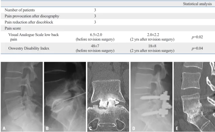
Table 4. Results of Discoblock and Revision Surgery in Low Back Pain (+) of Non-Union Group
Statistical analysis
Number of patients 3
Pain provocation after discography 3
Pain reduction after discoblock 3
Pain score
Visual Analogue Scale low back
pain 6.5±2.0
(before revision surgery) 2.0±2.2
(2 yrs after revision surgery) p=0.02
Oswestry Disability Index 48±7
(before revision surgery) 18±8
(2 yrs after revision surgery) p=0.04
A
A B C
B C D E
Fig. 1. A 35-year-old man with LBP and nonunion after ALIF surgery. Plain X-ray film images showing non-union after ALIF surgery at L5/S1 level (A). The pa- tient showed LBP. Pain was both provoked during the discography and decreased after the discoblock (B: X-ray film, and C: CT). 2 years after revision sur- gery, complete union was achieved and the patient did not show pain (D: X-ray film, and E: CT). LBP, low back pain; ALIF, anterior lumbar interbody fusion.
Fig. 2. A 28-year-old woman without LBP and nonunion after ALIF surgery. (A and B) Before surgery at L5-S1. (C) Nonunion after ALIF surgery at L5-S1. LBP, low back pain; ALIF, anterior lumbar interbody fusion.
diographic fusion. In the current study, 3 patients showed LBP after ALIF surgery where there was nonunion. They showed pain relief after injection of bupivacaine into the disc space, and good results after revision surgery. We con- cluded that injection of bupivacaine into the disc space of patients with discogenic LBP and nonunion after ALIF sur- gery is useful for diagnosing the origin of their pain.
However, bupivacaine has been found to be chondrotoxic in vitro and several studies have suggested a dose and time- dependent chondrotoxicity of bupivacaine.15-17 Furthermore, an apparently toxic effect of 0.5% bupivacaine on disc cells and articular chondrocytes in vitro has been reported.18 These reports indicated that 0.5% bupivacaine had an apparently toxic effect on disc cells and articular chondrocytes of ani- mals and humans in vitro.15-19 An effect of 0.5% bupiva- caine on articular chondrocytes in an in vivo model has been reported; however, the effect was somewhat different from the effects obtained in vitro.20 Rats receiving 0.5% bu- pivacaine into the knee joint were observed for 6 months.
The articular surfaces of joints injected with bupivacaine remained intact on gross and histological evaluation. It has been reported that radiological and MRI findings did not show acceleration of intervertebral disc degeneration within 5 years of a single injection of bupivacaine into human discs.21 Therefore, we conclude that there is a difference in the effect of bupivacaine on discs in vitro and clinically, and that injection of bupivacaine into the disc space is safe for patients with discogenic LBP.
This study has several limitations. First, we examined only a limited number of patients. Second, 3 patients showed LBP and pain relief after injection of bupivacaine into their disc space, and good results after revision sur- gery; however, the number of patients is quite small, and is probably not sufficient to make the current conclusions.
Third, the effect of single or multiple injections of bupiva- caine, the volume of bupivacaine, and its concentration were not examined in the current study. Fourth, there was no control group in the current study. It is desirable to per- form posterior fusion in patients with painful pseudarthro- sis without bupivacaine injection. Thus, larger numbers of patients, a control group, and the effects of bupivacaine ad- ministration need to be evaluated to strengthen the current findings.
In conclusion, it is difficult to detect whether pseudoarthro- sis is painful. Injection of bupivacaine into the disc space of patients with discogenic LBP and nonunion after ALIF sur- gery is useful for diagnosing the origin of their pain.
DISCUSSION
In the current study, 17% of patients (7) with discogenic LBP showed nonunion after ALIF surgery. Of these 7 pa- tients, 3 showed LBP and pain relief after injection of bupi- vacaine into their disc space, and good results after revision surgery. We concluded that injection of bupivacaine into the disc space of patients with nonunion after ALIF surgery and discogenic LBP is useful for diagnosis of the origin of their pain.
In the current study, VAS score and ODI in the union group were significantly better than those in the nonunion group 2 years after their first surgery. Some authors have reported better surgical results for back pain in patients with union compared with nonunion.8,9,14 By contrast, however, others have reported a discrepancy between bone union and clinical results.11,12
Fischgrund, et al.11 reported that there was no association with successful fusion and surgical outcome after surgery of degenerative lumbar spondylolisthesis. Fifty patients who had degenerative lumbar spondylolisthesis underwent fusion surgery. Thirty-six % of patients showed non-union after surgery. However, they showed better clinical results compared decompressive laminectomy alone.12
In patients with lumbar spinal stenosis, LBP may origi- nate from the intervertebral disc, facet joints, or spinal nerve roots. Therefore, it is difficult to compare postsurgi- cal LBP between the union and nonunion groups. In the current study, the patient subjects only had discogenic LBP, and this LBP was thought to originate only from interverte- bral discs. The current findings led us to conclude that union leads to superior surgical results in discogenic LBP patients, because other sites of back pain origin were ex- cluded.
Ploumis, et al.13 have reported a case of chronic back pain following two-level interbody and posterolateral fu- sion in the lumbar spine. They injected 5 mL of the contrast agent iohexol (240 mg/mL), followed by 2 mL of 0.5% bu- pivacaine into the L4-L5 interspace where there was uncer- tain radiographic fusion, and the patient noticed, essentially, complete resolution of his pain for the duration of the anes- thesia. The patient underwent revision surgery, and subse- quently achieved pain relief. These investigators concluded that injection of bupivacaine into the disc space could be used for diagnosis in cases of previous anterior interbody fusion with a cage, continuing back pain, and uncertain ra-
spective, randomized study comparing decompressive laminecto- my and arthrodesis with and without spinal instrumentation. Spine (Phila Pa 1976) 1997;22:2807-12.
12. Herkowitz HN, Kurz LT. Degenerative lumbar spondylolisthesis with spinal stenosis. A prospective study comparing decompres- sion with decompression and intertransverse process arthrodesis. J Bone Joint Surg Am 1991;73:802-8.
13. Ploumis A, Pinto MR, Schellhas KP. Disc space injection with marcaine as a method to evaluate painful nonunion of an inter- body fusion device: a case report. Spine J 2007;7:74-8.
14. Vamvanij V, Fredrickson BE, Thorpe JM, Stadnick ME, Yuan HA. Surgical treatment of internal disc disruption: an outcome study of four fusion tec hniques. J Spinal Disord 1998;11:375-82.
15. Chu CR, Izzo NJ, Papas NE, Fu FH. In vitro exposure to 0.5%
bupivacaine is cytotoxic to bovine articular chondrocytes. Arthros- copy 2006;22:693-9.
16. Dogan N, Erdem AF, Erman Z, Kizilkaya M. The effects of bupi- vacaine and neostigmine on articular cartilage and synovium in the rabbit knee joint. J Int Med Res 2004;32:513-9.
17. Gomoll AH, Kang RW, Williams JM, Bach BR, Cole BJ. Chon- drolysis after continuous intra-articular bupivacaine infusion: an experimental model investigating chondrotoxicity in the rabbit shoulder. Arthroscopy 2006;22:813-9.
18. Lee H, Sowa G, Vo N, Vadala G, O’Connell S, Studer R, et al. Ef- fect of bupivacaine on intervertebral disc cell viability. Spine J 2010;10:159-66.
19. Wang D, Vo NV, Sowa GA, Hartman RA, Ngo K, Choe SR, et al.
Bupivacaine decreases cell viability and matrix protein synthesis in an intervertebral disc organ model system. Spine J 2011;11:139-46.
20. Chu CR, Coyle CH, Chu CT, Szczodry M, Seshadri V, Karpie JC, et al. In vivo effects of single intra-articular injection of 0.5% bu- pivacaine on articular cartilage. J Bone Joint Surg Am 2010;92:
599-608.
21. Ohtori S, Inoue G, Orita S, Eguchi Y, Ochiai N, Kishida S, et al.
No acceleration of intervertebral disc degeneration after a single injection of bupivacaine in young age group with follow-up of 5 years. Asian Spine J 2013;7:212-7.
REFERENCES
1. Nachemson AL. The lumbar spine, an orthopaedic challenge.
Spine 1976;1:59-71.
2. Mooney V. Presidential address. International Society for the Study of the Lumbar Spine. Dallas, 1986. Where is the pain com- ing from? Spine (Phila Pa 1976) 1987;12:754-9.
3. Deyo RA, Weinstein JN. Low back pain. N Engl J Med 2001;344:
363-70.
4. Pauza KJ, Howell S, Dreyfuss P, Peloza JH, Dawson K, Bogduk N. A randomized, placebo-controlled trial of intradiscal electro- thermal therapy for the treatment of discogenic low back pain.
Spine J 2004;4:27-35.
5. Shuff C, An HS. Artificial disc replacement: the new solution for discogenic low back pain? Am J Orthop (Belle Mead NJ) 2005;
34:8-12.
6. Buenaventura RM, Shah RV, Patel V, Benyamin R, Singh V. Sys- tematic review of discography as a diagnostic test for spinal pain:
an update. Pain Physician 2007;10:147-64.
7. Carragee EJ, Lincoln T, Parmar VS, Alamin T. A gold standard evaluation of the “discogenic pain” diagnosis as determined by provocative discography. Spine (Phila Pa 1976) 2006;31:2115-23.
8. Ohtori S, Kinoshita T, Yamashita M, Inoue G, Yamauchi K, Koshi T, et al. Results of surgery for discogenic low back pain: a ran- domized study using discography versus discoblock for diagnosis.
Spine (Phila Pa 1976) 2009;34:1345-8.
9. Ohtori S, Koshi T, Yamashita M, Yamauchi K, Inoue G, Suzuki M, et al. Surgical versus nonsurgical treatment of selected patients with discogenic low back pain: a small-sized randomized trial.
Spine (Phila Pa 1976) 2011;36:347-54.
10. Mirza SK, Deyo RA. Systematic review of randomized trials comparing lumbar fusion surgery to nonoperative care for treat- ment of chronic back pain. Spine (Phila Pa 1976) 2007;32:816-23.
11. Fischgrund JS, Mackay M, Herkowitz HN, Brower R, Montgom- ery DM, Kurz LT. 1997 Volvo Award winner in clinical studies.
Degenerative lumbar spondylolisthesis with spinal stenosis: a pro-