1286 www.eymj.org
INTRODUCTION
Severe fever with thrombocytopenia syndrome (SFTS) is a fa- tal infectious disease caused by the SFTS virus (SFTSV), which is a novel Phlebovirus in the family Bunyavirida.1 The major clinical features of patients with SFTS are high fever, thrombo- cytopenia, leukopenia, and gastrointestinal symptoms. Ele- vated serum levels of alanine aminotransferase, aspartate ami- notransferase, blood urea nitrogen, lactate dehydrogenase, creatine kinase, and ferritin are also common laboratory find- ings in patients with SFTS. However, the pathological mecha- nism of thrombocytopenia and leukopenia in patients with
SFTS is not fully understood until now; it is unclear whether production failure or peripheral destruction/sequestration is the main mechanism of cytopenia in these patients.2-4
In the present study, therefore, we investigated the bone mar- row (BM) findings of patients with SFTS to understand the patho- genesis of SFTS.
CASE REPORT
Case 1
Abdominal pain developed in a 73-year-old man. He was trans- fused with packed red blood cells due to low hemoglobin (Hb) (7.6 g/dL) at a local hospital 10 days later. Fever, neutropenia, and elevated liver enzymes were observed. He was referred to our hospital for further evaluation and treatment. SFTSV was confirmed by reverse-transcription polymerase chain reaction (RT-PCR) analysis5 at Division of Arboviruses, National Institute of Health, Korea Centers for Disease Control and Prevention.
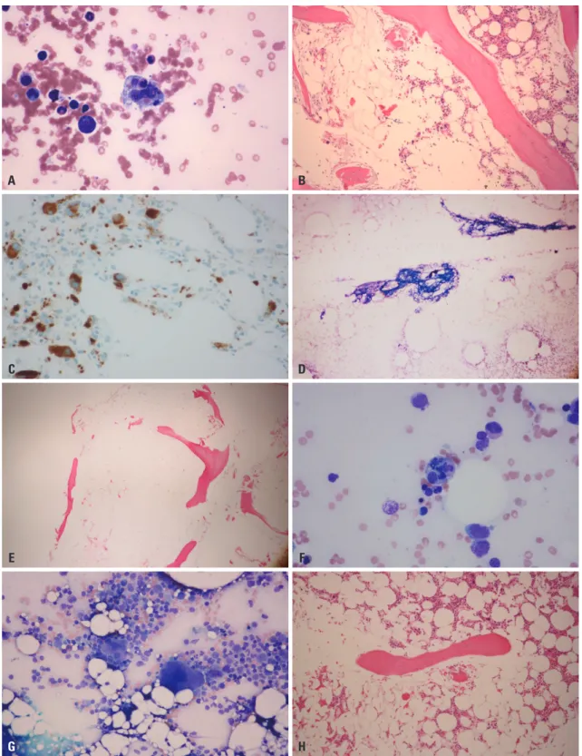
The laboratory and clinical findings are summarized in Table 1 and 2. A BM biopsy was performed. Hemophagocytic histio- cytes were observed in an aspirate (Fig. 1A), and hypocellular marrow was noted in the BM section (Fig. 1B). However, mega-
Bone Marrow Suppression and Hemophagocytic Histiocytes Are Common Findings in Korean Severe Fever with Thrombocytopenia Syndrome Patients
Sang-Yong Shin1, Oh-Hyun Cho2, and In-Gyu Bae2
1Department of Laboratory Medicine, Center for Diagnostic Oncology, Hospital and Research Institute, National Cancer Center, Goyang;
2Department of Internal Medicine, Gyeongsang National University School of Medicine, Jinju, Korea.
The causes of cytopenia in patients with severe fever with thrombocytopenia syndrome (SFTS) are not fully understood until now.
We reviewed the bone marrow (BM) findings of patients with SFTS to unravel the cause of the cytopenia. Three Korean SFTS were enrolled in this study. Thrombocytopenia, neutropenia, and anemia were detected in all three patients. Severe hypocellular mar- row (overall cellularity <5%) and a decreased number of megakaryocytes were noted in one patient, and hypo-/normocellular marrow and an increased number of hemophagocytic histiocytes were observed in two patients. Megakaryocytes were relatively preserved in two patients. Although a limited number of cases are available, our observations suggest that both BM suppression and peripheral destruction or sequestration are causes of cytopenia of patients with SFTS. To the best of our knowledge, this is the first well documented pathologic evaluation of Korean SFTS.
Key Words: Severe fever with thrombocytopenia syndrome bunyavirus, bone marrow, Korea
Case Report
pISSN: 0513-5796 · eISSN: 1976-2437
Received: February 12, 2016 Revised: April 2, 2016 Accepted: April 6, 2016
Corresponding author: Dr. Sang-Yong Shin, Department of Laboratory Medicine, Center for Diagnostic Oncology, Hospital and Research Institute, National Cancer Center, 323 Ilsan-ro, Ilsandong-gu, Goyang 10408, Korea.
Tel: 82-31-920-1793, Fax: 82-31-920-1268, E-mail: [email protected]
•The authors have no financial conflicts of interest.
© Copyright: Yonsei University College of Medicine 2016
This is an Open Access article distributed under the terms of the Creative Com- mons Attribution Non-Commercial License (http://creativecommons.org/licenses/
by-nc/3.0) which permits unrestricted non-commercial use, distribution, and repro- duction in any medium, provided the original work is properly cited.
Yonsei Med J 2016 Sep;57(5):1286-1289 http://dx.doi.org/10.3349/ymj.2016.57.5.1286
1287
Sang-Yong Shin, et al.
http://dx.doi.org/10.3349/ymj.2016.57.5.1286
karyocytes were relatively preserved in the section (Fig. 1C).
He was treated with antibiotics and plasmapheresis. However, he died 3 weeks after the initial symptoms (3 days after BM bi- opsy) due to metabolic acidosis and multi-organ failure.
Case 2
Fever and enlargement of the left inguinal lymph node devel- oped in a 53-year-old man, and he was treated with antibiot- ics at a local hospital. However, pancytopenia was detected (Hb, 12.9 g/dL; white blood cell, 3.77×10⁹/L; platelet, 24.0×
109/L) at local hospital, and he was referred to our hospital for further evaluation and treatment. SFTSV was confirmed by RT-PCR analysis 5 at Division of Arboviruses, National Insti- tute of Health, Korea Centers for Disease Control and Preven- tion. The laboratory and clinical findings at our hospital are summarized in Table 1 and 2. Severe hypocellular marrow was noted in an aspirate and section (Fig. 1D and E). Megakaryo- cytes were rarely found. He was treated with antibiotics; howev- er, he died 10 days after admission due to multi-organ failure.
Case 3
An 86-year-old woman was admitted to our hospital for a 3 day fever. She had been with antibiotics at local hospital; how- ever, pancytopenia was detected, and she was referred to our hospital for further evaluation and treatment. SFTSV was con- firmed by RT-PCR analysis5 at Division of Arboviruses, Na- tional Institute of Health, Korea Centers for Disease Control and Prevention. The laboratory and clinical findings at our hos- pital are summarized in Table 1 and 2. Hemophagocytic his- tiocytes were observed in an aspirate (Fig. 1F). The megakary- ocytes were normally observed in an aspirate (Fig. 1G). Nor- mocellular marrow with focal hypocellular area was noted in the BM section (Fig. 1H). The patient was successfully treated with antibiotics and ribavirin.
DISCUSSION
Thrombocytopenia and leukopenia are prominent features in Table 1. Basic Characteristics of the Patients with Severe Fever with Thrombocytopenic Syndrome (SFTS)
Case 1* Case 2* Case 3*
Age, yrs 73 53 86
Sex Male Male Female
Fever (°C) 38.0 38.1 37.7
Organomegaly/lymphadenpathy - Inguinal lymph node -
Hemorrhage - Melena -
Central nervous system symptom/sign - - -
Gastro-intestinal symptom/sign Abdominal pain, diarrhea Abdominal pain, melena, diarrhea Diarrhea
Clinical course Death (hospital day 4) Death (hospital day 10) Alive
*SFTS virus was confirmed by reverse-transcription polymerase chain reaction analysis.
Table 2. Laboratory Findings of the Patients with Severe Fever with Thrombocytopenia Syndrome
Case 1 Case 2 Case 3
White blood cell (×109/L) (4.0–10.0) 0.76 1.69 1.14
Neutrophil (×109/L) (1.5–7.0) 0.44 1.13 0.50
Hemoglobin (g/dL) (12.0–16.0) 9.7 14.0* 10.6
Platelet (×109/L) (130–400) 115 15 121
Reticulocyte (%) (0.5–2.0) 0.65 - 1.62
Prothrombin time (sec) (11.9–14.3) 14.4 12.9 14.2
Activated partial thromboplastin time (sec) (29.1–43.5) 47.9 46.6 43.6
Ferritin (ng/mL) (30.0–400.0) >2000 >2000 316
Fibrinogen (mg/dL) (200–450) 108 221 200
D-dimer [fibrinogen equivalent units (FEU) ug/mL] (0–0.5) 3.16 19.15 1.22
Alanine aminotransferase (U/L) (0–41) 352 49 23
Aspartate aminotransferase (U/L) (0–37) 781 117 64
Lactate dehydrogenase (U/L) (135–225) 740 560 259
Total bilirubin (mg/dL) (0–1.2) 0.23 0.23 0.32
C-reactive protein (mg/L) (0–5) 1.3 30.0 0.8
Erythrocyte sedimentation rate (sec) (0–9) 35 27 4
Proteinuria (-) + + +
Urine blood (-) + + -
*Hemoglobin was increased after transfusion of packed red blood cells.
1288
Cytopenia in SFTS Patients
http://dx.doi.org/10.3349/ymj.2016.57.5.1286 patients with SFTS. Viral replication in a mouse model mainly
occurs in splenic macrophages.4 However, SFTSV is not found in mice BM,4 but the numbers of megakaryocytes increase in
the spleen and BM of mice.4 In vitro cell assays show that SFTSV adheres to mouse platelets and facilitates phagocytosis of platelets by primary macrophages, suggesting that the cause
Fig. 1. Findings of bone marrow (BM) aspirate and section of case 1 (A, B, and C), 2 (D and E), and 3 (F, G, and H). (A) The hemophagocytic histiocytes were increased in the aspirate [Wright-Giemsa (W-G), ×400]. (B) Hypocellular area was noted in the BM section [hematoxylin and eosin (H&E), ×100]. (C) The number of dysplastic megakaryocytes increased slightly in the cellular area (CD61 immunohistochemistry, ×400). (D) Hypocellular particles (W-G, ×40) are noted. (E) Severe hypocellular marrow is noted (H&E, ×40). (F) The hemophagocytic histiocytes are increased in the aspirate (W-G, ×400). (G) Mega- karyocytes are normally observed in the aspirate (W-G, ×200). (H) Normocellular marrow for age (86 years) with a focally hypocellular area is noted (H&E,
×100).
A B
C D
E F
G H
1289
Sang-Yong Shin, et al.
http://dx.doi.org/10.3349/ymj.2016.57.5.1286
of the thrombocytopenia is destruction by splenic macroph- ages.4 QuanTai, et al.2 compared the BM findings of five Chi- nese patients with SFTS with those of patients with aplastic an- emia and normal healthy volunteers, and found no significant differences in cell morphology, cellularity, or numbers of me- gakaryocytes between them. They concluded that peripheral blood thrombocytopenia and leukopenia in patients with SFTS result from increased peripheral organ damage or circulating anti-platelet antibodies.2
However, hypocellular marrow with an increased number of hemophagocytic histiocytes is observed in Japanese patients with SFTS,3 whereas megakaryocytes are relatively preserved in BM.3 Consistent with these findings, our Korean patients also showed moderate to severe hypocellular marrow with an in- creased number of hemophagocytic histiocytes and/or rela- tively preserved megakaryocytes. The reason for the different BM findings between Chinese and Japanese or Korean patients with SFTS is unclear. The Chinese patients with SFTS were relatively young age (30–50 years) and all of them recovered successfully.2 However, our Korean patients with SFTS (53–86 years) and the Japanese patients with SFTS were older (>50 years), and two of our Korean patients died. Therefore, age and clinical status/severity may be the cause of the different BM findings.6,7
Deng, et al.8 also observed that two patients expired of SFTS presented with empty marrow. These two8 cases and our cases (case 1 and case 2) suggest that BM hypocellularity is associat- ed with severity of SFTS. However, further studies are needed.
Considering the results of animal experiments4 and those of pathological examinations of patients with SFTS,2,3 hemoph- agocytosis appears to be common in patients with SFTS, and the laboratory findings of most patients with SFTS are compati- ble with hemophagocytic lymphohistocytosis (fever, cytopenia, high ferritin level, etc.).7,9 Moreover, one study revealed that increased cytokine levels are correlated with viral load/clinical parameters in patients with SFTS.10 Since dysregulation of the immune system with hypercytokinemia is an underlying me- chanism of hemophagocytic lymphohistocytosis,11 SFTSV may produce hemophagocytic lymphohistocytosis.9 Therefore, cy- topenia in patients with SFTS may result from both peripheral destruction/sequestration and BM suppression.
Based on the “cytokine storm” and “immune-mediated plate- let consumption in the spleen” concepts, some authors have reported cases treated with plasmapheresis to reduce cytokine levels as well as other pathological immune-mediating agents.12 They reported two patients treated with ribavirin and plasma-
pheresis and they recovered from SFTS.12 However, plasma- pheresis does not have demonstrated therapeutic efficacy until now. Our Patient 1 died after a plasmapheresis treatment. Th- erefore, further studies are needed to define the exact patho- genesis and the therapeutic implications.
In conclusion, our results together with other studies indi- cate that BM suppression and hemophagocytic histiocytes are common findings in patients with SFTS. Although a limited number of cases were available, our observations may help un- derstand the pathogenic mechanism of SFTSV and aid in fu- ture therapeutic applications.
REFERENCES
1. Yu XJ, Liang MF, Zhang SY, Liu Y, Li JD, Sun YL, et al. Fever with thrombocytopenia associated with a novel bunyavirus in China.
N Engl J Med 2011;364:1523-32.
2. QuanTai X, FengZhe C, XiuGuang S, DongGe C. A study of cytolog- ical changes in the bone marrow of patients with severe fever with thrombocytopenia syndrome. PLoS One 2013;8:e83020.
3. Takahashi T, Maeda K, Suzuki T, Ishido A, Shigeoka T, Tominaga T, et al. The first identification and retrospective study of Severe Fe- ver with Thrombocytopenia Syndrome in Japan. J Infect Dis 2014;
209:816-27.
4. Jin C, Liang M, Ning J, Gu W, Jiang H, Wu W, et al. Pathogenesis of emerging severe fever with thrombocytopenia syndrome virus in C57/BL6 mouse model. Proc Natl Acad Sci U S A 2012;109:10053-8.
5. Kim WY, Choi W, Park SW, Wang EB, Lee WJ, Jee Y, et al. Nosoco- mial transmission of severe fever with thrombocytopenia syndrome in Korea. Clin Infect Dis 2015;60:1681-3.
6. Ding S, Niu G, Xu X, Li J, Zhang X, Yin H, et al. Age is a critical risk factor for severe fever with thrombocytopenia syndrome. PLoS One 2014;9:e111736.
7. Gai ZT, Zhang Y, Liang MF, Jin C, Zhang S, Zhu CB, et al. Clinical progress and risk factors for death in severe fever with thrombo- cytopenia syndrome patients. J Infect Dis 2012;206:1095-102.
8. Deng B, Zhou B, Zhang S, Zhu Y, Han L, Geng Y, et al. Clinical fea- tures and factors associated with severity and fatality among pa- tients with severe fever with thrombocytopenia syndrome Bunya- virus infection in Northeast China. PLoS One 2013;8:e80802.
9. Jordan MB, Filipovich AH. Hematopoietic cell transplantation for hemophagocytic lymphohistiocytosis: a journey of a thousand miles begins with a single (big) step. Bone Marrow Transplant 2008;
42:433-7.
10. Sun Y, Jin C, Zhan F, Wang X, Liang M, Zhang Q, et al. Host cytokine storm is associated with disease severity of severe fever with throm- bocytopenia syndrome. J Infect Dis 2012;206:1085-94.
11. Usmani GN, Woda BA, Newburger PE. Advances in understand- ing the pathogenesis of HLH. Br J Haematol 2013;161:609-22.
12. Oh WS, Heo ST, Kim SH, Choi WJ, Han MG, Kim JY. Plasma ex- change and ribavirin for rapidly progressive severe fever with throm- bocytopenia syndrome. Int J Infect Dis 2014;18:84-6.