Lab Anim Res 2018: 34(4), 216-222 https://doi.org/10.5625/lar.2018.34.4.216
ISSN 2233-7660 (Online)
In vitro and in vivo inhibition of Helicobacter pylori by Lactobacilllus paracasei HP7
Seong-Soo Hong
1, Hyun-A Lee
2, Joo Yun Kim
3, Ji-Woong Jeong
3, Jae-Jung Shim
3, Jung Lyoul Lee
3, Jae-Hun Sim
3, Yungho Chung
4, Okjin Kim
2,*
1
Division of Gastroenterology, Vievisnamuh Hospital, Seoul, Korea
2
Center for Animal Resource Development, Wonkwang University, Iksan, Korea
3
R & BD Center, Korea Yakult Co., Ltd., Yongin, Korea
4
Department of Companion Animal and Animal Resources Science, Joongbu University, Geumsan-gun, Korea
The efficacy of standard therapeutic strategies for Helicobacter pylori (H. pylori) infection is decreasing over time due to the emergence of drug-resistant strains. As an alternative, the present study investigated the capacity of Lactobacilllus paracasei (L. paracasei) HP7, isolated from kimchi, to inhibit H. pylori growth. The effects of L. paracasei HP7 on H. pylori adhesion and H. pylori-induced inflammation were examined in AGS human gastric adenocarcinoma epithelial cells and a mouse model of H. pylori SS1 infection. L. paracasei HP7 reduced H. pylori adhesion to AGS cells and suppressed the inflammatory response in infected cells by downregulating interleukin-8. H. pylori colonization in the stomach of C57BL/6 mice was demonstrated by rapid urease test, and results showed significant decrease in mice post-treated with L. paracasei HP7. Additionally, L. paracasei HP7 decreased gastric inflammation and epithelial lesions in the stomach of H. pylori-infected mice. These results demonstrate that L. paracasei HP7 treatment can inhibit H. pylori growth and is thus a promising treatment for patients with gastric symptoms such as gastritis that are caused by H. pylori infection.
Keywords: Lactobacilllus paracasei, HP7, Helicobacter pylori, AGS cells, Kimchi
Received 24 November 2018; Revised version received 2 December 2018; Accepted 3 December 2018
Helicobacter pylori is a Gram-negative, spiral-shaped bacterium in stomach that is the major pathogen of chronic gastric inflammation [1] and stomach ulcers [2]
and is related to increased risk of stomach cancer [3,4].
Removing H. pylori in the stomach by inoculating antibiotis can reduce H. pylori-related gastrointestinal diseases [5,6] and alleviate the risk of stomach cancers [7]. The standard recommended treatment for H. pylori therapy is triple combination therapy with two antibiotics- usually clarithromycin with amoxicillin or metronidazole- and a proton pump inhibitor, which reveals a successful eradication result in the beginning [8,9]. However, the efficacy of this triple therapy has decreased over time;
the recent therapy rate of <80% is mainly due to an
increase in the prevalence of H. pylori strains resistant to metronidazole and clarithromycin [10-12]. Furthermore, some patients reveal allergic side effects to antibiotics, which can occasionally cause adverse effects while failing to treat H. pylori [13]. Long-term inoculation of antibiotics to prevent H. pylori infection cannot be recommended. It is therefore important to develop new non-antimicrobial agents to treat H. pylori [14].
Lactic acid bacteria (Lactobacillus spp.) have been recommended as an additive agent in the standard recommended treatment for H. pylori therapy and can improve patient compliance by decreasing antimicrobial agents-associated side effects [15,16]. Lactobacillus salivarius was reported to inhibit H. pylori colonization
*Corresponding author: Okjin Kim, Center for Animal Resource Development, Wonkwang University, 460 Iksandaero, Iksan, Jeonbuk 54538, Korea
Tel: +82-63-850-6668; Fax: +82-63-850-7308; E-mail: [email protected]
This is an Open Access article distributed under the terms of the Creative Commons Attribution Non-Commercial License (http://creativecommons.org/licenses/
by-nc/3.0) which permits unrestricted non-commercial use, distribution, and reproduction in any medium, provided the original work is properly cited.
in a mice experiment as evidenced by a decrease in H.
pylori-specific IgG concentrations, while negative control mice were infected by H. pylori and revealed gastritis lesions [17]. In another study, intragastic treatment of a culture supernatant of Lactobacillus acidophilus revealed to inhibit Helicobacter felis infection [18,19]. Additionally, L. acidophilus culture supernatant had a partial but long- term inhibiting effect on H. pylori infection in humans [20].
In the present study, we are aimed to study that the lactic acid bacterium Lactobacillus paracasei HP7 isolated from kimchi, a fermented vegetable dish widely consumed in Korea, has inhibitory effects against H.
pylori in vitro and in vivo.
Materials and Methods
Bacterial strains and culture conditions
L. paracasei HP7 was cultured at 35
oC for 24 h in Man-Rogosa-Sharpe broth (Difco Laboratories, Detroit, MI, USA) composed of 0.2% dipotassium hydrogen phosphate, 0.5% sodium acetate, 0.8% meat extract, 0.1% Tween 80, 0.4% yeast extract, 2% D (þ)-glucose, 0.02% magnesium sulfate, 1% peptone from casein, 0.2% diammonium hydrogen citrate, and 0.004%
manganese sulfate. H. pylori strain SS1 (B0890; Korean Collection for Type Cultures, Jeongeup, Korea) was cultured overnight at 37
oC under microaerophilic conditions in brain-heart infusion broth containing 10%
fetal bovine serum (FBS) and was allowed to grow to a density of ~2.0×10
9CFU/mL. The cultured bacteria were then transferred to phosphate-buffered saline (PBS) before they were used to infect cells.
Cell culture
AGS human gastric adenocarcinoma epithelial cells (CRL-1739; American Type Culture Collection, Manassas, VA, USA) were cultured in Roswell Park Memorial Institute 1640 medium (Sigma-Aldrich, St. Louis, MO, USA) supplemented with 10% heat-inactivated FBS (Invitrogen, Carlsbad, CA, USA). Antibiotic-antimycotic (Gibco, Grand Island, NY, USA) was added if needed.
For analysis of H. pylori-induced interleukin (IL)-8 production, antibiotics were not added to the culture medium.
Inhibition of H. pylori adhesion to AGS cells
AGS cells were seeded in 6-well tissue culture plates
at 1×10
6cells/mL in Ham’s F-12 medium (Sigma- Aldrich) supplemented with 10% FBS and 1%
antibiotic-antimycotic solution and cultured at 37
oC in a humidified atmosphere of 95% air/5% CO
2(v/v) for 16 h. When the cells reached 90% confluence, the medium was replaced with serum- and antibiotic-free F- 12 medium. An overnight culture of H. pylori SS1 and L. paracasei HP7 was washed twice in sterile PBS and resuspended in Ham’s F-12 medium. For co-culture of bacteria and gastric epithelial cells, H. pylori SS1 cells (1×10
7CFU/mL) were added to wells containing 1×10
6AGS cells at a cell ratio of 10:1 and incubated for 4 h in the absence or presence of L. paracasei HP7.
RNA preparation and real-time (RT)-PCR
Total cellular RNA was extracted using TRIzol reagent (Sigma-Aldrich), and 2 µg were reverse-transcribed using murine leukemia virus reverse transcriptase, 1 mM dNTP, and 0.5 µg/µL oligo (dT12-18). The cDNA was used as a template for RT-PCR to detect H. pylori 16S RNA as a measure of the H. pylori infection rate. The reaction was carried out on a QuantStudio 6 Real-Time PCR system (Applied Biosystems, Foster City, CA, USA) using SYBR Premix Ex Taq (Takara Bio, Otsu, Japan), with glyceraldehyde 3-phosphate serving as an internal standard. Relative mRNA levels at each time point were compared with those in H. pylori-infected control AGS cells. Forward and reverse sequences of primers for amplifying the H. pylori 16S RNA gene were as follows: 5'-TCG GAA TCA CTG GGC GTA A- 3' and 5'-TTC TAT GGT TAA GCC ATA GGA TTT CAC-3' [21].
Measurement of IL-8 levels
IL-8 released by AGS cells infected with H. pylori was detected by enzyme-linked immunosorbent assay (ELISA).
AGS cells (2×10
4cells/well) were seeded in 96-well plates; L. paracasei HP7 cells were added to the cell culture medium 30 min before H. pylori infection for 24 h. AGS cells cultured in the absence of L. paracasei HP7 cells served as a control. The culture supernatant was collected and IL-8 levels were measured with a sandwich ELISA kit (R&D Systems, Minneapolis, MN, USA), according to the manufacturer’s instructions.
Each sample was tested in triplicate.
Animals
Specific pathogen-free (SPF) male C57BL/6 mice
weighing 20-24 g were purchased from Samtako Co.
(Osan, Korea) and were maintained at the inspection facility of Wonkwang University (Iksan, Korea) for 1 week before experiments. Thereafter, the mice were maintained in an SPF barrier room with regulated temperature (23
oC±1
oC) and humidity (50±5%) on a 12:12-h light/dark cycle. The animals were fed a sterilized pellet diet (2 Mrad radiation) (Purina, Seoul, Korea) and sterilized water ad libitum. All studies were performed in accordance with the Guide for Animal Experimentation of Wonkwang University and were approved by the Institutional Animal Care and Use Committee of Wonkwang University (approval no.
WKU 16-44).
Bacterial inoculation
H. pylori SS1 was incubated in brain-heart infusion broth containing 10% FBS overnight at 37
oC under a micro-aerophilic atmosphere and allowed to grow to a density of ~2.0×10
9CFU/mL. Animals were intragastrically inoculated three times at 3-day intervals with H. pylori at 1.0×10
9CFU in 0.5 mL broth. The challenged animals were confirmed as H. pylori-positive by stool antigen analysis using the Bioline H. pylori Ag kit (Standard Diagnostics, Suwon City, Korea) as previously described [22].
In vivo study protocol
The inhibition of H. pylori growth by L. paracasei HP7 was also investigated in a mouse model. Mice were divided into four groups: negative control (group I, n=10); H. pylori-infected without L. paracasei HP7 treatment (group II, n=10); L. paracasei HP7-treated without H. pylori infection (group III, n=10); and H.
pylori-infected with L. paracasei HP7 treatment (group IV, n=10). L. paracasei HP7 was orally administered at a daily dose of 2.0×10
7CFU/kg/day/day during a 4- week treatment period. Animals were then sacrificed and their stomachs were dissected after euthanasia with ether.
The stomach was opened along the greater curvature and washed with saline, and half of the glandular mucosa was scraped off for detection of colonizing H. pylori, while the residual portion was formalin-fixed and embedded in paraffin for histological analysis. H. pylori colonization was confirmed by the rapid urease test CLO as previously described [23]. Mucosal damage was evaluated according to established criteria [24].
Blood analysis
Blood samples were collected from the heart of sacrificed animals and centrifuged at 1000×g for 15 min at 4
oC; the plasma was stored at 80
oC until analysis.
Serum titers of anti-H. pylori antibody were measured using the mouse anti-H. pylori antibody (IgG-1) ELISA kit (Cusabio Biotech, Wuhan, China) according to the manufacturer’s instructions.
Statistical analysis
Values for all parameters under study were recorded for each experimental unit, and statistical analysis was performed using a general linear model. Values are reported as mean±standard deviation where appropriate.
The Student’s t test was used for pairwise comparisons.
The incidence with 95% confidence interval was calculated with MiniTab (State College, PA, USA) statistical software package. A P value <0.05 was considered significant.
Results
L. paracasei HP7 inhibits H. pylori adhesion to AGS cells
L. paracasei HP7 was screened with the agar well diffusion assay and was shown to have anti-microbial activity against H. pylori SS1 (inhibition zone diameter:
11.5-12 mm). To determine whether L. paracasei HP7 affects the adhesion of H. pylori to AGS cells, we examined H. pylori 16S RNA gene expression in AGS cells. HP7 reduced H. pylori adhesion by 65% relative to the control (P<0.05; Figure 1). These results demonstrate
Figure 1. Inhibition of H. pylori adhesion to AGS cells by L.
paracasei HP7 (HP7). AGS cells pre-treated with L. paracasei
HP7 showed lower expression of H. pylori 16S RNA.
that HP7 can inhibit bacterial adhesion to gastric epithelial cells.
L. paracasei HP7 suppresses H. pylori-induced IL-8 production
To determine whether L. paracasei HP7 can block H.
pylori-induced IL-8 production, AGS cells were left untreated or were pre-treated with L. paracasei HP7 prior to H. pylori infection, and IL-8 production was measured by ELISA. Pre-treatment of H. pylori-infected AGS cells with L. paracasei HP7 for 24 h decreased IL- 8 level by 65.9% (from 410 to 270 pg/mL) relative to control cells (Figure 2).
L. paracasei HP7 decreases anti-H. pylori antibody titer in serum
To confirm H. pylori colonization in mice, we measured serum levels of anti-H. pylori IgG-1, since the serological absorbance index of IgG against H. pylori is related to the degree of H. pylori colonization [25].
Serum antibody titers were elevated 4 weeks after H.
pylori inoculation, with values of 1.33±0.04 and 0.76±
0.02 pg/mL in the H. pylori infection (Group II) and H.
pylori infection/L. paracasei HP7 (Group IV) pre- treatment groups, respectively, as compared to 0.25±
0.005 in control animals (Group I) (Figure 3). These results indicate that H. pylori infection is reduced by pre- treatment with L. paracasei HP7.
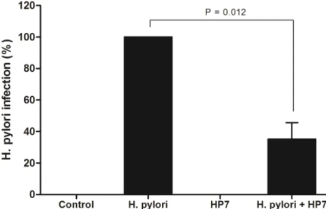
L. paracasei HP7 reduces H. pylori colonization Repeated intragastric inoculation of C57BL/6 mice with H. pylori (1.0×10
9CFU/mouse, three times) yielded a positive reaction in the campylobacter-like organism (CLO) test of gastric mucosa (Table 1). The stomachs of H. pylori-infected mice orally treated with L. paracasei HP7 at a dose of 2.0×10
7CFU/kg/day during a 4-week period showed a positive reaction rate of 50%. CLO scores were decreased by L. paracasei HP7 pre-treatment (Group IV) relative to H. pylori- infected animals without pre-treatment (Group II) (P<0.05; Figure 4). Thus, L. paracasei HP7 can decrease the rate of H. pylori colonization.
Figure 2. Inhibitory effect of L. paracasei HP7 (HP7) on H.
pylori-induced IL-8 production. L. paracasei HP7 was added to a confluent layer of AGS cells in a 96-well plate 30 min before adding H. pylori. After incubation for 24 h, the culture supernatant was collected to measure the amount of released IL-8.
Figure 3. Suppression of H. pylori infection in C57BL/6 mice by treatment with L. paracasei HP7. Serum samples collected after sacrifice were evaluated for H. pylori IgG-1 by ELISA. H.
pylori IgG-1 levels were decreased in the H. pylori/L. paracasei HP7 group as compared to the H. pylori infection group.
Table 1. Reactivity in the CLO test of gastric mucosa from mice infected with H. pylori followed by treatment with Lactobacilllus paracasei HP7 or vehicle
Group Treatment n Positive %
aTherapeutic %
I No treatment 10 0% (CI
b0-27.6) -
II H. pylori 10 100% (CI 72.2-100) 0%, CI (0-27.6)
III HP7 10 0% (CI 0-27.6) -
IV H. pylori+HP7 10 50% (CI 23.7-76.3) 50% (CI 23.7-76.3)
a
A positive percentage reflects H. pylori colonization, which was observed as medium color change from yellow to red.
b