◇ 위장관 ◇
1
The Genotypes of Helicobacter pylori, Gastric Epithelial Cell Proliferation and Apoptosis in Children
Ji Ah Jung, M.D., Mi Ae Lee, M.D.* and Jeong Wan Seo, M.D.† Department of Pediatrics, Hallym University College of Medicine,
Departments of *Laboratory Medicine and †Pediatrics, Ewha Womans University College of Medicine, Seoul, Korea
Purpose: To investigate the relation of the gastric epithelial cell proliferation, apoptosis and genotypes of H. pylori in children.
Methods: Histologic grading by updated Sydney system, PCNA immunostaining, TUNEL method and the genotypes (cagA, picB and iceA) by PCR were performed in H. pylori positive (N=20) and negative (N=20) gastric biopsy specimens.
Results: PCNA index was significantly different between H. pylori positive children (77.4±13.12) and H. pylori negative children (52.3±12.20) (p=0.000). There were positive correlations between PCNA index and H. pylori density (r=0.624, p=0.000), polymorphonuclear neutrophil activity (r=0.460, p=0.005) and chronic inflammation (r=0.433, p=0.009). Apoptosis index of H. pylori positive children (0.70±0.411) was significantly higher than of H. pylori negative children (0.14±
0.201) (p=0.000). Positive correlations between apoptosis index and H. pylori density (r=0.691, p=0.000), polymorphonuclear neutrophil activity (r=0.585, p=0.000) and chronic inflammation (r=0.535, p=0.001) were noted. As PCNA index increased, apoptosis index significantly increased (r=0.527, p=0.001). The positive rates of genotypes were cagA 90%, picB 75%, iceA1 60% and iceA2 15%, respectively. There were no significant correlations between the status of the genotypes
ꠏꠏꠏꠏꠏꠏꠏꠏꠏꠏꠏꠏꠏꠏꠏꠏꠏꠏꠏꠏꠏꠏꠏꠏꠏꠏꠏꠏꠏꠏꠏꠏꠏꠏꠏꠏꠏꠏꠏꠏꠏꠏꠏꠏꠏ 접수:2003년 3월 2일, 승인:2003년 3월 14일
책임저자:서정완, 158-710 서울시 양천구 목동 911-1, 이화여자대학교 의과대학 소아과학교실 Tel: 02-2650-5573, 5141, Fax: 02-2653-3718, E-mail: [email protected]
본 논문의 요지는 2002년도 대한소아과학회 추계학술대회에서 발표되었음.
소아 Helicobacter pylori 감염에서 균의 유전형, 위 상피세포의 증식과 세포사
한림대학교 의과대학 소아과학교실, 이화여자대학교 의과대학
*진단검사의학과학교실, †소아과학교실
정 지 아․이 미 애*․서 정 완
†서 론
Helicobacter pylori (H. pylori)감염의 병인으로 균 의 병독 인자, 숙주 요인 및 환경 등이 연구되고 있다. 균의 병독 인자로는 urease, 편모, adhesin 등과 cagA, pic B, iceA 등의 유전자가 있으며, 숙주 요인 으로는 위점막 방어벽과 위산도의 항상성, cyto- kines, mucin, gastrin, somatostatin, ABO 혈액형과 HLA형 등이다. 최근에는 위 상피세포 증식(proli- feration)과 세포사(apoptosis)의 변화가 H. pylori의 병 인으로 중요시되고 있으며1,2) 이러한 변화와 H.
pylori 유전형과의 관련성에 대한 연구들이 보고되 었다3∼6).
위 상피세포는 정상적으로 세포 증식과 세포사 의 과정을 거쳐 약 5일만에 새로운 세포로 대치되 는데7), H. pylori균에 감염되면 세포 증식과 세포사 의 변화가 유발된다. H. pylori에 의해 세포사가 증 가하면서 보상적으로 세포 증식이 증가하고1∼3), 박멸치료 후에는 세포사와 세포 증식이 동시에 감 소하므로 이는 가역적인 반응으로 생각된다1,2). 그 러나 H. pylori 감염 초기에는 세포사의 증가에도 불구하고 세포 증식이 없으면 위축, 궤양 등의 점 막 손상이 생기며8,9), H. pylori 감염이 장기간 지속 되어 세포사보다 세포 증식이 더 증가하거나8,9), 세 포사의 조절불능, 세포사의 저하로 암이 발생한다 고 추정되고 있다4,5,10).
H. pylori균의 유전형은 매우 다양하다. cagA (cytotoxin-associated gene A)와 picB (permits the induction of cytokines) 유전자는 cag pathogenicity island (PAI)의 일부로서 interleukin-8 (IL-8)의 분비 를 유도하여 점막 손상을 일으키는데 관여한다11,12). iceA (induced by contact with epithelium) 유전자는 iceA1과 iceA2의 대립 유전자가 있는데, iceA1이 H.
pylori가 위 상피세포에 접촉 시에 상향조정(upre- gulation)되어 염증반응을 유도하여 소화성 궤양의 표지자라고 한다13,14).
위 상피세포 증식과 세포사 및 H. pylori 유전형 과의 관련성에 대한 연구는 주로 성인을 대상으로 한 것이었다3∼6). 위 상피세포 증식과 세포사는 cagA와는 관련이 없다고 하였으며15,16), Moss 등3)은 세포사의 증가는 cagA, cag PAI와 관련성이 있으 나, 세포 증식은 cagA와는 관련이 없다고 하였으 며, Rokkas 등4)은 cagA 양성에서 세포 증식이 증가 하였으나 세포사와의 균형을 이루지 못하여 위암 의 위험요소가 된다고 하였다. 소아에서는 Jones 등15)이 소아기에 H. pylori에 감염되면 세포 증식과 세포사가 유의하게 증가하며, 이는 cagA 유전자와 는 관련이 없다고 하였다.
저자들은 H. pylori 감염 소아에서 위 상피세포 증식과 세포사가 증가함을 보고한 바 있다17). H.
pylori 감염 소아에서 위 상피세포 증식과 세포사 의 정도를 보고, 병독 인자인 cagA, picB 및 iceA 유전형과의 관련성을 보고자 본 연구를 시행하였다.
and PCNA index, apoptosis index, the endoscopic findings and the histologic findings.
Conclusion: PCNA index and apoptosis index in H. pylori positive children were higher than in H. pylori negative children but were not related to H. pylori genotypes. This study sug- gested that correlatively increased gastric epithelial cell proliferation and apoptosis are impor- tant to pathogenesis of H. pylori infection in children. (Korean J Pediatr Gastroenterol Nutr 2003; 6: 1∼9)
ꠏꠏꠏꠏꠏꠏꠏꠏꠏꠏꠏꠏꠏꠏꠏꠏꠏꠏꠏꠏꠏꠏꠏꠏꠏꠏꠏꠏꠏꠏꠏꠏꠏꠏꠏꠏꠏꠏꠏꠏꠏꠏꠏꠏꠏꠏꠏꠏꠏꠏꠏꠏꠏꠏꠏꠏꠏꠏꠏꠏꠏꠏꠏꠏꠏꠏꠏꠏꠏꠏꠏꠏꠏꠏꠏꠏꠏꠏꠏꠏꠏ Key Words: Helicobacter pylori, Gastric epithelial cell, Proliferation, Apoptosis, cagA, picB,
iceA
대상 및 방법
1. 대상
1999년 8월부터 2001년 6월까지 이화여자대학교 목동병원 소아과에서 심와부 동통, 토혈, 만성 복 부 불쾌감, 구토 등의 증상으로 상부위장관 내시경 을 시행하여 H. pylori 감염으로 진단된 20예와 무 작위로 추출한 감염 음성 20예를 대상으로 하였다.
H. pylori 감염 양성은 조직학적으로 H. pylori 균이 관찰되고, CLO 검사와 ureC PCR18)이 전부 양성인 경우로 하였다. 대상 환아의 연령과 성별은 감염 양성은 연령 5∼15세(정중값 10세), 남아 6예, 여아 14예이었으며, 감염 음성은 연령 5∼15세(정중값 10세), 남아 11예, 여아 9예이었다.
2. 방법
위생검 조직에서 개정된 시드니 체계를 이용하 여 조직 소견을 분석하고, 2002년 논문17)에서 기술 한 방법으로 proliferating cell nuclear antigen (PCNA) 발현으로 위 상피세포 증식의 정도를, in situ ter- minal deoxynucleotidyl transferase-mediated dUTP nick-end labeling (TUNEL) 방법으로 세포사의 정도 를 조사하였다.
1) H. pylori 유전자 중합효소연쇄반응: 영하 70oC
에 냉동 보관해 둔 위생검조직에서 QIAamp DNA Mini Kit (Qiagen Inc., CA, USA)를 이용하여 설명 서에 따라 DNA를 분리하였다. 추출한 DNA는 Beckman DU spectrophotometer (Beckman Coulter Inc., California, USA)로 각 검체마다 추출된 DNA 의 양을 측정하였다.
ureC18), cagA19), picB19,20) 및 iceA13) PCR의 시발체 의 염기서열은 표에 기술하였다(Table 1). 주형 DNA 10μL, 증류수, 10×PCR buffer, 1.5 mM MgCl2, 0.2 mM dNTP (dATP, dCTP, dGTP, dTTP), 0.6μL의 각 시발체, Taq polymerase 2.5 U의 총 50μL로 하였 다. PCR은 GeneAmp PCR System 9600 (Perkin- Elmer Cetus, CA, USA)를 사용하였고, PCR의 조건 은 Table 1과 같다. H. pylori DNA 양성대조는 ATCC 700392 균주에서 분리한 DNA를 이용하였고, 1×TE 완충액을 음성대조로 사용하였다. PCR을 실시한 후 반응산물 10μL를 취하여 전기영동 완 충액 1μL와 섞은 다음 ethidium bromide가 함유된 2% NuSieve 3:1 agarose gel (FMC BioProducts, USA)에 10μL씩 가하여 100 V의 직류로 20분간 전기영동시킨 후 UV transilluminator (Spectroline TVC-312A, Spectronics Co, NY, USA)와 폴라로이 드 사진기로 촬영하였다. 이때 증폭된 DNA는 DNA molecular marker VI (Boehringer-Mannheim, Germany)와 비교하여 ureC는 294 bp band, cagA는
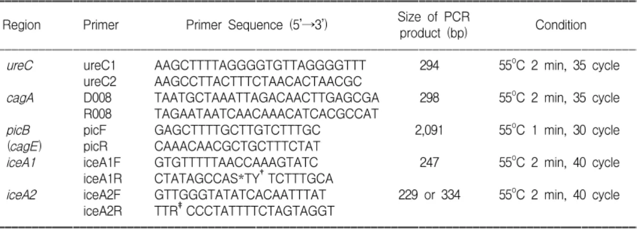
Table 1. Methods of PCR Assay for ureC, cagA, picB and iceA Genotypes
ꠚꠚꠚꠚꠚꠚꠚꠚꠚꠚꠚꠚꠚꠚꠚꠚꠚꠚꠚꠚꠚꠚꠚꠚꠚꠚꠚꠚꠚꠚꠚꠚꠚꠚꠚꠚꠚꠚꠚꠚꠚꠚꠚꠚꠚꠚꠚꠚꠚꠚꠚꠚꠚꠚꠚꠚꠚꠚꠚꠚꠚꠚꠚꠚꠚꠚꠚꠚꠚꠚꠚꠚꠚꠚꠚꠚꠚꠚꠚꠚꠚꠚꠚꠚꠚꠚꠚꠚꠚꠚꠚꠚꠚꠚ Size of PCR
Region Primer Primer Sequence (5’→3’) Condition
product (bp)
ꠏꠏꠏꠏꠏꠏꠏꠏꠏꠏꠏꠏꠏꠏꠏꠏꠏꠏꠏꠏꠏꠏꠏꠏꠏꠏꠏꠏꠏꠏꠏꠏꠏꠏꠏꠏꠏꠏꠏꠏꠏꠏꠏꠏꠏꠏꠏꠏꠏꠏꠏꠏꠏꠏꠏꠏꠏꠏꠏꠏꠏꠏꠏꠏꠏꠏꠏꠏꠏꠏꠏꠏꠏꠏꠏꠏꠏꠏꠏꠏꠏꠏꠏꠏꠏꠏꠏꠏꠏꠏꠏꠏꠏꠏ
ureC ureC1 AAGCTTTTAGGGGTGTTAGGGGTTT 294 55oC 2 min, 35 cycle
ureC2 AAGCCTTACTTTCTAACACTAACGC
cagA D008 TAATGCTAAATTAGACAACTTGAGCGA 298 55oC 2 min, 35 cycle
R008 TAGAATAATCAACAAACATCACGCCAT
picB picF GAGCTTTTGCTTGTCTTTGC 2,091 55oC 1 min, 30 cycle
(cagE) picR CAAACAACGCTGCTTTCTAT
iceA1 iceA1F GTGTTTTTAACCAAAGTATC 247 55oC 2 min, 40 cycle
iceA1R CTATAGCCAS*TY†TCTTTGCA
iceA2 iceA2F GTTGGGTATATCACAATTTAT 229 or 334 55oC 2 min, 40 cycle
iceA2R TTR‡CCCTATTTTCTAGTAGGT
ꠚꠚꠚꠚꠚꠚꠚꠚꠚꠚꠚꠚꠚꠚꠚꠚꠚꠚꠚꠚꠚꠚꠚꠚꠚꠚꠚꠚꠚꠚꠚꠚꠚꠚꠚꠚꠚꠚꠚꠚꠚꠚꠚꠚꠚꠚꠚꠚꠚꠚꠚꠚꠚꠚꠚꠚꠚꠚꠚꠚꠚꠚꠚꠚꠚꠚꠚꠚꠚꠚꠚꠚꠚꠚꠚꠚꠚꠚꠚꠚꠚꠚꠚꠚꠚꠚꠚꠚꠚꠚꠚꠚꠚꠚ
*S is C or G, †Y is C or T, ‡R is A or G.
298 bp band, picB는 2,091 bp band, iceA1은 247 bp band 및 iceA2는 229 혹은 334 bp band가 확인되면 양성으로 하였다(Fig. 1).
2) 통계분석: PC-SAS Release 6.12Ⓡ (SAS Institute Inc. Cary, NC)를 이용하여 Fisher's exact 검정, Wil- coxon rank sum 검정, Spearman 상관 분석으로 p 값 이 0.01 미만인 경우에 유의성이 있다고 판정하였다.
결 과
1. 내시경 소견
상부 위장관 내시경 소견은 H. pylori 감염 양성 소아에서 결절성 위염이 13예(65%)로 가장 많았 고, 위궤양 3예, 십이지장 궤양 2예, 발적성 위염 1 예, 정상 1예이었다. H. pylori 감염 음성 소아에서 는 정상 소견이 6예(30%)로 가장 많았고, 미란성 위염 5예, 발적성 위염 4예, 십이지장염 2예, 식도 염 1예, 십이지장 궤양 1예, 결절성 위염 1예 순이 었다(p=0.000).
2. 세포 증식 지표와 세포사 지표
세포 증식 지표는 H. pylori 감염 양성에서 77.4
±13.12, 음성에서 52.3±12.20로 감염 양성에서 음 성보다 유의하게 높았다(p=0.000, Table 2). 세포 증 식 지표는 H. pylori 밀도가 증가할수록(r=0.624, p=
0.000), 다핵형 중성구의 활동성이 증가할수록(r=
0.460, p=0.005), 만성염증이 증가할수록(r=0.433, p=0.009) 증가하였다(Table 3). 세포사 지표는 H.
pylori 감염 양성에서 0.70±0.411, 음성에서 0.14±
0.201로 감염 양성에서 음성보다 유의하게 높았다 (p=0.000, Table 2). 세포사 지표는 H. pylori 밀도가 증가할수록(r=0.691, p=0.000), 다핵형중성구의 활 동성이 증가할수록(r=0.585, p=0.000), 만성염증이 증가할수록(r=0.535, p=0.001) 증가하였다(Table 3).
Fig. 1. PCR results of the ureC, cagA, picB, iceA1 and iceA2 genes. (A) Lane 1; DNA molecular-weight marker VI, 3, 5-9; positive results, 2, 4, 10; negative results, 11; positive control, 12; negative control. (B) Lane 1; DNA molecular-weight marker VI, 3-5, 7, 9, 10, 12, 13; positive results, 2, 6, 8, 11; negative results, 14; positive control, 15; negative control. (C) Lane 1; DNA molecular-weight marker VI, 8-10, 12; positive results, 2-7, 11; negative results, 13; positive control, 14; negative control. (D) Lane 1; DNA molecular-weight marker VI, 2-4; positive results of iceA1, 5; negative results of iceA1, 6; positive control of iceA1, 7; negative control of iceA1, 8-10; positive results of iceA2, 11; positive control of iceA2, 12; negative control of iceA2.
1 2 3 4 5 6 7 8 9 10 11 12 13 14
←2,091 bp
C. picB
1 2 3 4 5 6 7 8 9 10 11 12
A. ureC
←294 bp
1 2 3 4 5 6 7 8 9 10 11 12 13 14 15
← 298 bp
B. cagA D. iceA1 and iceA2
←247 or 229 bp 1 2 3 4 5 6 7 8 9 10 11 12
Table 2. PCNA Index and Apoptosis Index of H. Pylori Positive and Neagtive Children
ꠚꠚꠚꠚꠚꠚꠚꠚꠚꠚꠚꠚꠚꠚꠚꠚꠚꠚꠚꠚꠚꠚꠚꠚꠚꠚꠚꠚꠚꠚꠚꠚꠚꠚꠚꠚꠚꠚꠚꠚꠚꠚꠚꠚꠚ H. pylori (+) H. pylori (-) p value ꠏꠏꠏꠏꠏꠏꠏꠏꠏꠏꠏꠏꠏꠏꠏꠏꠏꠏꠏꠏꠏꠏꠏꠏꠏꠏꠏꠏꠏꠏꠏꠏꠏꠏꠏꠏꠏꠏꠏꠏꠏꠏꠏꠏꠏ
PCNA 77.4±13.12 52.3±12.20 0.000
index Apoptosis
0.70±0.411 0.14±0.201 0.000 index
ꠚꠚꠚꠚꠚꠚꠚꠚꠚꠚꠚꠚꠚꠚꠚꠚꠚꠚꠚꠚꠚꠚꠚꠚꠚꠚꠚꠚꠚꠚꠚꠚꠚꠚꠚꠚꠚꠚꠚꠚꠚꠚꠚꠚꠚ Data are presented mean±SD.
세포 증식 지표가 증가할수록 세포사 지표는 증가 하여 유의한 상관관계가 있었다(r=0.527, p=0.001).
3. H. pylori 유전형의 양성률
H. pylori 감염 양성에서 유전형의 양성률은 cagA 90%, picB 75%, iceA1 60% 및 iceA2 15%이었다 (Table 4).
4. H. pylori 유전형에 따른 세포 증식 지표와 세포사 지표
세포 증식 지표와 세포사 지표는 cagA, picB, iceA 유전형에 따른 유의한 차이가 없었다(Table 5). 또한, H. pylori 유전형에 따른 내시경 소견, 조 직 소견에는 유의한 차이가 없었다.
고 찰
H. pylori의 유전형은 매우 다양하다. cagA는 중 성구의 이동에 관여하는 interleukin-8의 분비를 유 도하고, 점막의 손상을 일으킨다. 서구에서는 cagA 유전자와 소화성 궤양이나 위암과의 관련성이 있 다고 보고하였고13,21,22), 우리나라를 비롯한 동북아 시아 등지에서는 질병과는 관련이 없어, 지역적인 분포의 차이로 추정되고 있다23). cagA는 cag patho- genicity island (PAI)의 일부이다. cag PAI는 적어도 16개의 유전자로 이루어진 오른쪽 분절인 cagI 과 적어도 14개의 유전자로 이루어진 왼쪽 분절인 cagII와 그 사이를 잇는 insertion sequence (IS605) 등 으로 이루어져 있으며, 이러한 cag PAI가 부분적 결실이 없이 완전할 때 가장 병독성이 있다고 하
였다12,24,25). cag PAI는 cagA에 상관없이 상피세포
Table 5. PCNA Index and Apoptosis Index according to Genotypes of H. pylori
ꠚꠚꠚꠚꠚꠚꠚꠚꠚꠚꠚꠚꠚꠚꠚꠚꠚꠚꠚꠚꠚꠚꠚꠚꠚꠚꠚꠚꠚꠚꠚꠚꠚꠚꠚꠚꠚꠚꠚꠚꠚꠚꠚꠚꠚꠚꠚꠚꠚꠚꠚꠚꠚꠚꠚꠚꠚꠚꠚꠚꠚꠚꠚꠚꠚꠚꠚꠚꠚꠚꠚꠚꠚꠚꠚꠚꠚꠚꠚꠚꠚꠚꠚꠚꠚꠚꠚꠚꠚꠚꠚꠚꠚꠚ
Genotypes N PCNA index p value Apoptosis index p value
ꠏꠏꠏꠏꠏꠏꠏꠏꠏꠏꠏꠏꠏꠏꠏꠏꠏꠏꠏꠏꠏꠏꠏꠏꠏꠏꠏꠏꠏꠏꠏꠏꠏꠏꠏꠏꠏꠏꠏꠏꠏꠏꠏꠏꠏꠏꠏꠏꠏꠏꠏꠏꠏꠏꠏꠏꠏꠏꠏꠏꠏꠏꠏꠏꠏꠏꠏꠏꠏꠏꠏꠏꠏꠏꠏꠏꠏꠏꠏꠏꠏꠏꠏꠏꠏꠏꠏꠏꠏꠏꠏꠏꠏꠏ
cagA (+) 18 76.0±13.16 0.67±0.405
0.127 0.171
cagA (-) 2 89.5±1.27 1.00±0.467
picB (+) 15 75.8±13.88 0.62±0.412
0.342 0.062
picB (-) 5 83.7±8.19 1.00±0.269
iceA1 12 78.2±10.51 0.77±0.590
iceA2 3 84.8±10.92 0.290 1.00±0.692 0.423
iceA1/iceA2 (-/-) 5 69.7±20.30 0.50±0.196
ꠚꠚꠚꠚꠚꠚꠚꠚꠚꠚꠚꠚꠚꠚꠚꠚꠚꠚꠚꠚꠚꠚꠚꠚꠚꠚꠚꠚꠚꠚꠚꠚꠚꠚꠚꠚꠚꠚꠚꠚꠚꠚꠚꠚꠚꠚꠚꠚꠚꠚꠚꠚꠚꠚꠚꠚꠚꠚꠚꠚꠚꠚꠚꠚꠚꠚꠚꠚꠚꠚꠚꠚꠚꠚꠚꠚꠚꠚꠚꠚꠚꠚꠚꠚꠚꠚꠚꠚꠚꠚꠚꠚꠚꠚ Table 3. Correlation between Histologic Findings
and PCNA Index and Apoptosis Index
ꠚꠚꠚꠚꠚꠚꠚꠚꠚꠚꠚꠚꠚꠚꠚꠚꠚꠚꠚꠚꠚꠚꠚꠚꠚꠚꠚꠚꠚꠚꠚꠚꠚꠚꠚꠚꠚꠚꠚꠚꠚꠚꠚꠚꠚ
Histologic PCNA Apoptosis
parameters Index Index
ꠏꠏꠏꠏꠏꠏꠏꠏꠏꠏꠏꠏꠏꠏꠏꠏꠏꠏꠏꠏꠏꠏꠏꠏꠏꠏꠏꠏꠏꠏꠏꠏꠏꠏꠏꠏꠏꠏꠏꠏꠏꠏꠏꠏꠏ H. pylori density 0.624 (0.000) 0.691 (0.000) PMN* neutrophil
0.460 (0.005) 0.585 (0.000) activity
Chronic inflammation 0.433 (0.009) 0.535 (0.001) ꠚꠚꠚꠚꠚꠚꠚꠚꠚꠚꠚꠚꠚꠚꠚꠚꠚꠚꠚꠚꠚꠚꠚꠚꠚꠚꠚꠚꠚꠚꠚꠚꠚꠚꠚꠚꠚꠚꠚꠚꠚꠚꠚꠚꠚ
*PMN: polymorphonuclear, Data are presented as r value (p value).
Table 4. Positive Rate of cagA, picB and iceA Genotypes
ꠚꠚꠚꠚꠚꠚꠚꠚꠚꠚꠚꠚꠚꠚꠚꠚꠚꠚꠚꠚꠚꠚꠚꠚꠚꠚꠚꠚꠚꠚꠚꠚꠚꠚꠚꠚꠚꠚꠚꠚꠚꠚꠚꠚꠚ Genotype No. of positive patients (%) ꠏꠏꠏꠏꠏꠏꠏꠏꠏꠏꠏꠏꠏꠏꠏꠏꠏꠏꠏꠏꠏꠏꠏꠏꠏꠏꠏꠏꠏꠏꠏꠏꠏꠏꠏꠏꠏꠏꠏꠏꠏꠏꠏꠏꠏ
cagA 18/20 (90%)
picB 15/20 (75%)
iceA1 12/20 (60%)
iceA2 3/20 (15%)
iceA1/iceA2 (-/-) 5/20 (25%)
ꠚꠚꠚꠚꠚꠚꠚꠚꠚꠚꠚꠚꠚꠚꠚꠚꠚꠚꠚꠚꠚꠚꠚꠚꠚꠚꠚꠚꠚꠚꠚꠚꠚꠚꠚꠚꠚꠚꠚꠚꠚꠚꠚꠚꠚ
에서 interleukin-8 (IL-8)의 분비를 유도하는 NF-κB 를 활성화시킨다고 하였다26). 성인에서 위염보다 십이지장 궤양에서 전체 cag PAI가 더 많이 검출 되나, 임상질환과의 뚜렷한 연관성은 밝히지 못하
였다25,27). cag PAI의 대부분이 결손되어도 cagA가
존재할 수 있어 cagA가 cag PAI를 완전히 대변하 지는 못하는 것으로 되어 있다25). 적어도 40개의 cag PAI의 유전자 중 picB (permits the induction of cyto- kines), cagT, virD4 등은 interleukin-8의 분비에 필 수적인 것으로 알려져 있으며28), 이중 picB (cagE) 는 cagA처럼 cag PAI의 cagI에 위치한 유전자로 소 아에서 십이지장 궤양과 관련이 있다는 보고가 있
다12,29). 본 연구에서는 cagA와 picB에 따른 내시경
소견과 조직 소견의 차이를 발견할 수 없었으며, 이는 우리나라 성인과 소아에서의 기존의 연구와
일치한다30∼32). 또한 세포 증식 지표와 세포사 지
표는 cagA와 picB 양성과 음성 환아에서 차이가 없 었다.
iceA (induced by contact with epithelium) 유전자 는 iceA1과 iceA2의 두 가지 대립 유전자가 있으며 기능은 잘 알려져 있지 않다. iceA1은 type II re- striction endonuclease와 비슷하며, H. pylori가 위 상 피세포에 접촉 시 상향조정(upregulation)되어 염증 반응을 유도하므로 소화성 궤양의 표지자라고 하 며, iceA2는 비궤양성 소화불량과 관련이 있다고
한다13,14). van Doorn 등13)은 소화성 궤양과 iceA1과
의 연관을 주장하였고, Yamaoka 등23)은 질병과는 관련이 없고, 지역적인 분포의 차이가 있다고도 하 였다. 본 연구에서는 iceA 유전형에 따른 질환의 차이가 없었다. iceA 유전형과 세포 증식 및 세포 사와의 관계에 관한 보고는 없었는데, 본 연구에서 는 iceA 유전형에 따른 세포 증식 및 세포사에는 차이가 없었다.
위 상피세포 증식과 세포사에 관한 성인에서의 보고는 대부분이 H. pylori 감염 시에 세포사에 대 한 보상 작용의 결과로 세포 증식이 증가한다고 하였다3,5,8,28,33,34∼37)
. 국내 성인을 대상으로 한 연구 에서는 이 등38)은 H. pylori 감염 유무에 따라 PCNA 발현의 차이가 없었다고 하였고, 정 등37)은
세포사만 유의하게 증가하고 세포 증식에서는 차 이를 볼 수 없었다고 하였다. 방 등40)은 H. pylori 감염 양성에서 세포사와 세포 증식이 유의하게 증 가함을 보고하였다. 소아에서 H. pylori에 의한 위 상피세포 증식과 세포사에 대한 연구는 거의 없다.
Jones 등15)이 소아에서 세포 증식과 세포사가 H.
pylori 감염 시에 유의하게 증가한다고 하였다.
본 연구에서는 저자들의 이전 연구17)와 동일하 게 소아 H. pylori 감염 양성에서 세포 증식 지표와 세포사 지표가 유의하게 증가함을 관찰하여, H.
pylori 감염 소아에서 위 상피세포 증식과 세포사 가 증가함을 알 수 있었다. 또한, 세포 증식 지표와 세포사 지표는 유의한 상관성을 보여 위 상피세포 증식과 세포사는 균형을 이루면서 증가함을 알 수 있었다. 세포 증식과 세포사는 내시경 소견(위염, 궤양성 질환, 정상 소견)에 따른 차이가 없었다. 이 는 소아에서 궤양성 질환의 유병률이 적어서일 것 으로 생각되며, 더 많은 수의 환아를 대상으로 연 구되어야 할 것이다.
위 상피세포 증식, 세포사와 조직 소견과의 관계 에 대해서는 Lynch 등28)은 위 상피세포의 증식이 증가할수록 H. pylori 밀도, 다핵형중성구의 활동 성, 만성염증이 증가하여, 세포 증식을 H. pylori에 의한 염증 반응의 결과로 보았다. Moss 등1)은 세 포사 지표와 조직 소견과는 상관성이 없었다고 하 였다. 본 연구에서는 저자들의 이전 연구17)와 동일 하게 세포 증식 지표와 세포사 지표 모두 H. pylori 밀도와 다핵형중성구의 활동성, 만성염증이 증가 할수록 증가하여, 소아에서 증가된 세포 증식과 세 포사는 H. pylori에 의한 염증 반응의 결과로 생각 된다.
본 저자들은 H. pylori 감염 소아에서 세포 증식 지표와 세포사 지표가 균형을 이루면서 증가함을 확인하고, 위 상피세포 증식과 세포사가 균형을 이 루면서 증가하므로 성인에 비해 질병의 정도가 약 하지 않나 추정해 보았다. 또한 위 상피세포 증식 과 세포사에 cagA, picB 및 iceA 유전형에 따른 유 의한 차이가 없음을 알 수 있었다. 이는 소아에서 위 상피세포 증식과 세포사가 H. pylori의 병인에
중요함을 시사하며, 앞으로 더 많은 환아를 대상으 로 성인과의 비교 연구 등이 필요하리라 생각된다.
요 약
목 적: Helicobacter pylori (H. pylori)에 의한 질병 발생의 병독 인자로 cagA, picB 및 iceA 등의 유전 형이 연구되고 있으며, 최근에는 위 상피세포의 증 식(proliferation)과 세포사(apoptosis)의 불균형이 중 요시되고 있다. 이에 H. pylori 감염 소아에서 위 상피세포 증식과 세포사 및 cagA, picB 및 iceA 유 전형의 관련성을 알아보고자 하였다.
방 법: 1999년 8월부터 2001년 6월까지 이화여자 대학교 목동병원 소아과에서 소화기 증상으로 내 시경을 시행하여 H. pylori 감염으로 진단된 20예 와 감염 음성 20예를 대상으로 하였다. H. pylori 감염 양성은 조직학적으로 H. pylori 균이 관찰되 고, CLO 검사와 ureC PCR이 전부 양성인 경우로 하였다. 위생검 조직에서 개정된 시드니 체계를 이 용하여 조직 소견을 분석하고, proliferating cell nuclear antigen (PCNA) 발현으로 위 상피세포 증식 의 정도를, in situ terminal deoxynucleotidyl transfer- ase-mediated dUTP nick-end labeling (TUNEL) 방법 으로 세포사의 정도를 조사하였다. cagA, picB 및 iceA 유전자에 대해 중합효소연쇄반응을 시행하였다.
결 과:
1) 세포 증식 지표는 H. pylori 감염 양성에서 77.4±13.12로, 음성 52.3±12.20에 비하여 유의하 게 높았다(p=0.000). 세포 증식 지표는 H. pylori 밀 도가 증가할수록(r=0.624, p=0.000, 다핵형 중성구 의 활동성이 증가할수록(r=0.460, p=0.005), 만성 염 증이 증가할수록(r=0.433, p=0.009) 증가하였다.
2) 세포사 지표는 H. pylori 감염 양성에서 0.70
±0.411, 음성에서 0.14±0.201로 감염 양성에서 음 성보다 유의하게 높았다(p=0.000). 세포사 지표는 H.
pylori 밀도가 증가할수록(r=0.691, p=0.000), 다핵형 중성구의 활동성이 증가할수록(r=0.585, p=0.000), 만 성 염증이 증가할수록(r=0.535, p=0.001) 증가하였다.
3) 세포 증식 지표가 증가할수록 세포사 지표는
유의하게 증가하였다(r=0.527, p=0.001).
4) H. pylori 감염 양성에서 유전형의 양성률은 cagA 90%, picB 75%, iceA1 60% 및 iceA2 15%였으 며, cagA, picB, 및 iceA 유전형에 따른 세포 증식 지표, 세포사 지표, 내시경 소견 및 조직 소견에는 유의한 차이가 없었다.
결 론: H. pylori 감염 소아에서 위 상피세포 증 식 지표와 세포사 지표가 균형을 이루면서 증가하 였으며, cagA, pic B 및 iceA 유전형에 따른 유의한 차이는 없었다. 이는 위 상피세포 증식과 세포사가 H. pylori의 병인에 중요함을 시사하며, 앞으로 세 포 증식과 세포사의 기전과 유발 요인, 여러 유전 형과의 관계, 성인과의 비교 연구 등이 필요하리라 생각된다.
참 고 문 헌
1) Moss SF, Calam J, Agarwal B, Wang S, Holt PR.
Induction of gastric epithelial apoptosis by Helico- bacter pylori. Gut 1996;38:498-501.
2) Jang TJ, Lee JI, Kim JR, Kim DH, Bae SH. Decreased gastric proliferation of foveolar epithelial cells after the eradication of Helicobacter pylori. J Korean Med Sci 1997;12:421-6.
3) Moss SF, Sordillo EM, Abdalla AM, Makarov V, Hanzely Z, Perez-Perez GI, et al. Increased gastric epithelial cell apoptosis associated with colonization with cagA+ Helicobacter pylori strains. Cancer Res 2001;61:1406-11.
4) Rokkas T, Ladas S, Liatsos C, Petridou E, Papatheo- dorou G, Theocharis S, et al. Relationship of Helico- bacter pylori CagA status to gastric cell proliferation and apoptosis. Dig Dis Sci 1999;44:487-93.
5) Peek RM Jr, Moss SF, Tham KT, Perez-Perez GI, Wang S, Miller GG, et al. Helicobacter pylori cagA+ strains and dissociation of gastric epithelial cell pro- liferation from apoptosis. J Natl Cancer Inst 1997;
89:863-8.
6) Takagi A, Watanabe S, Igarashi M, Koike J, Hasumi K, Deguchi R, et al. The effect of Helicobacter pylori on cell proliferation and apoptosis in gastric epithelial cell lines. Aliment Pharmacol Ther 2000;14:188-92.
7) Crawford JM. The gastrointestinal tract: Stomach. In:
Cotrans RS, Qumar V, Robbins SL, eds. Pathologic basis of disease. 5th ed. Philadelphia: WB Saunders, 1994;767-83.
8) Jang TJ, Kim JR. Proliferation and apoptosis in gastric antral epithelial cells of patients infected with Helico- bacter pylori. J Gastroenterol 2000;35:265-71.
9) Suzuki H, Ishii H. Role of apoptosis in Helicobacter pylori-associated gastric mucosal injury. J Gastroen- terol Hepatol 2000;15(Suppl):D46-54.
10) Thompson CB. Apoptosis in the pathogenesis and treatment of disease. Science 1995;267:1456-62.
11) Akopyants NS, Clifton SW, Kersulyte D, Crabtree JE, Youree BE, Reece CA, et al. Analyses of the cag pathogenicity island of Helicobacter pylori. Mol Microbiol 1998;28:37-53.
12) Censini S, Lange C, Xiang Z, Crabtree JE, Ghiara P, Borodovsky M, et al. cag, a pathogenicity island of Helicobacter pylori, encodes type I-specific and dis- ease-associated virulence factors. Proc Natl Acad Sci USA 1996;93:14648-53.
13) van Doorn LJ, Figueiredo C, Sanna R, Plaisier A, Schneeberger P, de Boer W, et al. Clinical relevance of the cagA, vacA, and iceA status of Helicobacter pylori. Gastroenterology 1998;115:58-66.
14) van Doorn LJ, Henskens Y, Nouhan N, Verschuuren A, Vreede R, Herbink P, et al. The efficacy of laboratory diagnosis of Helicobacter pylori infections in gastric biopsy specimens is related to bacterial den- sity and vacA, cagA, and iceA genotypes. J Clin Mi- crobiol 2000;38:13-7.
15) Jones NL, Shannon PT, Cutz E, Yeger H, Sherman PM. Increase in proliferation and apoptosis of gastric epithelial cells early in the natural history of Helico- bacter pylori infection. Am J Pathol 1997;151:
1695-703.
16) 김정목, 김주성, 정현채, 고은주, 송인성, 김정룡. Helicobacter pylori 병독인자에 의한 인체 위상피세 포의 친염증성 Cytokine 발현과 Apoptosis 유도 및 임 상 질환과의 관계. 대한소화기학회지 2000;36:583-96.
17) 정지아, 김 철, 한운섭, 서정완. 소아 Helicobacter pylori 감염에서 위 상피세포의 증식과 세포사. 대한 소아소화기영양학회지 2002;1:1-10.
18) Labigne A, Cussac V, Courcoux P. Shuttle cloning and nucleotide sequences of Helicobacter pylori genes responsible for urease activity. J Bacteriol 1991;173:
1920-31.
19) Slater E, Owen RJ, Williams M, Pounder RE. Conser- vation of the cag pathogenicity island of Helicobacter pylori: associations with vacuolating cytotoxin allele and IS605 diversity. Gastroenterology 1999;117:1308-15.
20) Tomb JF, White O, Kerlavage AR, Clayton RA, Sut- ton GG, Fleischmann RD, et al. The complete genome sequence of the gastric pathogen Helicobacter pylori.
Nature 1997;388:539-47.
21) Blaser MJ, Perez-Perez GI, Kleanthous H, Cover TL, Peek RM, Chyou PH, et al. Infection with Helico- bacter pylori strains possessing cagA is associated with an increased risk of developing adenocarcinoma of the stomach. Cancer Res 1995;55:2111-5.
22) Holtmann G, Talley NJ, Mitchell H, Hazell S.
Antibody response to specific H. pylori antigens in functional dyspepsia, duodenal ulcer disease, and health. Am J Gastroenterol 1998;93:1222-7.
23) Yamaoka Y, Kodama T, Gutierrez O, Kim JG, Ka- shima K, Graham DY. Relationship between Helico- bacter pylori iceA, cagA, and vacA status and clinical outcome: studies in four different countries. J Clin Microbiol 1999;37:2274-9.
24) Israel DA, Salama N, Arnold CN, Moss SF, Ando T, Wirth HP, et al. Helicobacter pylori strain-specific differences in genetic content, identified by micro- array, influence host inflammatory responses. J Clin Invest 2001;107:611-20.
25) Maeda S, Yoshida H, Ikenoue T, Ogura K, Kanai F, Kato N, et al. Structure of cag pathogenicity island in Japanese Helicobacter pylori isolates. Gut 1999;
44:336-41.
26) Keates S, Hitti YS, Upton M, Kelly CP. Helicobacter pylori infection activates NF-kappa B in gastric epi- thelial cells. Gastroenterology 1997;113:1099-109.
27) Jenks PJ, Megraud F, Labigne A. Clinical outcome after infection with Helicobacter pylori does not appear to be reliably predicted by the presence of any of the genes of the cag pathogenicity island. Gut;43:752-8.
28) Lynch DA, Mapstone NP, Clarke AM, Jackson P, Moayyedi P, Dixon MF, et al. Correlation between epithelial cell proliferation and histological grading in gastric mucosa. J Clin Pathol 1999;52:367-71.
29) Day AS, Jones NL, Lynett JT, Jennings HA, Fallone CA, Beech R, et al. cagE is a virulence factor associated with Helicobacter pylori-induced duodenal ulceration in children. J Infect Dis 2000;181:1370-5.
30) Kim SY, Woo CW, Lee YM, Son BR, Kim JW, Chae HB, et al. Genotyping cagA, vacA subtype, iceA1, and babA of Helicobacter pylori isolates from Korean patients, and their association with gastroduodenal diseases. J Korean Med Sci 2001;16:579-84.
31) Kim JM, Kim JS, Jung HC, Song IS, Kim CY.
Virulence factors of Helicobacter pylori in Korean isolates do not influence proinflammatory cytokine gene expression and apoptosis in human gastric epi- thelial cells, nor do these factors influence the clinical outcome. J Gastroenterol 2000;35:898-906.
32) 고재성, 정주영, 배선환, 김의종, 서정기. 소아에서 반 복성 복통증과 Helicobacter pylori 감염의 관계와 Helicobacter pylori 감염에서 CagA와 VacA의 역할. 대한소화기학회지 2001;37:167-72.
33) Yamaguchi T, Nakajima N, Kuwayama H, Ito Y, Iwasaki A, Arakawa Y. Gastric epithelial cell pro- lieration and apoptosis in Helicobacter pylori-infected mice. Aliment Pharmacol Ther 2000;14:68-73.
34) Piotrowski J, Piotrowski E, Skrodzka D, Slomiany A,
Slomiany BL. Induction of acute gastritis and epi- thelial apoptosis by Helicobacter pylori lipopolysac- charide. Scand J Gastroenterol 1997;32:203-11.
35) von Herbay A, Rudi J. Role of apoptosis in gastric epithelial turnover. Microsc Res Tech 2000;48:303-11.
36) Carson DA, Ribeiro JM. Apoptosis and disease. Lancet 1993;341:1251-4.
37) Cover TL, Blaser MJ. Helicobacter pylori infection, a paradigm for chronic mucosal inflammation: patho- genesis and implications for eradication and preven- tion. Adv Intern Med 1996;41:85-117.
38) 이옥재, 최종상, 이대일. 위점막 조직에서 Helico- bacter pylori 감염과 PCNA 발현 및 림프여포의 형성 과의 관계. 대한소화기학회지 1997;29:17-24.
39) 정상수, 박효진, 정병천, 채보원, 이관식, 이상인 등. Helicobacter pylori 감염과 세포증식 및 세포사멸간 의 관계. 대한소화기학회지 1998;32:427-34.
40) 방춘상, 최명규, 김진일, 한석원, 정인식, 박두호 등. Helicobacter pylori 감염이 아포토시스, Bcl-2 및 세포 증식에 미치는 영향. 대한소화기학회지 1999;34:10-20.