Intravitreal Bevacizumab for Treatment of Refractory Central Serous Choroidoretinopathy
4
0
0
전체 글
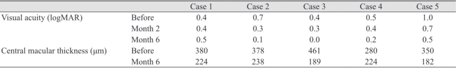
(2) Korean J Ophthalmol Vol.26, No.2, 2012. Case Report This prospective interventional case series was approved by the review board/ethics committee of the Ophthalmic Research Center of the university. An informed consent was obtained from each patient. Five eyes of 5 patients with diagnosis of refractory CSC (lasting more than 1 year) were included in this study. Diagnosis was made by the history of recurrent blurred vision and metamorphopsia for more than one year, detection of neurosensory detachment in ophthalmoscopy and optical coherence tomography (OCT), and observation of active RPE leakage in flourscein angiography. Exclusion criteria consisted of any accompanying macular disease, severe media haziness which precludes OCT evaluation, and noncompliance. Each participant underwent a thorough ophthalmic examination. All eyes received a single injection of 0.05 mL (1.25 mg) intravitreal bevacizumab (Avastin; Genentech Inc., South San Francisco, CA, USA [made for F. Haffmann-La Roche Ltd., Basel, Switzerland]) performed by a 30-guage needle through supratemporal quadrant 4 mm from the limbus under sterile condition. All patients underwent a through ophthalmic examination 1 day, 1 week, and 1, 2, and 6 months after the injection. Best corrected visual acuity (BCVA) of the eyes was checked by a masked optometrist. It was changed to the logarithm of minimum angle of resolution (logMAR) scale for statistical purposes and compared at months 2 and 6 with the baseline values. Central macular thickness (CMT) measured by OCT (3D OCT-1000; Topcon Corporation, Tokyo, Japan) was performed at presentation and repeated 6 months after the intervention. It was measured in a 1-mm circle centered on the fovea by an optician who was masked to the study. The data were analyzed by paired t-. test. Statistical level of significance was preset at 0.05. The initial characteristics are listed in Table 1. None of the patients had history of intraocular surgery, diabetes mellitus, hypertension, cardiovascular disease and smoking. An increase of BCVA was noticed during the followup in all eyes except case 1. Mean BCVA at baseline was 0.60 ± 0.25 that improved to 0.42 ± 0.16 and 0.24 ± 0.21 logMAR up to the month 2 and 6, respectively. This improvement at 2 months did not reach to a meaningful level ( p = 0.064); however, it was statistically significant at 6 months ( p = 0.025). No recurrence was observed in any of the eyes during the follow-up period. Central macular thickness decreased significantly from 370 ± 65 µm at baseline to 210 ± 24 µm at 6 months after injections ( p = 0.009) (Table 2). Figure 1 demonstrates the OCT pictures of 4 cases before and after treatment. No major ocular or systemic complication was encountered in this study. None of the eyes had intraocular pressure rise (>21 mmHg) or cataract progression during the follow-up period.. Discussion Various medications have been suggested for the treatment of CSC by different authors. They include acetazolamide, beta-blockers, vitamins, and non-steroidal antiinflammatory drugs. None of them has been proved to be beneficial. On the other hand, there are some controversial recommendations in the literature on the use of laser photocoagulation in this field. Some authors reporting that laser photocoagulation shortens the duration of disease and reduces recurrence rate, while others maintain that it does not affect final vision and recurrence rate. Furthermore, laser may be associated with permanent scotoma which may enlarge over time with RPE scar expansion, as the possible development of CNV [2,3].. Table 1. Initial characteristics of the cases Sex Age (yr) Laterality Duration of symptoms (yr) No. of attacks Previous fellow eye involvement. Case 1 Male 36 Left 6 4 -. Case 2 Male 47 Left 2 5 -. Case 3 Male 40 Left 2 3 +. Case 4 Male 36 Left 1 2 +. Case 5 Female 39 Right 3 2 -. Table 2. Best corrected visual acuity and central macular thickness changes during follow-up Visual acuity (logMAR). Central macular thickness (μm). 140. Before Month 2 Month 6 Before Month 6. Case 1 0.4 0.4 0.5 380 224. Case 2 0.7 0.3 0.1 378 238. Case 3 0.4 0.3 0.0 461 189. Case 4 0.5 0.4 0.2 280 224. Case 5 1.0 0.7 0.5 350 182.
(3) M Entezari, et al. Intravitreal Bevacizumab for CSC. Before. After. A. Case 1. B. Case 2. C. Case 3. D. Case 4. E. Case 5. Fig. 1. OCT pictures of 4 cases before and after treatment by IVB showing nearly complete (cases 1 [A], 2 [B], and 3 [C]) and partial (cases 4 [D] and 5 [E]) resolutions of subretinal fluid.. Photodynamic therapy has also been attempted with some success for treatment of refractory CSC. It may hasten resolution of exudation by reducing choroidal blood f low and hence favoring cessation of leakage [10]. Most recently, several case series have reported the use of indocyanine green guided PDT in the treatment of chronic CSC [11]. Ober et al. [12] reported the successful treatment of focal RPE leaks in CSC by PDT in a small pilot series which showed resolution and visual improvement. Cardillo Piccolino et al. [6] performed indocyanine green guided PDT in 16 eyes with chronic CSC and treatment resulted in complete resolution of serous retinal detachment 1 month after treatment in 75% of eyes. At 3 months after PDT, 69% of eyes had visual improvement of 1 or more lines. However, 31% of their cases developed secondary RPE changes at the site of PDT, which were thought to be due to hypoxic damage caused by choriocapillaris occlusion. Moreover, PDT is an expensive treatment and may cause CNV formation [13]. Our study showed a significant visual improvement and CMT reduction following single injection of IVB (1.25 mg) in 5 cases suffering from refractory CSC for more than one year. In a similar study on 5 cases with CSC, Torres-. Soriano et al. noticed an improvement in BCVA, fluorescein angiographic leakage, and reduced or resolved neurosensory detachment. However, they injected 2.5 mg IVB and included cases with history of decreased visual acuity more than 3 months, recurrent episodes of CSC or even acute cases with excessive discomfort about visual acuity [7]. In a case series on 12 eyes, Schaal et al. [9] showed that in cases with chronic CSC IVB injection improved BCVA and reduced CMT. However, they performed multiple injections of 2.5 mg IVB at 6 to 8 week intervals (range, 1 to 4 weeks). However, recurrence did not occur in any case of our study during follow up period. In summery, the present study demonstrated a promising effect of IVB in the treatment of refractory CSC; however, we can not make specific treatment recommendations based on this small, uncontrolled case series. Further clinical trials with control group are warranted. Further studies with large sample size are warranted.. Conflict of Interest No potential conflict of interest relevant to this article was reported.. References 1. Wang M, Munch IC, Hasler PW, et al. Central serous chorioretinopathy. Acta Ophthalmol 2008;86:126-45. 2. Marmor MF, Tan F. Central serous chorioretinopathy: bilateral multifocal electroretinographic abnormalities. Arch Ophthalmol 1999;117:184-8. 3. Burumcek E, Mudun A, Karacorlu S, Arslan MO. Laser photocoagulation for persistent central serous retinopathy: results of long-term follow-up. Ophthalmology 1997;104:616-22. 4. Spitznas M. Pathogenesis of central serous retinopathy: a new working hypothesis. Graefes Arch Clin Exp Ophthalmol 1986;224:321-4. 5. Marmor MF. New hypotheses on the pathogenesis and treatment of serous retinal detachment. Graefes Arch Clin Exp Ophthalmol 1988;226:548-52. 6. Cardillo Piccolino F, Eandi CM, Ventre L, et al. Photodynamic therapy for chronic central serous chorioretinopathy. Retina 2003;23:752-63. 7. Torres-Soriano ME, Garcia-Aguirre G, Kon-Jara V, et al. A pilot study of intravitreal bevacizumab for the treatment of central serous chorioretinopathy (case reports). Graefes Arch Clin Exp Ophthalmol 2008;246:1235-9. 8. Von Graefe A. Ueber centrale recidivierende retinitis. Graefes Arch Clin Exp Ophthalmol 1866;12:211-5. 9. Schaal KB, Hoeh AE, Scheuerle A, et al. Intravitreal bevacizumab for treatment of chronic central serous chorioretinopathy. Eur J Ophthalmol 2009;19:613-7. 10. Chan WM, Lam DS, Lai TY, et al. Choroidal vascular remodelling in central serous chorioretinopathy after indocyanine green guided photodynamic therapy with verteporfin: a novel treatment at the primary disease level. Br J Ophthalmol 2003;87:1453-8.. 141.
(4) Korean J Ophthalmol Vol.26, No.2, 2012. 11. Battaglia Parodi M, Da Pozzo S, Ravalico G. Photodynamic therapy in chronic central serous chorioretinopathy. Retina 2003;23:235-7. 12. Ober MD, Yannuzzi LA, Do DV, et al. Photodynamic therapy for focal retinal pigment epithelial leaks second-. 142. ary to central serous chorioretinopathy. Ophthalmology 2005;112:2088-94. 13. Colucciello M. Choroidal neovascularization complicating photodynamic therapy for central serous retinopathy. Retina 2006;26:239-42..
(5)
수치
![Fig. 1. OCT pictures of 4 cases before and after treatment by IVB showing nearly complete (cases 1 [A], 2 [B], and 3 [C]) and partial (cases 4 [D] and 5 [E]) resolutions of subretinal fluid.](https://thumb-ap.123doks.com/thumbv2/123dokinfo/5465211.658330/3.892.78.434.112.571/pictures-treatment-showing-nearly-complete-partial-resolutions-subretinal.webp)
관련 문서
Time series of vertical cross section of potential vorticity and wind vector calculated by Case 1 along the A-A' line indicated at Fig.. Same
USB 연결 케이블을 이용하여 라즈베리 파이와 센서보드 연결 라즈베리 파이의 USB 포트와 WeDo 의 컴퓨터 연결 허브를 연결하 면 된다...
Results : The expression of p21 was increased in boderline serous tumor and serous cystadenocarcinoma in contrast to benign serous tumors. The expression of
그늘 에 주차된 자동차의 온도와 햇빛이 비치는 곳에 주차된 자동차의 온도가 다르다.. 불과 가까운 쪽에서 불에서 먼
Risk factors related to fixed airway obstruction in patients with asthma after antiasthma treatment. Identification of sub- types of refractory asthma in Korean patients by
In this group of high-risk patients with severe COVID-19 pneumonia, treatment with lenzilumab was associated with a significantly shorter time to clinical improvement compared
[r]
[r]
