• 검색 결과가 없습니다.

Analysis of Treatment Response in Patients with Oral Lichen Planus JOMP

N/A
N/A
Protected

Academic year: 2021

Share "Analysis of Treatment Response in Patients with Oral Lichen Planus JOMP"

Copied!
8
0
0

로드 중.... (전체 텍스트 보기)

전체 글

(1)

pISSN 2288-9272 eISSN 2383-8493 J Oral Med Pain 2021;46(2):41-48 https://doi.org/10.14476/jomp.2021.46.2.41

Analysis of Treatment Response in Patients with Oral Lichen Planus

Hye-Min Ju 1 , Kyung-Hee Kim 2 , Hye-Mi Jeon 3 , Yong-Woo Ahn 4 , Soo-Min Ok 4 , Sung-Hee Jeong 4

1

Department of Oral Medicine, Pusan National University Dental Hospital, Dental Research Institute, Yangsan, Korea

2

Department of Oral Medicine, Inje University Busan Paik Hospital, Busan, Korea

3

Dental Clinic Center, Pusan National University Hospital, Busan, Korea

4

Department of Oral Medicine, Pusan National University, School of Dentistry, Dental Research Institute, Dental and Life Science Institute, Yangsan, Korea

Received May 28, 2021 Revised June 16, 2021 Accepted June 16, 2021

Purpose:

Purpose: To evaluate compliance by analyzing and comparing treatment duration, degree of improvement after treatment and treatment response of oral lichen planus (OLP) patients according to characteristics of them and the severity of the lesion.

Methods:

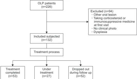
Methods: According to treatment process, 132 subjects with OLP who first visited the De- partment of Oral Medicine at the Pusan National University Dental Hospital from January 2017 to December 2020were classified into three groups: Treatment completed (CT) group, Under treatment (UT) group, and Dropped out during follow-up (DT) group. The reticula- tion/keratosis, erythema, and ulceration (REU) scoring system was used to assess the sever- ity of OLP. The degree of improvement after treatment was evaluated in CT group.

Results:

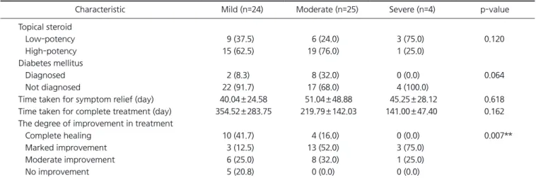
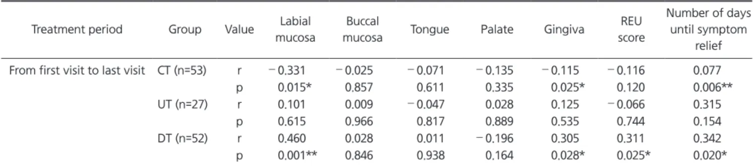
Results: There were 53 (40.15%) CT, 27 (20.45%) UT and 52 (39.39%) DT. In CT group, ac- cording to initial REU score there was a statistical difference in the degree of improvement, but not in the length of time to complete treatment. There was no statistical difference be- tween the days it took for patients to feel symptom relief, and the days of entire treatment among three groups. However, there was a positive correlation between the REU score of gingiva and duration of treatment in DT group. In the CT and DT groups, there was a corre- lation between the length of time taken to relieve symptoms and the duration of treatment.

Conclusions:

Conclusions: The severity of the gingival lesion and the initial response to treatment have a large effect on the entire treatment period and prognosis, so it should be considered when explaining the disease prognosis and treatment period to patients, and the clinician needs to focus on initial symptom relief.

Key Words:

Key Words: Long-term care; Oral lichen planus; Patient compliance

Correspondence to:

Sung-Hee Jeong

Department of Oral Medicine, Pusan National University, School of Dentistry, Dental Research Institute, Dental and Life Science Institute, 49 Busandaehak-ro, Mulgeum-eup, Yangsan 50612, Korea Tel: +82-55-360-5242

Fax: +82-55-360-5238 E-mail: drcookie@pusan.ac.kr

https://orcid.org/0000-0002-6296-4775 This work was supported by a 2-Year Research Grant of Pusan National University.

JOMP Journal of Oral Medicine and Pain

Copyright Ⓒ 2021 Korean Academy of Orofacial Pain and Oral Medicine. All rights reserved.

CC

This is an open-access article distributed under the terms of the Creative Commons Attribution Non-Commercial License (http://creativecommons.org/licenses/by-nc/4.0/), which permits unrestricted non-commercial use, distribution, and reproduction in any medium, provided the original work is properly cited.

INTRODUCTION

Oral mucosal diseases (OMD) include many types of dis- eases caused by various etiologies. Since oral mucosa show rapid recovery from damage, the treatment period is not long if the cause is removed [1]. In this instance, if the di- agnosis is correct, the patient’s discomfort can be quickly removed through appropriate treatment and medication provided and there is a high degree of patient cooperation

and compliance. Conversely, OMD that require long-term treatment often develop into more serious OMD due to low patient compliance and poor treatment results [2].

Most of the OMD that require long-term treatment in-

clude autoimmune diseases such as oral lichen planus (OLP),

mucosal pemphigoid, and mucosal pemphigus [3]. Of these,

OLP is the most common disease affecting patient requiring

long-term treatment for mucosal disease by the Department

of Oral Medicine. It occurs mainly bilaterally and can occur

(2)

anywhere in the oral cavity such as the buccal mucosa, tongue, and gingiva. Almost all lesions of OLP have a re- ticular form of white striae. Therefore, striae are also ob- served around erosive or ulcerative form. The clinical pre- sentation of OLP occurs in a wide spectrum from patients showing only reticular form without any symptoms, to pa- tients with multiple erythema and ulceration with severe symptoms [4,5]. Whilst there is a high correlation between the severity of OLP and the level of pain experienced by the patients, pain symptoms are not always present [6]. Many OLP patients are concerned about the possibility of malig- nancy in the lesion and the possibility of disease transmis- sion. Although the actual rate of malignant transformation of OLP is only as low as 0% to 2%, continuous follow-up is very important as there is still a risk of conversion [7].

Steroids are the main option used in the treatment of OLP and are available in various dosage forms such as gargles, ointments, lesional injections, and pills [8]. Topical agents are most commonly used to treat, however, they present a challenge for patient compliance as the treatment require patients to keep their mouth closed post application. In clinically, this requires a high degree of patient cooperation and diligence, especially when socializing. Therefore, dif- ferences in compliance to treatment in OLP patients lead to differences in treatment outcomes [2].

This study aims to evaluate compliance by analyzing and comparing treatment duration, the degree of improvement after treatment, and treatment response according to the

characteristics of the patient’s initial visit and the severity of the lesion.

MATERIALS AND METHODS

1. Subjective

Patients with OLP were recruited from those who first vis- ited the Department of Oral Medicine at the Pusan National University Dental Hospital from January 2017 to December 2020. Patients who visited more than 3 times were includ- ed. The following set of exclusion criteria was then applied to the potential cohort of 226 patients: subjects with other oral lesions, those who are taking corticosteroids or immu- nosuppressive medications at that time due to OLP or other systemic diseases, or had a record of taking them within 6 months, patients who could not confirm treatment results due to no clinical photo, and patients with dysplasia as a result of biopsy. As a result, a total of 132 patients were se- lected for the study (Fig. 1). The patients were grouped ac- cording to the following characteristics: sex, age, systemic disease of the patients, and the severity of OLP, which was analyzed using clinical photo through the reticulation/kera- tosis, erythema, and ulceration (REU) scoring system. The medicines applied at each visit were investigated, and the total treatment period of the patients as well as the time period until the subjective symptoms of the patient re- solved were investigated. This study was approved by the Institutional Review Board of Pusan National University

Treatment completed (n=53)

Under treatment

(n=27)

Dropped out during follow up

(n=52)

Excluded (n=94) Other oral lesion Taking corticosteroid or

immunosuppressive medicine at first visit

No clinical photo Dysplasia

Treatment process Included subjected

(n=132) OLP patients

(n=226)

Fig. 1.

Fig. 1. Subject flow chart. OLP, oral

lichen planus.

(3)

Dental Hospital (IRB No. PNUDH-2021-019). Written in- formed consent was obtained from all patients at the initial visit.

2. Reticulation/Keratosis, Erythema, and Ulceration Scoring System and Grouping

Based on the REU criteria, the oral cavity was divided into 10 sites (upper/lower labial mucosa, right buccal muco- sa, left buccal mucosa, dorsal tongue, ventral tongue, floor of the mouth, hard palate mucosa, soft palate/tonsillar pil- lars, maxillary gingiva, and mandibular gingiva), and 0 or 1 point was given to each part for the existence or nonexis- tence of a reticular/hyperkeratotic form. In addition, if there was no erythema or ulcer, 0 points were given. One point was given for lesions smaller than 1 cm

2

, 2 points for 1 to 3-cm

2

lesions, and 3 points for lesions greater than 3 cm

2

. The final score was calculated by summing the total scores of reticular, erythema, and ulcer for each clinical sign, with weights of 1, 1.5, and 2 given to them, respectively [9].

Based on the REU score, OLP severity was divided into three groups: Mild (0 to 5), Moderate (5.5 to 15), and Severe (15.5 or more). According to the REU scoring system, 5 points or less can be scored only when there is an erythema or ulcer smaller than 1 cm

2

with a reticular form, hence, this group was called mild. The severe group with a score of 15.5 points or more must have at least one reticular form site with at least two erythema with a size of 3 cm

2

or more and an ulcer with a size of 3 cm

2

or more in at least one site. Treatment response analysis was evaluated by dividing into three groups according to the score.

According to the treatment process, 132 subjects were classified into three groups: Treatment completed (CT), Under treatment (UT), and Dropped out during follow-up (DT) (Fig. 1). CT in this study refers to a case where erosive and ulcer lesions do not exist, stating that the patient is us- ing only the steroid gargle solution and is only required to have a follow-up assessment every 3 months or more and that the patient has no discomfort in their daily life.

3. Criteria for Evaluating the Degree of Improvement in Treatment

Various types of steroid agents such as gargles, ointments, lesional injections (triamcinolone injection 40 mg/mL) and

pills (prednisolone) have been used for the treatment of OLP patients. As for topical ointments, clobetasol propionate 0.05% and fluocinonide 0.05% were used as high potency, dexamethasone sodium phosphate 0.1% as low potency [10].

T

0

refers to the REU score at the initial visit and T

F

is the score after the final treatment. The efficacy index of clini- cal REU improvement was calculated using the formula T (%)= [(T

0

–T

F

)÷T

0

]×100. The efficacy index was classified as follows to determine the level of improvement: (1) Complete healing: efficacy index of 100%; (2) Marked improvement:

70%<efficacy index<100%; (3) Moderate improvement:

30%<efficacy index<70%; (4) No improvement: efficacy in- dex<30% [11].

4. Statistical Analysis

All statistical analyses were performed using IBM Sta- tistical Package for the Social Sciences (SPSS) version 22.0 (IBM Corp., Armonk, NY, USA). A chi-squared test was used to determine distribution differences in sex, ointment used, and diabetes mellitus (DM) status between groups. After T

F

in the treated group, a chi-squared test was also used to de- termine whether there was a difference in the degree of im- provement after treatment between the three groups. If the expected frequency of the cell was lower than 5, Fisher’s exact test was used. Kruskall–Wallis was used to determine the difference in age, the period until treatment completion, the REU score at the initial visit, and the REU score at the time of treatment completion between groups. The ANOVA test was used to determine the difference in REU scores, time taken to symptom relief, and the time taken until the final visit between CT, UT and DT groups at the time of in- vestigation. The Spearman rho correlation was used to in- vestigate the correlation between treatment duration and REU score at the initial visit. p<0.05 was considered to be statistically significant.

RESULTS

Using the REU score at the initial visit, the 132 eligible

OLP patients were grouped as follow: 58 in the mild group

(43.94%), 58 in the moderate group (43.94%), and 16 in the

severe group (12.12%). There were no differences in the age,

sex, and proportions of diabetic patient between groups

(4)

(Table 1). There was no statistical difference between groups in systemic diseases (data not shown). There were statistical differences in REU scores for sites and the total mean be- tween the groups divided by mild, moderate, and severe ac- cording to the REU score (p<0.001; Table 1).

Of the 132 patients, 53 (40.15%) CT, 27 (20.45%) of the remaining 79 were UT, and 52 (39.39%) were DT. Of the 53 CT, 24 patients (45.28%) were in the mild, 25 (47.17%) were in the moderate, and 4 (7.55%) were in the severe group ac- cording to the REU score at the initial visit. There was no statistically significant difference in the efficacy of the oint- ment used in the patients and whether or not the patients ware diabetic. In addition, there was no statistical difference

in the time taken to relieve symptoms by group, and there was no statistical difference in the time taken to complete treatment (Table 2). However, there was a statistical differ- ence in the degree of improvement after treatment at com- pletion (p<0.01; Table 2). The mild group showed a high percentage of complete healing at 41.7%, but no improve- ment was also 20.8% compared to the moderate and se- vere groups. In the moderate group, the degree of complete healing was 16.0%, which was lower than that of the mild group, but the patient with no improvement did not exist, and complete healing patients did not exist in the severe group (Table 2).

When comparing the characteristics between the CT, UT

Table 1.

Table 1. Characteristics of the patients at T

0

Characteristic Total (n=132) Mild (n=58) Moderate (n=58) Severe (n=16) p-value

Age (y) 59.63±10.63 58.18±11.8 60.57±9.25 61.50±10.66 0.388

Sex 0.691

Female 97 (73.5) 41 (70.7) 43 (74.1) 13 (81.3)

Male 35 (26.5) 17 (29.3) 15 (25.9) 3 (18.8)

Diabetes mellitus 0.265

Diagnosed 24 (18.2) 7 (12.1) 13 (22.4) 4 (25.0)

Not diagnosed 108 (81.8) 51 (87.9) 45 (77.6) 12 (75.0)

Labial mucosa 0.38±0.84 0.22±0.63 0.32±0.60 1.16±1.60 0.000***

Buccal mucosa 4.18±4.18 1.57±1.27 4.81±3.12 11.34±5.18 0.000***

Tongue 1.08±1.87 0.28±0.56 1.42±2.07 2.78±2.67 0.000***

Palate 0.16±1.01 0.00±0.00 0.09±0.40 1.00±2.74 0.001**

Gingiva 2.62±3.66 0.91±1.40 2.87±2.72 7.91±6.32 0.000***

REU score 8.26±7.08 2.97±1.35 9.42±2.81 23.19±7.05 0.000***

REU, reticulation/keratosis, erythema, and ulceration.

Values are presented as mean±standard deviation or number (%). p-values were obtained by one-way Kruskal–Wallis and chi-squared test.

**p<0.01; ***p<0.001.

Table 2.

Table 2. Characteristics of the treatment completed group patients (n=53) with stable sign and symptom after treatment

Characteristic Mild (n=24) Moderate (n=25) Severe (n=4) p-value

Topical steroid

Low-potency 9 (37.5) 6 (24.0) 3 (75.0) 0.120

High-potency 15 (62.5) 19 (76.0) 1 (25.0)

Diabetes mellitus

Diagnosed 2 (8.3) 8 (32.0) 0 (0.0) 0.064

Not diagnosed 22 (91.7) 17 (68.0) 4 (100.0)

Time taken for symptom relief (day) 40.04±24.58 51.04±48.88 45.25±28.12 0.618

Time taken for complete treatment (day) 354.52±283.75 219.79±142.03 141.00±47.40 0.162 The degree of improvement in treatment

Complete healing 10 (41.7) 4 (16.0) 0 (0.0) 0.007**

Marked improvement 3 (12.5) 13 (52.0) 3 (75.0)

Moderate improvement 6 (25.0) 8 (32.0) 1 (25.0)

No improvement 5 (20.8) 0 (0.0) 0 (0.0)

Values are presented as number (%) or mean±standard deviation. p-values were obtained by one-way Kruskal–Wallis and chi-squared test.

**p<0.01.

(5)

and DT groups, there was no statistical difference between groups in REU scores for other intraoral sites and the total score at the initial visit, but the REU score for tongue in UT group was higher on average (p<0.01; Table 3). There was no statistical difference between the days it took for pa- tients to feel symptom relief and the final treatment period between the three groups.

When examining the correlation between the total treat- ment duration of patients, REU score and duration of symp- tom relief, there was a negative correlation between REU scores of labial mucosa and gingiva and duration of treat- ment in the CT group, and the UT group had no correlation between duration and score. There was a positive correla- tion between the REU score of labial mucosa and the du- ration of treatment in the DT group, and there was also a correlation with the REU score of gingiva. In the CT and DT groups, there was a correlation between the time taken to relieve symptoms and the duration of treatment (Table 4).

DISCUSSION

Patients visited the hospital with various degrees of dis- ease severity, and because one or more treatment methods were applied based on the severity of the disease, the ef- fectiveness of a specific treatment could not be compared so this could not be used as an evaluation item. Therefore, characteristics were compared by classifying them accord- ing to the initial severity, not according to the treatment type, and the characteristics of patients according to the difference in the treatment process at the time of the final investigation were compared. This study was conducted over four-year period from 2017 to 2020, and at the time of the final investigation, patients who were still undergo- ing treatment or did not visit the hospital during treatment might have the possibility of completing treatment or re- visit in the future. In other words, these patients were more likely to return for follow-up but could not be included in CT group, thus limitations that may have had an effect on

Table 3.

Table 3. Differences in characteristics of patients according to treatment progress

Characteristic CT group

(n=53)

UT group (n=27)

DT group

(n=52) p-value Post-Hoc

result

Labial mucosa 0.43±0.88 0.28±0.59 0.37±0.92 0.735

Buccal mucosa 4.08±3.85 4.17±3.67 4.29±4.78 0.967

Tongue 0.81±1.49 2.15±2.74 0.81±1.47 0.004** 2>1, 3 (Scheffe)

Palate 0.30±1.69 0.09±0.34 0.13±0.65 0.653

Gingiva 2.27±3.80 3.11±3.22 2.72±3.75 0.609

Total REU score 7.59±6.58 9.70±6.79 8.18±7.71 0.453

Time taken for symptom relief (day) 45.73±38.21 62.82±38.24 72.28±142.98 0.370 Length of time from first to last visit (day) 293.34±257.44 444.85±372.39 296.21±299.66 0.072

CT, Treatment completed; UT, Under treatment; DT, Drop out during follow-up; REU, reticulation/keratosis, erythema, and ulceration.

Values are presented as mean±standard deviation. p-values were obtained by ANOVA test.

**p<0.01.

Table 4.

Table 4. Correlations between REU score and treatment period

Treatment period Group Value Labial mucosa

Buccal

mucosa Tongue Palate Gingiva REU

score

Number of days until symptom

relief From first visit to last visit CT (n=53) r –0.331 –0.025 –0.071 –0.135 –0.115 –0.116 0.077

p 0.015* 0.857 0.611 0.335 0.025* 0.120 0.006**

UT (n=27) r 0.101 0.009 –0.047 0.028 0.125 –0.066 0.315

p 0.615 0.966 0.817 0.889 0.535 0.744 0.154

DT (n=52) r 0.460 0.028 0.011 –0.196 0.305 0.311 0.342

p 0.001** 0.846 0.938 0.164 0.028* 0.025* 0.020*

REU, reticulation/keratosis, erythema, and ulceration; CT, Treatment completed; UT, Under treatment; DT, Drop out during follow-up.

p-values were obtained by Spearman rho.

*p<0.05; **p<0.01.

(6)

the results. Nevertheless, there was no statistical difference in the duration for the final visit at the time of the previ- ous four years of investigation, so it is clinically meaning- ful to compare the patient groups according to the differ- ence in treatment process at the time of the investigation (Table 3). The characteristics of the patients at the initial visit showed the same results as previous studies in age and sex [12]. In addition, DM is considered a risk factor for OLP development, and It was reported that OLP patients have a significant relationship with non-insulin-dependent DM compared to normal population [13,14]. In previous study that analyzed the relationship between DM and OLP preva- lence, the prevalence of OLP in DM patients was 1.6% to 37.7%, and a 2.432-fold relative risk was reported, and the prevalence of DM in OLP patients was 0.5% to 6.1%, and the relative risk was 1.4 times [15]. In this study, there were approximately 18.2% of OLP patients with DM, which was a higher proportion than that of the general population of South Korea, which has a prevalence of diabetes in men of 2.26% and in women of 2.03% [16]. However, other studies reported that DM had no direct relationship with the cause of OLP, and the relationship between DM and OLP is still debated [17]. In this study, the ratio was higher than that of normal people, but there was no statistical difference be- tween the groups according to OLP severity and the ratio of diabetes patients and the same result was shown in the CT group (Table 1). A variety of other systemic diseases were also investigated, including hypertension, hyperlipidemia, cardiac disease, asthma, thyroid disease, hepatitis B and 52 (39.39%) of the 132 patients had one or more systemic dis- orders. Among them, there was one patient with hepatitis C virus infection, which was highly related to OLP. However, there was no statistical relationship between the type of systemic disease and the groups according to the severity of OLP (data not shown) [12].

OLP is a chronic disease that is not cured and requires continuous follow-up. However, patients demonstrating a high level of treatment compliance usually experience no sign and symptom of discomfort, and can easily man- age the disease with only intermittent use of topical medi- cations. When these patients were referred to as the CT group, the Moderate and Severe group showed a degree of improvement above the moderate level. However, the

complete healing rate was low or nonexistent, whereas the Mild group showed either complete healing or no improve- ment in some patients (Table 2). This was largely influenced by a reticular form, which is known to require no treat- ment if there are no symptoms [18]. However, in practice, there are some patients with reticular form who show im- provement with corticosteroid treatment, and others who do not respond at all [4,19]. The reason that the patient’s subjective symptom relief time showed similar results ac- cording to OLP severity is thought to be due to differences in patient expectations for treatment effect. Patients with mild conditions are more likely to expect complete heal- ing, and patients with moderate and severe conditions are considered to have improved by showing a certain level of improvement in treatment compared to before treatment.

In addition, there was no statistical difference in the actual mitigation period of sign and symptom, which is thought to be because the drug was selected and applied according to the severity of the patient’s physician and patient prefer- ence. The Moderate and Severe groups used a combination of strong steroids such as systemic corticosteroid and in- jection from the first visit, and the mild group used gargles and topical agents.

Symptoms of OLP according to the subjectiveness and

perception of the patient, resulting in some patients do not

recognizing lesion, whilst others complain of severe pain

[4]. The UT group in this study showed a higher REU score

of tongue compared to the other two groups (Table 3). The

tongue first contacts food and is a movable structure in the

oral cavity that has a major effect on the patient’s symp-

toms. Previous studies that investigated predictors of symp-

tomatic OLP also found that the probability of symptoms

increased by 2.3 times when the lesion was located on

the tongue [20]. OLP is most common in buccal mucosa,

but patients with gingival lesions are easily stimulated by

brushing or eating food, and it is difficult to control plaque,

which leads to a long treatment period (Table 4). As in this

study, if symptoms are not eased well or the treatment pe-

riod itself is prolonged, patients’ adherence to treatment

decreases, and they are likely to drop out during follow-up

[21]. However, it is not easy to explain long-term care to

patients who want a quick and complete cure of the disease

and make them visit the hospital regularly. The current goal

(7)

of OLP therapy is to treat mucosal erythema and ulceration, relieve symptoms, and reduce the risk of oral cancer [22].

OLP should also be aimed at maintaining improved condi- tions, such as diabetes and high blood pressure, rather than at the concept of complete cure, so that all patients should be educated on the characteristics of the disease at the first visit and the importance of periodic follow-ups should be emphasized to patients.

Patients with good treatment progress required an aver- age of one year for the total treatment period regardless of the severity of the lesion and commonly experienced symptom relief at the beginning of treatment. In particu- lar, symptom relief of gingival lesions had an important ef- fect on the treatment period. Most of the patients in the CT group took a short time to relieve symptoms even when the initial gingival lesion was severe. In the DT group, it took a long time for gingival lesions symptoms to be relieved during the treatment period. In actual clinical practice, le- sions of the buccal mucosa are the most common, but the response to treatment does not significantly affect the pa- tient’s compliance, whereas the response of the gingival le- sions affects the patient’s compliance. Therefore, since the severity of the gingival lesion and the initial response to treatment have a large effect on the treatment period and prognosis, so it should be considered when explaining the disease prognosis and treatment period to patients, and the clinician needs to focus on initial symptom relief.

CONFLICT OF INTEREST

No potential conflict of interest relevant to this article was reported.

ORCID

Hye-Min Ju

https://orcid.org/0000-0002-9252-6717 Kyung-Hee Kim

https://orcid.org/0000-0003-2922-6452 Hye-Mi Jeon

https://orcid.org/0000-0003-0007-5662 Yong-Woo Ahn

https://orcid.org/0000-0002-2197-0394

Soo-Min Ok

https://orcid.org/0000-0003-1776-371X Sung-Hee Jeong

https://orcid.org/0000-0002-6296-4775

REFERENCES

1. Stoopler ET, Sollecito TP. Oral mucosal diseases: evaluation and management. Med Clin North Am 2014;98:1323-1352.

2. Gonzalez-Moles MA, Bravo M, Gonzalez-Ruiz L, Ramos P, Gil- Montoya JA. Outcomes of oral lichen planus and oral lichenoid lesions treated with topical corticosteroid. Oral Dis 2018;24:573- 579.

3. Sciubba JJ. Autoimmune oral mucosal diseases: clinical, etio- logic, diagnostic, and treatment considerations. Dent Clin North Am 2011;55:89-103.

4. Chainani-Wu N, Silverman S Jr, Lozada-Nur F, Mayer P, Watson JJ. Oral lichen planus: patient profile, disease progression and treatment responses. J Am Dent Assoc 2001;132:901-909.

5. Sugerman PB, Savage NW. Oral lichen planus: causes, diagnosis and management. Aust Dent J 2002;47:290-297.

6. Park HK, Hurwitz S, Woo SB. Oral lichen planus: REU scoring system correlates with pain. Oral Surg Oral Med Oral Pathol Oral Radiol 2012;114:75-82.

7. Shirasuna K. Oral lichen planus: malignant potential and diagno- sis. Oral Sci Int 2014;11:1-7.

8. Lodi G, Manfredi M, Mercadante V, Murphy R, Carrozzo M. Inter- ventions for treating oral lichen planus: corticosteroid therapies.

Cochrane Database Syst Rev 2020;2:CD001168.

9. Piboonniyom SO, Treister N, Pitiphat W, Woo SB. Scoring system for monitoring oral lichenoid lesions: a preliminary study. Oral Surg Oral Med Oral Pathol Oral Radiol Endod 2005;99:696-703.

10. Kwatra G, Mukhopadhyay S. Topical corticosteroids: pharmacol- ogy. In: Lahiri K, ed. A treatise on topical corticosteroids in der- matology. Singapore: Springer; 2018. pp. 11-22.

11. Liu J, Zeng X, Chen Q, et al. An evaluation on the efficacy and safety of amlexanox oral adhesive tablets in the treatment of recurrent minor aphthous ulceration in a Chinese cohort: a ran- domized, double-blind, vehicle-controlled, unparallel multicenter clinical trial. Oral Surg Oral Med Oral Pathol Oral Radiol Endod 2006;102:475-481.

12. Lavanya N, Jayanthi P, Rao UK, Ranganathan K. Oral lichen pla- nus: an update on pathogenesis and treatment. J Oral Maxillofac Pathol 2011;15:127-132.

13. Ahmed I, Nasreen S, Jehangir U, Wahid Z. Frequency of oral li- chen planus in patients with noninsulin dependent diabetes mel- litus. J Pak Assoc Dermatol 2012;22:30-34.

14. Romero MA, Seoane J, Varela-Centelles P, Diz-Dios P, Garcia- Pola MJ. Prevalence of diabetes mellitus amongst oral lichen pla- nus patients. Clinical and pathological characteristics. Med Oral 2002;7:121-129.

15. Otero Rey EM, Yáñez-Busto A, Rosa Henriques IF, López-López J,

Blanco-Carrión A. Lichen planus and diabetes mellitus: system-

(8)

atic review and meta-analysis. Oral Dis 2019;25:1253-1264.

16. Kim SA, Park WS, Ohrr HC, et al. Prevalence and management status of diabetes mellitus in Korea. Korean J Med 2005;68:10- 17.

17. Bagewadi A, Bhoweer AK. Oral lichen planus and its association with diabetes mellitus and hypertension. J Indian Acad Oral Med Radiol 2011;23:300-303.

18. Edwards PC, Kelsch R. Oral lichen planus: clinical presentation and management. J Can Dent Assoc 2002;68:494-499.

19. Fernández-González F, Vázquez-Álvarez R, Reboiras-López D, Gándara-Vila P, García-García A, Gándara-Rey JM. Histo- pathological findings in oral lichen planus and their correlation

with the clinical manifestations. Med Oral Patol Oral Cir Bucal 2011;16:e641-e646.

20. Osipoff A, Carpenter MD, Noll JL, Valdez JA, Gormsen M, Bren- nan MT. Predictors of symptomatic oral lichen planus. Oral Surg Oral Med Oral Pathol Oral Radiol 2020;129:468-477.

21. Graber AL, Davidson P, Brown AW, McRae JR, Woolridge K. Dropout and relapse during diabetes care. Diabetes Care 1992;15:1477-1483.

22. Boorghani M, Gholizadeh N, Taghavi Zenouz A, Vatankhah M,

Mehdipour M. Oral lichen planus: clinical features, etiology,

treatment and management; a review of literature. J Dent Res

Dent Clin Dent Prospects 2010;4:3-9.

수치

Fig.  1.  Subject  flow  chart.  OLP,  oral  lichen planus.
Table 1. Characteristics of the patients at T 0
Table 3. Differences in characteristics of patients according to treatment progress

참조

관련 문서

In this study, two types of treatment methods, ozone treatment and combination of alkaline and ozone treatment, was used for character's variation and

Considering the above conclusions, effect of 12 weeks Aerobic exercise program treatment in body composition by exercise and diet treatment program was

Bismuth-containing quadruple therapy as second-line treatment for Helico- bacter pylori infection: effect of treatment duration and antibiotic resistance on the eradication

Anoxic

The purpose of this study is to evaluate the effects of heat treatment on the load-deflection properties and transitional temperature range(TTR) of

Purpose: This study was designed to evaluate the clinical characteristics and the treatment of colonoscopic perforation and the availability of

Concentration of soil residual endosulfan-sulfate with days after treatment of endosulfan in different soils

Diagnosis and Treatment of Oral admidstrated Bisphosphonate-related osteonecrosis of the jaw.. and implantation