Apoptotic Effect of Co-Treatment with Valproic Acid and HS-1200 on Human Osteosarcoma Cells
Duck-Han Kim, D.D.S.,M.S.D., Kee-Hyun Lee, D.D.S.,M.S.D., In-Ryoung Kim, M.S.,Ph.D., Hyun-Ho Kwak, M.S.,Ph.D., Bong-Soo Park, D.D.S.,M.S.D.,Ph.D.,
Sung-Hee Jeong
1, D.D.S.,M.S.D.,Ph.D., Myung-Yun Ko
1, D.D.S.,M.S.D.,Ph.D., Yong-Woo Ahn
1, D.D.S.,M.S.D.,Ph.D.
Department of Oral Anatomy, School of Dentistry, Pusan National University
1
Department of Oral Medicine, School of Dentistry, Pusan National University
Valproic acid (VPA) is a well-known anticonvulsive agent and has been used in the treatment of epilepsy for almost 30 years. VPA emerged in 1997 as an antineoplastic agent as well, when findings indicated the substance inhibited proliferation and induced differentiation of primitive neuroectocdermal tumor cells in vivo (Cinatl et al., 1997). Antitmor activity of VPA is associated with its targeting histone deacetylases. Bile acids and their synthetic derivatives induced apoptosis in various kinds of cancer cells and anticancer effects. It has been reported that the synthetic chenodeoxycholic acid (CDCA) derivatives showed apoptosis-inducing activity on various cancer cells in vitro. This study was undertaken to investigate the synergistic apoptotic effect of co-treatment with the histone deacetylases inhibitor, VPA and a CDCA derivative, HS-1200 on human osteosarcoma (HOS) cells.
Cell viability was evaluated by trypan-blue exclusion. Induction and augmentation of apoptosis were confirmed by Hoechst staining, flow cytometry (DNA hypoploidy and MMP change), Westen blot analysis and immunofluorescent staining.
In this study, HOS cells co-treated with VPA and HS-1200 showed several lines of apoptotic manifestation such as nuclear condensations, the reduction of MMP, the decrease of DNA content, the release of cytochrome c into cytosol, the translocation of AIF onto nuclei, and activation of caspase-7, caspase-3 and PARP whereas each single treated HOS cells did not. Although the single treatment of 1 mM VPA or 25 μM HS-1200 for 48 h did not induce apoptosis, the co-treatment of them induced prominently apoptosis. Therefore our data provide the possibility that combination therapy of VPA and HS-1200 could be considered as a novel therapeutic strategy for human osteosarcoma.
Key words : Apoptosis, Valproic acid, HS-1200, Human osteosarcoma
Corresponding author : Yong-Woo Ahn
Associate Professor, Department of Oral Medicine, Schoool of Dentistry Pusan National University, Beomeo-Ri, Mulgeum-Eup, Yangsan-Si, Gyeongsangnam-Do, 626-870, Korea
Foot note : *This work was supported by for two years Pusan National University research grant.
Received: 2010-06-15 Accepted: 2010-07-20
Ⅰ. INTRODUCTION
The acetylation state of histone is reversibly
regulated by histone acetyltransferase (HAT) and
histone deacetylase (HDAC). An inappropriate
acetylation state of histones causes abnormal
outgrowth and the altered pattern of cell death,
which leads to neoplasmic transformation.
1)HDACs
are overexpressed under specific environmental
conditions, such as hypoxia, hypoglycemia, and serum deprivation.
2)Among these conditions, hypoxia is one of the key factors to trigger angiogenesis via the induction of angiogenic factors. Regulation of such gene expression through the acetylation of histone is highly involved in the control of angiogenesis.
2-4)HDAC inhibitors were known to cause growth arrest, differentiation, or apoptosis of a variety of transformed cells in culture, including human bladder, breast, prostate, lung, ovary, colon cancer cells.
5)Several classes of HDACIs have been identified, which include organic hydroxamic acids (e.g., TSA and suberoyl anilide bisydroxamine [SAHA]), short-chain fatty acides (e.g., butyrates and valproic acid [VPA]), cyclic tetrapeptides (e.g., MS-275).
6)Valproic acid (VPA) is a well-known anticonvulsive agent and has been used in the treatment of epilepsy for almost 30 years. VPA emerged in 1997 as an antineoplastic agent as well, when findings indicated the substance inhibited proliferation and induced differentiation of primitive neuroectocdermal tumor cells in vivo.
7)Antitmor activity of VPA is associated with its targeting histone deacetylases. VPA in particular, was able to down-regulate class II HDAC protein levels significantly in several cells in contrast to TSA, which implies that VPA might be a more selective HDAC inhibitor than TSA.
8-11)Bile acids are polar derivatives of cholesterol essential for the absorption of dietary lipids and regulate the transcription of genes that control cholesterol homeostasis. Different bile acids exhibit distinct biological effects. Importantly, natural bile salts were reported to inhibit cell proliferation and induce apoptosis in various cells.
12,13)Im et al.
14,15)developed several ursodeoxycholic acid (UDCA) and chenodeoxycholic acid (CDCA) derivatives, and it have been reported that they had apoptosis- inducing effect in various cancer cells.
16-21)Cells undergoing apoptosis usually develop characteristic changes, including nuclear condensation and degradation of DNA into oligonucleosomal fragments.
22)Apoptotic cell death
is thought to result ultimately from the proteolytic actions of caspase
23)and alterations in mitochondrial function play a key part in the regulation of apoptosis.
24)Moreover, the proteasome system has been shown to be implicated as a negative or positive mediator of apoptosis. The proteasome pathway is mostly known to work upstream of mitochondrial alterations and caspase activation.
25)Osteosarcoma is one of the most common primay malignant tumors of bone. Treatment of this tumor with systemic chemotherapy dramatically improves the prognosis. Numerous studies depicted that the therapeutic effect of a variety of chemotherapeutic agents on osteosarcoma depended on the induction of apoptosis.
26-28)To date, there is no report about the synergistic apoptotic effects of co-treatment with VPA and HS-1200 on human osteosarcoma cells. Therefore, this study was undertaken to investigate the synergistic apoptotic effect of co-treatment with VPA, and a representative of CDCA derivative, HS-1200, on human osteosarcoma (HOS) cells.
Ⅱ. MATERIALS AND METHODS 1. Reagents
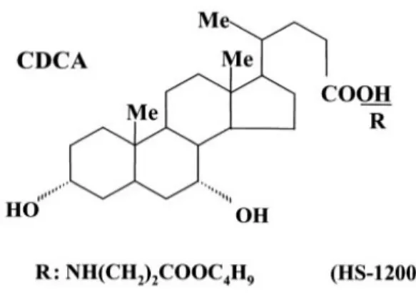
The synthetic bile acid derivative, HS-1200 was kindly provided by Professor Young-Hyun Yoo (Department of Anatomy, College of Medicine, Dong-A University, Busan, Korea). The structure and methods of the synthesis of the synthetic bile acid derivatives were described (Im EO et al., 2001). HS-1200 is a conjugate form of CDCA with β-alanine benzyl ester (N-[(3α, 5β, 7 α)-3,7-dihydroxyl-24-oxocholan -yl] β-alanine benzyl ester). The structures of CDCA and its conjugate form (HS-1200) are shown in Fig. 1.
The following reagents were obtained
commercially: 5,5',6,6'-tetrachloro-1,1',3,3'-tetra-
ethylbenzimidazol carbocyanine iodide (JC-1) was
from Molecular Probes (Eugene, USA). Suc-
LLVY-AMC was from Calbiochem (EMD
Biosciences, Germany). Dulbecco’s modified Eagle’s
Fig. 1. Chemical structures of CDCA and its derivative, HS-1200
medium (DMEM) and FBS were from Gibco (Gaithersburg, MD, USA). Dimethyl sulfoxide (DMSO), Hoechst 33342, RNase A, aprotinin, leupeptin, PMSF, thiazolyl blue tetrazolium bromide and propidium iodide (PI) were from Sigma (St.
Louis, MO, USA); SuperSignal West Pico enhanced chemiluminescence Western blotting detection reagent was from Pierce (Rockford, IL, USA).
2. Antibodies
Mouse monoclonal anti-human caspase-3, caspase-7, poly(ADP-ribose) polymerase (PARP), cytochrome c, apoptosis-inducing factor (AIF) antibodies, and FITC-conjugated goat anti-mouse and anti-rabbit IgGs were from Santa Cruz Biotechnology (Santa Cruz, CA, USA); HRP- conjugated sheep anti-mouse and anti-rabbit IgGs were from Amersham GE Healthcare (Little Chalfront, UK).
3. Cell culture
The HOS human osteosarcoma cell line was purchased from ATCC (Rockville, USA). Cells were maintained at 37℃ with 5% CO
2in air atmosphere in Dulbecco’s modified Eagle's medium (DMEM) with 4 mM L-glutamine, 1.5 g/L sodium bicarbonate, 4.5 g/L glucose and 1.0 mM sodium
pyruvate supplemented with 10% FBS.
4. Assessment of co-treatment of Valproic acid (VPA) and HS-1200
The stock solutions of VPA (2 M) made by dissolving the drug in PBS and HS-1200 (100 mM) made by dissolving the drug in ethanol were kept frozen at -20℃ until use. Twenty four hours after HOS cells were subcultured. the original medium was removed. The cells were washed with phosphate-buffered saline (PBS) and then incubated in the same fresh medium. Since 1 mM VPA or 25 μM HS-1200 is approximately the highest concentration not to induce HOS cell death, we determined to utilize this single concentration of each chemical for the combination treatment study.
HOS cells were co-treated with 1 mM VPA and 25 μM HS-1200 for 48 h. Cells were harvested, stained with trypan blue and then counted using a hemcytometer.
5. Hoechst staining
Cells were harvested and cell suspension was centrifuged onto a clean, fat-free glass slide with a cytocentrifuge. The samples were stained in 4 μ g/mL Hoechst 33342 for 30 min at 37℃ and fixed for 10 min in 4% paraformaldehyde.
6. Quantification of DNA hypoploidy by flow cytometry
After treatment for 48 h, cells were harvested by trypsinization and ice cold 95% ethanol with 0.5%
Tween 20 was added to the cell suspensions to a final concentration of 70% ethanol. Fixed cells were pelleted, and washed in 1% BSA-PBS solution.
Cells were resuspended in 1 mL PBS containing 20
μg/mL RNase A, incubated at 4℃ for 30 min,
washed once with BSA-PBS, and resuspended in
PI solution (10 μg/mL). After cells were incubated
at 4℃ for 5 min in the dark, DNA content were
measured on a CYTOMICS FC500 flow cytometry
system (Beckman Coulter, FL, CA, USA) and data was analyzed using the Multicycle software which allowed a simultaneous estimation of apoptosis.
7. Assay of mitochondrial membrane potential (MMP)
JC-1 was added directly to the cell culture medium (1 μM final concentration) and incubated for 15 min. The medium was then replaced with PBS.
Flow cytometry to measure MMP was performed on a CYTOMICS FC500 flow cytometry (Beckman Coulter, FL, CA, USA). Data were acquired and analyzed using CXP software version 2.2.
8. Immunofluorescent staining
Cells were cytocentrifuged and fixed for 10 min in 4% paraformaldehyde, incubated with each primary antibody for 1 h, washed 3 each for 5 min, and then incubated with FITC-conjugated secondary antibody for 1 h at room temperature.
Cells were mounted with PBS. Fluorescent images were observed and analyzed under Zeiss LSM 510 laser-scanning confocal microscope (Göettingen, Germany).
9. Western blot analysis
Cells (2 x 10
6) treated with VPA and/or HS-1200 were washed twice with ice-cold PBS, resuspended in 200 μL ice-cold solubilizing buffer [300 mM NaCl, 50 mM Tris-Cl (pH 7.6), 0.5% Triton X-100, 2 mM PMSF, 2 μL/mL aprotinin and 2 μL/mL leupeptin] and incubated at 4℃ for 30 min. The lysates were centrifuged at 14,000 revolutions per min for 15 min at 4℃. Protein concentrations of cell lysates were determined with Bradford protein assay (Bio-Rad, USA) and 50 μg of proteins were loaded onto 7.5-15% SDS/PAGE. The gels were transferred to Nitrocellulose membrane (Amersham Pharmacia Biotech, UK) and reacted with each antibody. Immunostaining with antibodies was performed using SuperSignal West Pico enhanced
chemiluminescence substrate and detected with Alpha Imager HP (Alpha Innotech, USA).
Ⅲ. RESULTS
1. Co-treatment of VPA and HS-1200 augmented the reduction in viability of HOS cells.
Single treatment of VPA at 1 mM or HS-1200 at 25 μM for 48 h reduced viability of HOS cells, slightly (VPA, 89.00% ; HS-1200, 83.84%).
Co-treatment of VPA and HS-1200 significantly reduced cell viability compared to the effect of each single treatment (co-treatment, 35.92%) (Fig. 2).
2. Co-treatment of VPA and HS-1200 augmented the nuclear condensation and fragmentation in HOS cells.
To explore whether nuclear condensation and fragmentation were induced, Hoechst staining which is a hallmark of apoptosis, was conducted.
0 20 40 60 80 100 120
Ct r l V H V +H
Cell viability (%)
Fig. 2. Co-treatment of VPA and HS-1200 signifi- cantly reduced cell viability in HOS cells.
Cell viability was determined by hemato-
cytometer. Three independent assays were
performed. Values are means ± SD of
triplicates of each experiment. (V, cells
treated with 1 mM VPA for 48 h; H, cells
treated with 25 μM HS-1200 for 48 h; V+H,
cells treated with 1 mM VPA plus 25 μM
HS-1200 for 48 h)
Fig. 3. Immunofluorescent micrographs showing nuclear morphology after Hoechst staining. Co- treatment of VPA and HS-1200 showed numerous condensed and fragmented nuclei in HOS cells compared to the single treatment (V, cells treated with 1 mM VPA for 48 h; H, cells treated with 25 μM HS-1200 for 48 h; V+H, cells treated with 1 mM VPA plus 25 μM HS-1200 for 48 h).
The co-treatment of VPA and HS-1200 showed a variety of condensed and fragmented nuclei compared to the single treatment (Fig. 3).
3. Augmentation of apoptosis by co- treatment of VPA and HS-1200 was demonstrated by the decrease of DNA content in HOS cells.
The flow cytometry showed that co-treatment of VPA and HS-1200 remarkably increased apoptotic cells with DNA hypoploidy compared to the single treatment (Fig. 4).
Fig. 4. The kinetic analysis of the effect of co- treatment on HOS cell cycle progression and induction of apoptosis by flow cytometry.
Co-treatment remarkably showed the increase of apoptotic cells with DNA hypoplpoidy compared to the single treatment (V, cells treated with 1 mM VPA for 48 h; H, cells treated with 25 μM HS-1200 for 48 h; V+H, cells treated with 1 mM VPA plus 25 μM HS-1200 for 48 h).
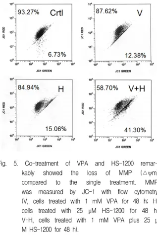
4. Augmentation of apoptosis by co-treatment of VPA and HS-1200 was demonstrated by reduction of mitochondrial membrane potential (MMP) in HOS cells.
The single treatment of VPA and HS-1200 did not show the loss of MMP compared to control group. But the co-treatment of VPA and HS-1200 remarkably reduced MMP compared to the single treatment (Fig. 5).
5. Efficient apoptotic effect of co-treatment of VPA and HS-1200 was demonstrated by Western blot assay.
The co-treatment of VPA and HS-1200 induced
the degradation of caspase-3, caspase-7 and PARP
whereas the single treatment did not (Fig. 6).
Fig. 5. Co-treatment of VPA and HS-1200 remar- kably showed the loss of MMP (△ψm) compared to the single treatmemt. MMP was measured by JC-1 with flow cytometry (V, cells treated with 1 mM VPA for 48 h; H, cells treated with 25 μM HS-1200 for 48 h;
V+H, cells treated with 1 mM VPA plus 25 μ M HS-1200 for 48 h).
Fig. 6. Western blot analysis showing that the co- treatment of VPA and HS-1200 in HOS cells remarkably induced caspase-3, caspase-7 and PARP degradations (V, cells treated with 1 mM VPA for 48 h; H, cells treated with 25 μM HS-1200 for 48 h; V+H, cells treated with 1 mM VPA plus 25 μM HS-1200 for 48 h).
Fig. 7. The confocal microscopy showed that AIF was evidently translocated onto nuclei in HOS cells co-treated with VPA and HS-1200 (V, cells treated with 1 mM VPA for 48 h; H, cells treated with 25 μM HS-1200 for 48 h; V+H, cells treated with 1 mM VPA plus 25 μM HS-1200 for 48 h).
6. Co-treatment of VPA and HS-1200 showed to lead to the translocation of AIF from mitochondria onto the nuclei.
The confocal microscopy showed that AIF was located at mitochondria in the single treatment of VPA or HS-1200 whereas AIF was evidently translocated onto nuclei in the co-treatment (Fig.
7).
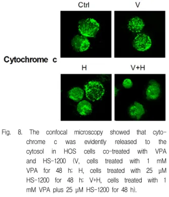
7. Co-treatment of VPA and HS-1200 showed to lead to the release of cytochrome c from mitochondria into the cytosol.
The confocal microscopy showed that
cytochrome c was located at mitochondria in the
single treatment of VPA or HS-1200 whereas
cytochrome c was evidently released into the
cytosol in the co-treatment (Fig. 8).
Fig. 8. The confocal microscopy showed that cyto- chrome c was evidently released to the cytosol in HOS cells co-treated with VPA and HS-1200 (V, cells treated with 1 mM VPA for 48 h; H, cells treated with 25 μM HS-1200 for 48 h; V+H, cells treated with 1 mM VPA plus 25 μM HS-1200 for 48 h).
Ⅳ. DISCUSSION
VPA was employed as monotherapy or combination therapy for various types of malignancy.
29-33)Those studies indicated that VPA as monotherapy or combination therapy showed antiproliferative activity on cancer cells. It is noticeable that VPA, at concentrations of clinical interest, significantly enhanced the antiproli- fearative activity.
31,34)This range of concentrations of VPA can be achieved in an patient's serum when receiving a daily dose of 20-30 mg/kg for epilepsy. Thus, VPA is being considerd as a promising potential therapeutic agent for cancers.
Combination anticancer therapies using VPA and other drugs, especially non-toxic drugs, may offer a substantial advantage over VPA monotherapy in a clinical setting. VPA combined with all-trans retinoic acid
35)or interferon alpha (IFN-α)
11)has been demonstrated to enhance the efficacy of each antitumor agent. We, in the present study, designed three VPA-based combination anticancer therapies using HS-1200 for HOS.
It has been reported the antiproliferative efficacy
of synthetic CDCA derivatives in various cancer cells by inducing apoptosis. Those studies demon- strated the decrease of proteasome activity, mitochondrial events, and nuclear condensation
16-18,36,37)
in synthetic CDCA derivatives-induced apoptosis. In addition, it has been demonstrated that a synthetic CDCA derivative, HS-1200 shows the strongest apoptosis-inducing effect among the synthetic CDCA derivatives.
16,18,38,39)Mitochondria plays an important role in apoptosis, induction of the mitochondrial permeability transition play a key part in the regulation of apoptosis.
24,40,41)Outer mitochondrial membrane becomes permeable to intermembrane space proteins such as cytochrome c and AIF (apoptosis inducing factor) during apoptosis.
42)Cytochrome c release and disruption of mitochondrial membrane potential (MMP) are in fact known features in apoptosis triggered by proteasome inhibition.
43,44)On induction of apoptosis, AIF translocates to the nucleus, resulting in chromatin condensation and large-scale DNA fragmentation.
45)This study evidently showed that co-treatment with CGM and HS-1200 in HOS cells results in significant decrease of MMP, the release of cytochrome c into cytosol and the translocation of AIF onto nuclei whereas the single treatment does not.
A common final event of apoptosis is nuclear condensation, which is controlled by caspases, DFF, and PARP. Caspases, the cysteinyl aspartate- specific intracellular proteinase, play an essential role during apoptotic death.
46)Once activated, the effector caspases (caspase-3, caspase-6 or caspase-7) are responsible for the proteolytic cleavage of a broad spectrum of cellular targets, leading ultimately to cell death. This study demonstrated that co-treatment with CGM and HS-1200 in HOS cells results in the degradation of caspase-3, caspase-7 and PARP whereas the single treatment does not.
In the study, HOS cells co-treated with VPA and
HS-1200 showed several lines of apoptotic
manifestation such as nuclear condensations, the
decrease of MMP, the decrease of DNA content, the release of cytochrome c into cytosol, the translocation of AIF, and degradation of caspase-7, caspase-3 and PARP whereas each single treated HOS cells did not.
In conclusion, combination therapy of VPA and HS-1200 could be considered, in the future, as an alternative therapeutic strategy for human osteosarcoma. Its clinical application awaits further extensive studies.
V. CONCLUSION
Valproic acid (VPA) is a well-known anticonvulsive agent and has been used in the treatment of epilepsy for almost 30 years. VPA emerged in 1997 as an antineoplastic agent as well, when findings indicated the substance inhibited proliferation and induced differentiation of primitive neuroectocdermal tumor cells in vivo (Cinatl et al., 1997). Antitmor activity of VPA is associated with its targeting histone deacetylases. Bile acids and their synthetic derivatives induced apoptosis in various kinds of cancer cells and anticancer effects.
It has been reported that the synthetic chenodeoxycholic acid (CDCA) derivatives showed apoptosis-inducing activity on various cancer cells in vitro. This study was undertaken to investigate the synergistic apoptotic effect of co-treatment with the histone deacetylases inhibitor, VPA and a CDCA derivative, HS-1200 on human osteosarcoma (HOS) cells.
Cell viability was evaluated by trypan-blue exclusion. Induction and augmentation of apoptosis were confirmed by Hoechst staining, flow cytometry (DNA hypoploidy and MMP change), Westen blot analysis and immunofluorescent staining.
In this study, HOS cells co-treated with VPA and HS-1200 showed several lines of apoptotic manifestation such as nuclear condensations, the reduction of MMP, the decrease of DNA content, the release of cytochrome c into cytosol, the translocation of AIF onto nuclei, and activation of
caspase-7, caspase-3 and PARP whereas each single treated HOS cells did not. Although the single treatment of 1 mM VPA or 25 μM HS-1200 for 48 h did not induce apoptosis, the co-treatment of them induced prominently apoptosis. Therefore our data provide the possibility that combination therapy of VPA and HS-1200 could be considered as a novel therapeutic strategy for human osteosarcoma.
REFERENCES
1. Marks PA, Rifkind RA. Erythroleukemic differenti- ation. Annu Rev Biochem 1978;47:419-448.
2. Kim MS, Kwon HJ, Lee YM, Baek JH, Jang JE, Lee SW, Moon EJ, Kim HS, Lee SK, Chung HY, Kim CW, Kim KW. Histone deacetylases induce angiogenesis by negative regulation of tumor suppressor genes. Nat Med 2001;7:437-443.
3. Kwon HJ, Kim MS, Kim MJ, Nakajima H, Kim KW.
Histone deacetylase inhibitor FK228 inhibits tumor angiogenesis. Int J Cancer 2002;97:290-296.
4. Deroanne CF, Bonjean K, Servotte S, Devy L, Colige A, Clausse N, Blacher S, Verdin E, Foidart JM, Nusgens BV and Castronovo V. Histone deacetylases inhibitors as anti-angiogenic agents altering vascular endothelial growth factor signaling. Oncogene 2002;21:427-436.
5. Marks PA, Richon VM, Rifkind RA. Histone deacetylase inhibitors: induces of differentiation or apoptosis of transformed cells. J Natl Cancer Inst 2000;92:1210-1216.
6. De Ruijter AJ, van Gennip AH, Caron HN, Kemp S, van Kuilenburg AB. Histone deacetylases (HDACs):
characterization of the classical HDAC family.
Biochem J 2003;370:737-749.
7. Cinatl J Jr, Cinatl J, Driever PH, Kotchetkov R, Pouckova P, Kornhuber B, Schwabe D. Sodium valproate inhibits in vivo growth of human neuroblastoma cells. Anticancer Drugs 1997;8:958- 963.
8. Gurvich N, Tsygankova OM, Meinkoth JL, Klein PS.
Histone deacetylase is a target of valproic acid-mediated cellular differentiation. Cancer Res 2004;64:1079-1086.
9. Kramer OH, Zhu P, Ostendorff HP, Golebiewski M,
Tiefenbach J, Peters MA, Brill B, Groner B, Bach I,
Heinzel T, Göttlicher M. The histone deacetylase
inhibitor valproic acid selectively induces proteasomal degradation of HDAC2. EMBO J 2003;22:3411-3420.
10. Blaheta RA, Cinatl J Jr. Anti-tumor mechanisms of valproate: a novel role for an old drug. Med Res Rev 2002;22:492-511.
11. Cinatl J Jr, Kotchetkov R, Blaheta R, Driever PH, Vogel JU, Cinatl J. Induction of differentiation and suppression of malignant phenotype of human neuroblastoma BE(2)-C cells by valproic acid:
enhancement by combination with interferon-alpha.
Int J Oncol 2002;20:97-106.
12. Blake J, Roberts PJ, Faubion WA, Kominami E, Gores GJ. Cystatin A expression reduces bile salt-induced apoptosis in a rat hepatoma cell line. Am J Physiol 1988;275:723-730.
13. Martinez JD, Stratagoules ED, LaRue JM, Powell AA, Gause PR, Craven MT, Payne CM, Powell MB, Gerner EW, Earnest DL. Different bile acids exhibit distinct biological effects: the tumor promoter deoxycholic acid induces apoptosis and the chemopreventive agent ursodeoxycholic acid inhibits cell proliferation. Nutr Cancer 1998;31:111-118.
14. Im EO, Lee S, Suh H, Kim KW, Bae YT, Kim ND.
A novel ursodeoxycholic acid derivative induces apoptosis in human MCF-7 breast cancer cells.
Pharm Pharmacol Commun 1999;5:293-298.
15. Im EO, Choi YH, Paik KJ, Suh H, Jin Y, Kim KW, Yoo YH, Kim ND. Novel bile acid derivatives induce apoptosis via a p53-independent pathway in human breast carcinoma cells. Cancer Lett 2001;163:83-93.
16. Choi YH, Im EO, Suh H, Jin Y, Yoo YH, Kim ND.
Apoptosis and modulation of cell cycle control by synthetic derivatives of ursodeoxycholic acid and chenodeoxycholic acid in human prostate cancer cells.
Cancer Lett 2003;199:157-167.
17. Jeong JH, Park JS, Moon B, Kim MC, Kim JK, Lee S, Suh H, Kim ND, Kim JM, Park YC, Yoo YH.
Orphan nuclear receptor Nur77 translocates to mitochondria in the early phase of apoptosis induced by synthetic chenodeoxycholic acid derivatives in human stomach cancer cell line SNU-1. Ann N Y Acad Sci 2003;1010:171-177.
18. Seo SY, Jun EJ, Jung SM, Kim KH, Lim YJ, Park BS, Kim JK, Lee S, Suh H, Kim ND, Yoo YH. Synthetic chenodeoxycholic acid derivative HS-1200-induced apoptosis of p815 mastocytoma cells is augmented by co-treatment with lactacystin. Anticancer Drugs 2003;14:219-225.
19. Park SE, Choi HJ, Yee SB, Chung HY, Suh H, Choi
YH, Yoo YH, Kim ND. Synthetic bile acid derivatives inhibit cell proliferation and induce apoptosis in HT-29 human colon cancer cells. Int J Oncol 2004;25:231-236.
20. Im EO, Choi SH, Suh H, Choi YH, Yoo YH, Kim ND.
Synthetic bile acid derivatives induce apoptosis through a c-Jun N-terminal kinase and NF- kappaB-dependent process in human cervical carcinoma cells. Cancer Lett 2005;229:49-57.
21. Kim ND, Im E, Yoo YH, Choi YH. Modulation of the cell cycle and induction of apoptosis in human cancer cells by synthetic bile acids. Curr Cancer Drug Targets 2006;6:681-689.
22. Williams GT. Programmed cell death: Apoptosis and oncogenesis. Cell 1991;65:1097-1098.
23. Yuan J. Evolutionary conservation of a genetic pathway of programmed cell death. J Cell Biochem 1996;60:4-11.
24. Susin SA, Lorenzo HK, Zamzami N, Marzo I, Snow BE, Brothers GM, Mangion J, Jacotot E, Costantini P, Loeffler M, Larochette N, Goodlett DR, Aebersold R, Siderovski DP, Penninger JM, Kroemer G. Molecular characterization of mitochondrial apoptosis-inducing factor. Nature 1999;397:441-446.
25. Orlowski RZ. The role of the ubiquitin-proteasome pathway in apoptosis. Cell Death Differ 1999;6:
303-313.
26. Lu Y, Yagi T. Apoptosis of human tumor cells by chemotherapeutic anthracyclines is enhanced by Bax overexpression. J Radiat Res(Tokyo) 1999;40:263-272.
27. Fellenberg J, Mau H, Nedel S, Ewerbeck V, Debatin KM. Drug-induced apoptosis in osteosarcoma cell lines is mediated by caspase activation independent of CD95-receptor/ligand interaction. J Orthop Res 2000;18:10-17.
28. Seki K, Yoshikawa H, Shiiki K, Hamada Y, Akamatsu N, Tasaka K. Cisplatin (CDDP) specifically induces apoptosis via sequential activation of caspase-8, 3 and -6 in osteosarcoma. Cancer Chemother Pharmacol 2000;45:199-206.
29. Kieslich M, Schwabe D, Cinatl J Jr, Driever PH.
Increase of fetal hemoglobin synthesis indicating differentiation induction in children receiving valproic acid. Pediatr Hematol Oncol 2003;20:15-22.
30. Li XN, Shu Q, Su JM, Perlaky L, Blaney SM, Lau CC.
Valproic acid induces growth arrest, apoptosis, and
senescence in medulloblastomas by increasing
histone hyperacetylation and regulating expression of
p21Cip1, CDK4, and CMYC. Mol Cancer Ther
2005;4:1912-1922.
31. Takai N, Desmond JC, Kumagai T, Gui D, Said JW, Whittaker S, Miyakawa I, Koeffler HP. Histone deacetylase inhibitors have a profound antigrowth activity in endometrial cancer cells. Clin Cancer Res 2004;10:1141-1149.
32. Witt O, Schweigerer L, Driever PH, Wolff J, Pekrun A. Valproic acid treatment of glioblastoma multiforme in a child. Pediatr Blood Cancer 2004;43:181.
33. Warrell RP Jr, He LZ, Richon V, Calleja E, Pandolfi PP. Therapeutic targeting of transcription in acute promyelocytic leukemia by use of an inhibitor of histone deacetylase. J Natl Cancer Inst 1998;90:
1621-1625.
34. Graziani G, Tentori L, Portarena I, Vergati M, Navarra P. Valproic acid increases the stimulatory effect of estrogens on proliferation of human endometrial adenocarcinoma cells. Endocrinology 2003;144:2822-2828.
35. Mongan NP, Gudas LJ. Valproic acid, in combination with all-trans retinoic acid and 5-aza-2'- deoxycytidine, restores expression of silenced RARbeta2 in breast cancer cells. Mol Cancer Ther 2005;4:477-486.
36. Choi YH, Im EO, Suh H, Jin Y, Lee WH, Yoo YH, Kim KW, Kim ND. Apoptotic activity of novel bile acid derivatives in human leukemic T cells through the activation of caspases. Int J Oncol 2001;18:
979-984.
37. Yoon HS, Rho JH, Yoo KW, Park WC, Rho SH, Choi YH, Suh H, Kim ND, Yoo KS, Yoo YH. Synthetic bile acid derivatives induce nonapoptotic death of human retinal pigment epithelial cells. Curr Eye Res 2001;22:367-374.
38. Kim GC, Her YS, Park JH, Moon YS, Yoo YH, Shin SH, Park BS. Synthetic Bile Acid Derivative HS-1200-induced Apoptosis of Human Osteosarcoma Cells. The Korean J Anat 2004;37:449-457.
39. Baek CJ, Min JH, Moon SH, Kim IR, Lee SE, Kim DH, Kim GC, Kwak HH, Park BS. Synthetic Chenodeoxycholic Acid Derivative HS-1200-induced Apoptosis of Human Melanoma Cells. Korean J Phys Anthropol 2007;20:363-373.
40. Kroemer G, Zamzami N, Susin SA. Mitochondrial control of apoptosis. Immunol Today 1997;18:44-51.
41. Green DR, Reed JC. Mitochondria and apoptosis.
Science 1998;281:1309-1312.
42. Golab J, Stoklosa T, Czajka A, Dabrowska A, Jakobisiak M, Zagozdzon R, Wojcik C, Marczak M, Wilk S. Synergistic antitumor effects of a selective proteasome inhibitor and TNF in mice. Anticancer Res 2000;20:1717-1721.
43. Wagenknecht B, Hermisson M, Groscurth P, Liston P, Krammer PH, Weller M. Proteasome inhibitor- induced apoptosis of glioma cells involves the processing of multiple caspases and cytochrome c release. J Neurochem 2000;75:2288-2297.
44. Marshansky V, Wang X, Bertrand R, Luo H, Duguid W, Chinnadurai G, Kanaan N, Vu MD, Wu J.
Proteasomes modulate balance among proapoptotic and antiapoptotic Bcl-2 family members and compromise functioning of the electron transport chain in leukemic cells. J Immunol 2001;166:
3130-3142.
45. Dauglas E, Susin SA, Zamzami N, Ferri KF, Irinopoulou T, Larochette N, Prevost MC, Leber B, Andrews D, Penninger J, Kroemer G. Mitochondrio -nuclear translocation of AIF in apoptosis and necrosis. FASEB J 2000;14:729-739.
46. Acehan D, Jiang X, Morgan DG, Heuser JE, Wang X,
Akey CW. Three-dimensional structure of the
apoptosome: Implications for assembly, procaspase-9
binding, and activation. Mol Cell 2002;9:423-432.
국문초록
Valproic acid와 HS-1200의 병용처리가 사람골육종세포에 미치는 세포자멸사 효과에 대한 연구
1