106 www.labmedonline.org eISSN 2093-6338 자가면역질환으로 알려져 있으며 관련된 자가항체로는 류마티스 인자, 항cyclic citrullinated peptide (CCP) 항체 등이 있다. 이들 자 가항체의 민감도와 특이도는 높지 않지만 질환의 발현에 선행하 여 나타나기도 하며 항체가 존재할 경우 좋지 않은 경과를 보일 수 있다[1, 2]. 이 외에도 antiperinuclear factor (APF), 항microtubule organizing center with microtubule (MTOC-MT) 항체 등도 류마 티스관절염과 관련이 있는 것으로 알려져 있다[3]. 류마티스관절염 환자에서 proliferating cell nuclear antigen (PCNA)에 대한 항체의 존재는 거의 보고 된 적이 없으며 기존에 보고된 4명의 환자도 모 두 낮은 역가를 보였다[4, 5].
본 증례에서 PCNA에 대한 항체를 검출하는데 이용한 autoim- mune target (AIT) 검사는 비종양성대식세포에서 유래된 IT-1 세 포주를 기질로 사용하여 간접면역형광법으로 환자의 혈청에서 자 가항체를 검출하며 HEp-2 세포를 이용한 항핵항체(antinuclear antibody, ANA) 검사의 문제점을 해결하기 위해 개발되었다.
서 론
류마티스관절염(rheumatoid arthritis, RA)은 관절의 부종, 통증, 윤활관절의 파괴를 특징으로 하는 만성염증질환으로 주로 대칭성 을 보이며 환자에 따라 다양한 경과를 보인다. 류마티스관절염은
항Proliferating Cell Nuclear Antigen 항체 양성의 류마티스관절염 환자 1예
Anti-Proliferating Cell Nuclear Antigen Antibodies Detected in Rheumatoid Arthritis:
A Case Report
송수덕·김신규
Su Dok Song, M.D., Think-You Kim, M.D.
한양대학교 의과대학 진단검사의학교실
Department of Laboratory Medicine, College of Medicine, Hanyang University, Seoul, Korea.
증례보고
Lab Med Online
Vol. 5, No. 2: 106-109, April 2015
http://dx.doi.org/10.3343/lmo.2015.5.2.106 진단면역학
Corresponding author: Think-You Kim
Department of Laboratory Medicine, Hanyang University Medical Center, 222 Wangsimni-ro, Seongdong-gu, Seoul 133-792, Korea
Tel: +82-2-2290-8975, Fax: +82-2-2298-1735, E-mail: [email protected] Received: June 18, 2014
Revision received: July 15, 2014 Accepted: August 7, 2014
This article is available from http://www.labmedonline.org 2015, Laboratory Medicine Online
This is an Open Access article distributed under the terms of the Creative Commons Attribution Non-Commercial License (http://creativecommons.org/licenses/by-nc/3.0/) which permits unrestricted non-commercial use, distribution, and reproduction in any medium, provided the original work is properly cited.
Rheumatoid arthritis (RA) is an autoimmune disease that results in a chronic inflammatory disorder, which principally attacks the small joints. Sev- eral autoantibodies, such as rheumatoid factor (RF) and anti-cyclic citrullinated peptide (CCP) antibody, are known to be associated with RA. Anti- proliferating cell nuclear antigen (PCNA) antibodies are mainly observed in patients with systemic lupus erythematosus (SLE). Indeed, a high titer of these antibodies is considered highly suggestive of SLE; however, anti-PCNA antibodies also appear in other autoimmune diseases. Two previ- ous reports described RA patients with low titers of anti-PCNA antibodies, respectively. In this report, we describe a case of an RA patient exhibit- ing a high titer (>1:2,560) of anti-PCNA antibodies. The 56-yr-old female patient, with no underlying disease or medication history, presented with multiple joint pain and morning stiffness that had begun 6 months prior. The erythrocyte sedimentation rate (ESR) and RF were elevated (102 mm/
hr and 77 IU/mL, respectively), and C-reactive protein (CRP) was 0.8 mg/dL. While the test for anti-CCP antibodies was negative, an anti-PCNA pattern (>1:2,560) and a homogeneous pattern (1:320) were detected by autoimmune target (AIT) test. The presence of anti-PCNA antibodies was subsequently confirmed using the double immunodiffusion method. The anti-dsDNA test was also positive (1:160). X-ray imaging showed soft tissue swelling of multiple joints of both hands and wrists. According to the 2010 American College of Rheumatology/European League Against Rheumatism (ACR/EULAR) classification criteria, the patient was classified as having RA. This is the first case to describe high titers anti-PCNA an- tibodies associated with RA.
Key Words: Autoantibody, Proliferating cell nuclear antigen, Rheumatoid arthritis
송수덕 외: High Titer Anti-PCNA Antibodies in RA
http://dx.doi.org/10.3343/lmo.2015.5.2.106 www.labmedonline.org 107
PCNA에 대한 항체는 전신홍반루푸스(systemic lupus erythe- matosus, SLE)에 특이적인 지표로 알려져 왔으나[6, 7], 근래에는 전 신홍반루스프뿐만 아니라 다른 자가면역질환, B형 간염, C형 간염 등에서도 보고되고 있으며[8, 9] SLE에 특이적이지 않다는 보고도 있었다[5]. 하지만 최근의 연구에서 PCNA에 대한 항체가 기존에 알려진 것보다 낮은 민감도나 특이도를 보이나 해당 연구에서 시 행한 메타분석에서 높은 역가의 PCNA에 대한 항체를 가진 환자 들은 모두 전신홍반루프스 환자였다고 보고했다[4]. 본 저자들은 류마티스관절염 환자에서 PCNA에 대한 항체가 높은 역가로 관찰 되어 보고하는 바이다.
증 례
56세 여자환자는 약 6개월 전부터 시작된 아침강직을 동반한 손, 손목, 팔꿈치, 어깨의 통증을 주소로 외래를 통해 입원하였다.
기저질환이나 약물복용력은 없었다. 혈액검사에서 적혈구침강속 도는 102 mm/hr로 증가해 있었다. 면역검사에서 류마티스인자는 77 IU/mL로 양성이었으며 항CCP 항체는 음성이었다. 면역혈청검 사에서 C-반응성단백은 0.8 mg/dL였다. 수부 X선 검사에서 손목, 중수지 관절, 근위 손가락사이 관절의 연부조직에 부종이 관찰되 었다(Fig. 1). AIT 검사에서 낮은 희석배수에서는 균일형(homoge- neous pattern)에 가려 PCNA형이 뚜렷하게 관찰되지 않았지만 (Fig. 2) 높은 희석배수에서 PCNA형이 확인되어(Fig. 3) PCNA형 (>1:2,560), 균일형(1:320)으로 판독하였다. 이중 면역확산(double immunodiffusion) 검사에서 PCNA에 대한 항체가 다시 한 번 확인
되었고, Crithidia luciliae immunofluorescence (CLIF)를 이용한 항 DNA 검사에서 이중가닥 DNA에 대한 항체 양성소견(1:160)을 보 였다. 환자는 2010년 American College of Rheumatology/Euro- pean League Against Rheumatism (ACR/EULAR) classification criteria for RA에서 1) 4-10개의 소관절 침범(3점), 2) 고역가의 류마 티스 인자 양성(3점), 3) 적혈구침강속도 증가(1점), 4) 6주 이상의 유병기간(1점)에 해당하여 총 8점으로 류마티스관절염으로 확진 되었다.
고 찰
PCNA에 대한 항체는 1978년 전신홍반루프스 환자에서 처음 발 견되었다[10]. 이후 항체의 표적이 34 kDa의 단백질이고[11] 핵 안 에 존재하며 PCNA-interacting proteins (PIPs)로 통칭되는 다양한 단백질들과 결합하여 DNA의 복제, 복구, 세포 주기조절, 유전자발 현, 세포자멸사 등에 관여하는 것으로 밝혀졌다[12-15]. PCNA에 대 한 항체의 검출은 double immunodiffusion, immunoblot, ELISA, line immunoassay (LIA) 등의 다양한 검사를 이용할 수 있으며 본 원에서 사용한 AIT 검사상에서는 세포 주기에 따라 다양한 형광 강도를 보이는 특징을 통해 감별할 수 있는데 이는 PCNA가 G1-S 기 동안 발현이 증가하기 때문에 나타나는 현상이다[4].
증례의 환자에서 고역가의 PCNA에 대한 항체가 관찰된 것은 두 가지의 가능성을 생각할 수 있는데 첫 번째는 환자가 순수한 류마 티스관절염 환자이면서 고역가의 PCNA에 대한 항체가 동반되었 을 가능성이다. PCNA에 대한 항체는 전신홍반루프스 환자에서 처
Fig. 1. X-ray image showing soft tissue swelling of multiple joints of hands and wrists.
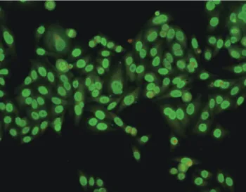
Fig. 2. AIT test of IT-1 cells using the indirect immunofluorescence method. Image depicts the pattern of anti-proliferating cell nuclear antigen (PCNA) antibody (1:20 dilution) binding, which is masked by the homogeneous pattern (×200).
송수덕 외: High Titer Anti-PCNA Antibodies in RA
http://dx.doi.org/10.3343/lmo.2015.5.2.106 108 www.labmedonline.org
음 발견되었고 전신홍반루프스에 특이적으로 생각되었으나, 전신 홍반루프스 환자에서도 발생빈도가 높지 않고(2-8%), 경피증, 자가 면역성 갑상샘염, 류마티스관절염, B형 간염, C형 간염 등에서도 나타나는 것이 보고되었으며[5, 9] Lee and Kim[16]도 PCNA에 대 한 항체가 검출된 소세포폐암 환자를 보고한 바가 있다. 이번 증례 역시 앞서 나타난 연구에서처럼 전신홍반루프스가 아닌 질환에서 고역가로 검출된 PCNA에 대한 항체라면 PCNA에 대한 항체가 전 신홍반루프스에 특이적이지 않다는 하나의 증거로 볼 수 있을 것 이다.
두 번째는 rhupus 증후군의 가능성으로, rhupus 증후군이란 환 자가 류마티스관절염과 전신홍반루프스의 특징을 모두 보이는 경 우를 말하며, 단일질환인지 아니면 단지 전신홍반루스프와 류마 티스관절염이 병발하는 것인지는 아직 명확히 밝혀지지 않은 상태 이다[17]. 증례의 환자는 2010년 ACR/EULAR classification criteria for RA에서 8점에 해당하여 명백한 류마티스관절염으로 진단할 수 있었는데 한편으로는 전신홍반루프스에 특이적인 자가항체로 알려진 이중가닥 DNA에 대한 항체가 양성이었으며 AIT 검사도 양 성이었다. 하지만 RA 환자에서도 약 5.5%에서 이중가닥 DNA에 대 한 항체가 양성으로 나타났다는 보고도 있었고[18], 역가도 높지 않았으며 전신홍반루프스에 해당하는 다른 임상양상을 보이지 않 았으므로 rhupus 증후군의 가능성은 낮다고 생각된다.
류마티스관절염 환자에서 항핵항체의 양성률은 연구자의 역가 기준에 따라 29-75%까지 다양하게 보고되고 있다[18]. Garcia 등 [19]은 균일형이 가장 흔하게 관찰되고 다음으로는 반점형(speck- led pattern), 중심립형(centromere pattern) 순의 빈도로 나타난다 고 보고하였으며, Nishimura 등[20]은 반점형, 균일형, 핵인형(nu-
cleolar pattern), 중십립형 순으로 보고하여 두 연구 간에 빈도의 차이를 보였으나 관찰되는 형은 유사하였다. 형 간의 임상적 차이 는 보고된 바가 없었다. 류마티스관절염 환자에서 PCNA형은 거의 보고된 경우가 없었기 때문에 증례의 환자에서 PCNA형이 발견된 것이 어떤 의미가 있는지 판단하기는 어려웠다.
PCNA의 구조나 역할은 많은 부분이 확인되었지만 아직 PCNA 에 대한 항체의 형성기전이나 병태생리적인 역할은 명확히 밝혀지 지 않은 상태이다. 기존에 PCNA에 대한 항체가 보고된 전신홍반 루스프, 자가면역질환, 간염 등에서도 발생 빈도만 보고되었을 뿐 항체를 가진 환자들이 임상적으로 어떤 차이를 보이는지는 거의 연구된 바가 없다. PCNA에 대한 항체의 전신홍반루프스에 대한 특이성이 의심받고 있는 상황인 만큼 항체 양성인 환자와 음성인 환자의 임상적 특성 등이 추가로 연구되어 PCNA에 대한 항체의 임상적 의의를 재평가할 필요가 있을 것이다.
결론적으로 본 증례는 류마티스관절염이 확진된 환자에서 고역 가의 PCNA에 대한 항체가 발견된 세계 최초의 증례로 PCNA에 대 한 항체의 임상적 유용성을 평가하는 데 도움을 줄 수 있을 것으 로 생각된다.
요 약
류마티스관절염(rheumatoid arthritis, RA)은 관절의 부종, 통증, 윤활관절의 파괴를 특징으로 하는 만성염증질환으로 관련된 자가 항체로는 류마티스인자, 항cyclic citrullinated peptide (CCP) 항체 등이 있다. Proliferating cell nuclear antigen (PCNA)에 대한 항체 는 주로 전신홍반루푸스(systemic lupus erythematosus, SLE) 환자
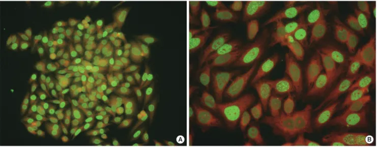
A B
Fig. 3. AIT test of IT-1 cells using the indirect immunofluorescence method. Images depict the pattern of anti-proliferating cell nuclear antigen (PCNA) antibody binding at a 1:1,280 dilution (A: ×200, B: ×400).
송수덕 외: High Titer Anti-PCNA Antibodies in RA
http://dx.doi.org/10.3343/lmo.2015.5.2.106 www.labmedonline.org 109
에서 발견되는데 특히 고역가의 항체는 전신홍반루프스 환자와 연 관성이 높은 것으로 알려져 있다. 전신홍반루프스 환자가 아닌 다 른 자가면역질환에서도 PCNA에 대한 항체가 발견되기도 하는데, Vermeersch 등과 Mahler 등은 PCNA에 대한 항체가 양성인 두 명 의 류마티스관절염 환자를 각각 보고하였으나 모두 낮은 역가를 보였다. 본원에서 시행한 AIT 검사를 통해 한 명의 류마티스관절 염 환자에서 고역가의 PCNA에 대한 항체가 발견되어 보고하는 바 이다. 56세 여자환자로 다른 기저질환, 약물복용력은 없었으며 약 6개월 전부터 아침강직을 동반한 다발성 관절통을 주소로 외래를 통해서 입원하였다. 적혈구침강속도는 102 mm/hr 였고 C-반응성 단백은 0.8 mg/dL (정상범위 0-0.8 mg/dL)였다. 류마티스인자는 77 IU/mL로 양성, 항CCP 항체검사는 음성이었다. AIT 검사에서 균일형(1:320), PCNA형(>1:2,560)이 관찰되었다. PCNA에 대한 항 체는 이중면역확산법을 통해 다시 한번 확인되었다. 항 DNA검사 는 양성이었다(1:160). 수부 X선 검사에서 손목, 중수지 관절, 근위 손가락사이 관절의 연부 조직에 부종이 관찰되었다. 2010년 ACR/
EULAR classification criteria for RA에서 8점에 해당되어 류마티스 관절염으로 진단되었다. 이는 류마티스관절염 환자에서 발견된 고 역가의 PCNA에 대한 항체를 보고한 최초의 증례로 향후에 PCNA 에 대한 항체의 임상적 유용성을 평가할 때 도움을 줄 수 있을 것 으로 생각된다.
REFERENCES
1. Nielen MM, van Schaardenburg D, Reesink HW, van de Stadt RJ, van der Horst-Bruinsma IE, de Koning MH, et al. Specific autoantibodies precede the symptoms of rheumatoid arthritis: a study of serial mea- surements in blood donors. Arthritis Rheum 2004;50:380-6.
2. Lee AN, Beck CE, Hall M. Rheumatoid factor and anti-CCP autoanti- bodies in rheumatoid arthritis: a review. Clin Lab Sci 2008;21:15-8.
3. Kim DA and Kim TY. Anti-microtubule organizing center with micro- tubule by autoimmune target test is also useful serological marker in rheumatoid arthritis evaluation. Rheumatol Int 2013;33:805-8.
4. Mahler M, Miyachi K, Peebles C, Fritzler MJ. The clinical significance of autoantibodies to the proliferating cell nuclear antigen (PCNA). Au- toimmun Rev 2012;11:771-5.
5. Vermeersch P, De Beeck KO, Lauwerys BR, Van den Bergh K, Devel- ter M, Marien G, et al. Antinuclear antibodies directed against prolifer- ating cell nuclear antigen are not specifically associated with systemic lupus erythematosus. Ann Rheum Dis 2009;68:1791-3.
6. Beyne-Rauzy O, Thébault S, Adoue D, Fortenfant F. Anti-PCNA antibod-
ies: prevalence and predictive value. Joint Bone Spine 2005;72:432-5.
7. Fritzler MJ, McCarty GA, Ryan JP, Kinsella TD. Clinical features of pa- tients with antibodies directed against proliferating cell nuclear anti- gen. Arthritis Rheum 1983;26:140-5.
8. Mahler M, Silverman ED, Fritzler MJ. Novel diagnostic and clinical as- pects of anti-PCNA antibodies detected by novel detection methods.
Lupus 2010;19:1527-33.
9. Tzang BS, Chen TY, Hsu TC, Liu YC, Tsay GJ. Presentation of autoanti- body to proliferating cell nuclear antigen in patients with chronic hep- atitis B and C virus infection. Ann Rheum Dis 1999;58:630-4.
10. Miyachi K, Fritzler MJ, Tan EM. Autoantibody to a nuclear antigen in proliferating cells. J Immunol 1978;121:2228-34.
11. Ogata K, Ogata Y, Nakamura RM, Tan EM. Purification and N-termi- nal amino acid sequence of proliferating cell nuclear antigen (PCNA)/
cyclin and development of ELISA for anti-PCNA antibodies. J Immunol 1985;135:2623-7.
12. Celis JE, Madsen P, Nielsen HV, Gesser B, Rasmussen HH, Cruger D.
Cyclin/PCNA is a cell cycle modulated nuclear protein with a role in DNA replication. Arch Biol Med Exp (Santiago) 1988;21:417-21.
13. Bravo R, Frank R, Blundell PA, Macdonald-Bravo H. Cyclin/PCNA is the auxiliary protein of DNA polymerase-delta. Nature 1987;326:515-7.
14. Moldovan GL, Pfander B, Jentsch S. PCNA, the maestro of the replica- tion fork. Cell 2007;129:665-79.
15. Maga G and Hubscher U. Proliferating cell nuclear antigen (PCNA): a dancer with many partners. J Cell Sci 2003;116:3051-60.
16. Lee ZY and Kim TY. A case of small cell lung carcinoma with coexist- ing autoantibodies against proliferating cell nuclear antigen and centri- ole detected using the autoimmune target test. Lab Med Online 2014;4:
218-21.
17. Simón JA, Granados J, Cabiedes J, Morales JR, Varela JA. Clinical and immunogenetic characterization of Mexican patients with ‘rhupus’. Lu- pus 2002;11:287-92.
18. Paulus HE, Wiesner J, Bulpitt KJ, Patnaik M, Law J, Park GS, et al. Au- toantibodies in early seropositive rheumatoid arthritis, before and dur- ing disease modifying antirheumatic drug treatment. J Rheumatol 2002;29:2513-20.
19. Garcia-de la Torre and Miranda-Mendez L. Studies of antinuclear anti- bodies in rheumatoid arthritis. J Rheumatol 1982;9:603-6.
20. Nishimura S, Nishiya K, Hisakawa N, Chikazawa H, Ookubo S, Naka- tani K, et al. Positivity for antinuclear antibody in patients with ad- vanced rheumatoid arthritis. Acta Med Okayama 1996;50:261-5.