436 http://www.jchestsurg.org
JCSJournal of Chest Surgery
Necrotizing Chest Wall Fasciitis Complicating Closed Tube Thoracostomy: Can It Be Avoided?
Abdel-Mohsen M. Hamad, M.D.1,2, Elsayed M. Elmistekawy, M.D.1,3, Ahmed F. Elmahrouk, M.D.1,4
1Department of Cardiothoracic Surgery, Tanta University, Tanta, Egypt; 2Department of Thoracic Surgery, King Fahd Specialist Hospital, Buraydah, Saudi Arabia;
3Cardiac Surgery Division, Department of Surgery, University of Ottawa Heart Institute, Ottawa, Canada; 4Department of Cardiovascular Surgery, King Faisal Specialist Hospital and Research Centre, Jeddah, Saudi Arabia
ARTICLE INFO
Received February 9, 2021, Revised March 9, 2021, Accepted March 10, 2021 Corresponding author
Abdel-Mohsen M. Hamad
Tel 966-565618155, Fax 966-565618155, E-mail [email protected], ORCID https://orcid.org/0000-0003-1303-0818
Copyright©2021, The Korean Society for Thoracic and Cardiovascular Surgery
This is an Open Access article distributed under the terms of the Creative Commons Attribution Non-Commercial License (http://creativecommons.org/licenses/
by-nc/4.0) which permits unrestricted non-commercial use, distribution, and reproduction in any medium, provided the original work is properly cited.
Necrotizing fasciitis (NF) remains a life-threatening sur- gical condition. In its classical form, NF usually involves the extremities. The underlying causes are usually related to trauma or minor injuries. The infectious process usually affects the deep fascial plane, with secondary necrosis of subcutaneous tissue and skin caused by the thrombosis of the subcutaneous and perforator vessels. The muscles are involved in the infectious process; however, myonecrosis typically rarely occurs.
We read with great interest the recent article by Chun et al. [1], who presented their experience with the manage- ment of a patient with massive chest wall fasciitis compli- cating empyema after the insertion of a chest tube. We congratulate the authors for their superb work and excel- lent results. However, we have some comments and would like to share our insights on the management of this case.
A MEDLINE search retrieved 7 reported cases of chest wall NF complicating tube thoracostomy [1-5] (Table 1).
Pleural empyema was a common denominator in almost all reported cases. One patient had pulmonary tuberculo- sis, and according to the authors, tube thoracostomy was inserted for spontaneously developed pneumothorax; how- ever, chest radiography showed obliteration of the costo- phrenic angle with air-fluid level [4]. When available, the bacteriological studies showed that the microorganisms isolated from the pleural fluid were also present in the chest wall purulent materials. These data support our con- cept that the source of infection is the empyema, rather than an external source introduced from thoracostomy
tube insertion. Moreover, some cases of chest wall NF complicating empyema were reported before the insertion of the drainage tube; some sort of empyema necessitans must therefore have initially developed and then pro- gressed to NF.
A possible mechanism for the development of pneumo- nia, lung abscess, and subsequent pleural empyema is the aspiration of infected debris from the mouth or secretions from the upper gastrointestinal tract. Abundant aerobic and anaerobic organisms are present in the normal flora of the mouth, gingival crevices, upper respiratory tract, and gastrointestinal tract. These mixed aerobic and anaerobic organisms have strong proteolytic activity and act in syn- ergy to produce liquefactive necrosis of the affected tissue, whether in the lung or the chest wall tissue. These organ- isms are aggressive and are capable of producing pneumo- nia (if not primary lung abscess) with a high incidence of pleural involvement. The resulting pleural inflammatory process is usually severe and has a rapidly progressive course.
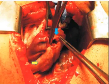
In our experience, during decortication in many cases of empyema thoracis, we observed the presence of ruptured small superficial abscesses (pustules) with resultant pyo-pneumothorax and bronchopleural fistula (Fig. 1). The isolation of Streptococcus constellatus (which is a normal component of the mouth flora) and anaerobic organisms from the pleural fluid of the reported case of Chun et al. [1]
supports this pathogenesis theory.
The technique of closed tube thoracostomy insertion en-
https://doi.org/10.5090/jcs.21.015 pISSN: 2765-1606 eISSN: 2765-1614 J Chest Surg. 2021;54(5):436-438
Brief
Communication or Correspondence
437
Abdel-Mohsen M. Hamad, et al. Necrotizing Chest Wall Fasciitis
http://www.jchestsurg.org
JCS
tails blunt dissection of the intercostal muscles deep to the pleural space. In addition to opening the fascial planes during dissection, the opening in the intercostal space may be wider than the diameter of the catheter, with subse- quent leakage of pleural fluid around the catheter. This leakage provides access for organisms to reach the chest wall with subsequent formation of a submuscular abscess or NF depending on the virulence of the pathogenic organ- isms. Clamping the intercostal tube for any reason increas- es the likelihood of this outcome. Therefore, we do not ad- vise clamping the thoracostomy tube in cases of empyema thoracis, as the lung collapse is usually of short duration and the possibility of pulmonary edema upon re-expansion is relatively low.
The insertion of a small-bore catheter is a good alterna- tive to ordinary tube thoracostomy; using the Seldinger technique during insertion makes the opening in the inter-
Fig. 1. Operative view showing a ruptured superficial small ab- scess (blue arrow) and fibro-purulent membrane covering the lung (yellow arrow).
Table 1. Reported cases of chest wall necrotizing fasciitis secondary to tube thoracostomy Author Patient
sex Age
(yr) Pathology Comorbidities Time of diagnosis
Organisms in pleura
Organisms in chest wall
Surgical
management Outcome Pingleton
et al. [2]
(1983)
Female 36 Empyema Smoking and ethanol consumption
Suspected on the 4th day
Bacteroides, S. viridans, Streptococcus group C
None Incision and
drainage (delayed)
Died on the 10th day (sepsis) Chen
et al. [3]
(1992)
Male 63 Empyema Lung cancer and obstructive pneumonia
3rd day Enterococcus group D
Enterococcus group D and Bacteroides
None Died on the
5th day (sepsis) Chen
et al. [3]
(1992)
Male 77 Empyema Not mentioned 3rd day E. coli;
Bacteroides buccae
E. coli, Bacteroides, Staphylococci, Enterococcus group D
Debridement (twice)
Died on the 24th day (ventricular tachycardia) Chen
et al. [3]
(1992)
Male 68 Empyema COPD 3rd day Pseudomonus
aerogenosa, Serratia marcescens, Bacteroides
Notavailable None Died on the 22nd day (sepsis)
Hsu et al. [4]
(2006)
Male 46 TB; pneu- mothoraxa)
DM 5th day S. viridans,
Klebsiella pneumoniae
Debridement (twice)
Recovery
Sokouti et al. [5]
(2016)
Female Empyema;
perforated esophagus
None NR NR NR Debridement
(twice)
Recovery
Chun et al. [1]
(2020)
Male 69 Empyemab) DM; distant history of stroke
S. constellatus, an anaerobe
Acinetobacter baumannii complex
Debridement, and negative- pressure dressings
Recovery
S. viridans, Streptococcus viridans; E. coli, Escherichia coli; COPD, chronic obstructive pulmonary disease; TB, tuberculosis; DM, diabetes mellitus;
NR, not reported.
a)The chest X-ray presented in the paper showed obliteration of the costophrenic angle with air-fluid level. b)The chest X-ray presented in the paper showed pyo-pneumothorax with air-fluid level.
438
https://doi.org/10.5090/jcs.21.015
http://www.jchestsurg.org
JCS
costal space just sufficient to accommodate the desired catheter and may eliminate the risk of purulent fluid leak- age into the inter-muscular and the subcutaneous planes.
The drawbacks of the catheter’s small lumen and liability to obstruction by thick pus can be overcome by using a catheter larger than 12F, in addition to frequent flushing of the catheter with normal saline.
Rib resection drainage is seldom performed nowadays for the management of empyema, but it was used frequent- ly during the early era of thoracic surgery. It has the advan- tages of making it possible to break the loculation, clean the empyema cavity, and adjust the drainage tube in the most dependent position. In this procedure, it is advised to keep the skin opened or just approximated with wide su- tures to allow drainage of leaking purulent materials to the outside and to prevent submuscular or subcutaneous col- lection.
In summary, in cases of chest wall NF complicating tho- racostomy tube placement, unlike NF of the extremities, the source of infection is usually from the pleural space.
The use of a small-bore catheter or minimal dissection during ordinary closed tube thoracostomy and the avoid- ance of tube clamping are important tools to avoid chest wall NF related to pleural drainage. Needless to say, ag- gressive control of general predisposing factors, together with early recognition and prompt management of infec- tion, is also of paramount importance.
Conflict of interest
No potential conflict of interest relevant to this article was reported.
ORCID
Abdel-Mohsen M. Hamad: https://orcid.org/0000-0003-1303-0818 Elsayed M. Elmistekawy: https://orcid.org/0000-0001-7088-1614 Ahmed F. Elmahrouk: https://orcid.org/0000-0002-7678-5700
References
1. Chun S, Lee G, Ryu KM. Massive necrotizing fasciitis of the chest wall: a very rare case report of a closed thoracostomy complication.
Korean J Thorac Cardiovasc Surg 2020 Dec 9 [Epub]. https://doi.
org/10.5090/kjtcs.20.125.
2. Pingleton SK, Jeter J. Necrotizing fasciitis as a complication of tube thoracostomy. Chest 1983;83:925-6.
3. Chen YM, Wu MF, Lee PY, Su WJ, Perng RP. Necrotizing fasciitis:
is it a fatal complication of tube thoracostomy?: report of three cases.
Respir Med 1992;86:249-51.
4. Hsu SP, Wang HC, Huang IT, Chu KA, Chang HC. Tube thoracosto- my-related necrotizing fasciitis: a case report. Kaohsiung J Med Sci 2006;22:636-40.
5. Sokouti M, Ghaffari MR, Sokouti M, Rahimi-Rad MH. Chest tube insertion in the delayed esophageal perforation phenomenon: a tragic or beneficial outcome? Chirurgia (Bucur) 2016;111:259-62.