www.krspine.org
Synchronous Development of Spinal Cord Tumor:
Meningioma and Schwannoma - A Case Report -
Hak-Jin Min, M.D., Jin-Soo Kim, M.D., Ju-Pil Seok, M.D.
J Korean Soc Spine Surg 2011 Dec;18(4):263-267.
Originally published online December 31, 2011;
http://dx.doi.org/10.4184/jkss.2011.18.4.263
Korean Society of Spine Surgery
Department of Orthopedic Surgery, Inha University School of Medicine
#7-206, 3rd ST. Sinheung-Dong, Jung-Gu, Incheon, 400-711, Korea Tel: 82-32-890-3044 Fax: 82-32-890-3467
©Copyright 2011 Korean Society of Spine Surgery pISSN 2093-4378 eISSN 2093-4386
The online version of this article, along with updated information and services, is located on the World Wide Web at:
http://www.krspine.org/DOIx.php?id=10.4184/jkss.2011.18.4.263
This is an Open Access article distributed under the terms of the Creative Commons Attribution Non-Commercial License (http://
creativecommons.org/licenses/by-nc/3.0) which permits unrestricted non-commercial use, distribution, and reproduction in any medium, provided the original work is properly cited.
Journal of Korean Society of
Spine Surgery
J Korean Soc Spine Surg. 2011 Dec;18(4):263-267.
http://dx.doi.org/10.4184/jkss.2011.18.4.263
Case Report
pISSN 2093-4378 eISSN 2093-4386Received: Juyl 25, 2011 Revised: November 9, 2011 Accepted: November 25, 2011 Published Online: December 31, 2011 Corresponding author: Jin-Soo Kim, M.D.
Department of Orthopaedic Surgery, Seoul Medical Center, 316, Sinnae-dong, Jungnang-gu, Seoul 131-865, Korea.
TEL: 82-2-2276-8607, FAX: 82-2-539-1262 E-mail: [email protected]
“This is an Open Access article distributed under the terms of the Creative Commons Attribution Non-Commercial License (http://
creativecommons.org/licenses/by-nc/3.0/) which permits unrestricted non-commercial use, distribution, and reproduction in any medium, provided the original work is properly cited.”
본 논문의 요지는 2011년도 대한척추외과학회 춘계학술대회에서 포스터로 발 표되었음.
Synchronous Development of Spinal Cord Tumor:
Meningioma and Schwannoma - A Case Report -
Hak-Jin Min, M.D., Jin-Soo Kim, M.D., Ju-Pil Seok, M.D.
Department of Orthopedic Surgery, Seoul Medical Center, Seoul, Korea
Study Design:
A case report.
Objectives:
To report a case of thoracic spinal meningioma and lumbar spinal schwannoma found in one patient.
Summary of Literature Review:
patients with different types of spinal cord tumor, specifically meningioma and schwannoma, are rare in medical literature.
Materials and Methods:
A 66 year-old female presented with complaints of walking difficulty. She had masses on the thoracic and lumbar spine and underwent open excision and biopsy.
Results:
Three months after operation, the patient could walk independently and no recurrence was found at 1-year follow up.
Conclusions:
Thoracic spinal meningioma and lumbar spinal schwannoma occurring in one individual were treated successfully by operative management.
Key Words:
Spinal cord, Meningioma, Schwannoma
서 론
수막종은 가장 흔한 경막내 척수외 종양으로 약 75% 이상이 여성에서 호발하며, 주로 흉추부에서 발생한다. 수막종은 척수 의 치상인대(dentate ligament) 주위의 경막에 존재하는 지주막 모자세포(arachnoid cap cell)에서 유래되며 대개의 경우 종양 이 척수의 측면에 위치한다. 신경초종은 경막내 척수외 종양으 로 신경막 종양의 약 85%를 차지한다. 신경초종은 주로 배측 감 각 신경근에서 발생하며 피막에 둘러싸인 단단한 종물로 인접한 신경을 압박하는 소견을 보이지만 신경근을 침윤하지는 않는다.
문헌에 따르면 한 환자에서 다른 종류의 척수 종양의 발생은 매 우 드문 것으로 보고되어왔다. 저자들은 한 환자에서 동시에 발 견된 흉추 수막종과 요추 신경초종의 임상 증례를 경험하여 문 헌고찰과 함께 보고하고자 한다.
증례보고
66세 여자 환자로 내원 2개월 전부터 시작된 허리 통증과 양 측 다리에 근력 약화로 인한 보행장애 및 배뇨 장애를 내원하였 다. 이학적 검사상 양측 하지의 근력이 3등급으로 감소되어 있 었으며 제1요추 신경 이하 신경 분포 영역의 감각 이상을 보였 다. 배뇨 장애로 시행한 소변검사 상 요 저류로 인한 요로 감염 증을 확인하였다. 흉, 요추 단순 방사선 검사상 특이 소견 발견
되지 않았다. 흉추 와 요추부에 시행한 자기 공명 영상 검사에 서 제 3-4 흉추 및 제 1-2 요추부 척추강내 종괴가 확인되었다.
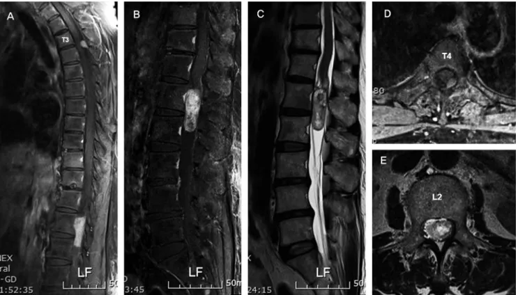
자기 공명 영상상 제 3-4 흉추부 척추강내 종괴는 T1, T2 영상 모두에서 저신호 강도(low-signal)를 보였으며 gadolinium 조 영 영상에서 종괴 전체가 고신호 강도(high-signal)의 조영 증가 소견을 보였다. 또한 축상면 조영 영상에서 경막에 기저부를 형 성하는 dural tail sign을 확인할 수 있었다. 제 1-2 요추부 종괴 는 자기 공명 영상상 T1 영상에서 저신호 강도의 소견을 보였으
Hak-Jin Min et al Volume 18 • Number 4 • December 2011
www.krspine.org
264
며, T2 영상에서 고신호 강도 및 gadolinium 조영 영상에서 불균 질한(heterogenous) 조영 증강 소견을 보였다(Fig. 1). 전산화 단 층촬영 검사상 제 3-4 흉추부 종괴 내부의 석회화를 확인 할 수 있었으며(Fig. 2), 제 1-2 요추부 종괴에서는 석회화 소견이 관 찰 되지 않았다. 수술은 제 3-4 흉추부 종괴의 경우 제 3 흉추 후 궁 절제술을 시행한 뒤 경막에 종적 절개를 가하여 종괴를 노출 시키고 경막과 유착 부위를 박리하여 약 1.2×0.6×0.8cm의 경 막내 종괴를 제거하였다(Fig. 3). 제 1-2 요추부 종괴의 경우 제 1, 2 요추의 후궁 절제술 및 제 12 흉추 및 제 3 요추의 부분 후궁 절제술을 시행 후 경막에 종적 절개를 시행하여 종괴를 제거하 였다. 제거된 종괴는 약 4.1×1.9×1.7cm 크기의 황색을 띄며 피 막이 잘 발달되어 있었다(Fig. 4). 흉추부 종괴는 주변 척수와 신 경들로부터 잘 박리 되었으며 경막에 유착되어있었으나 경막 결 손을 남기지 않고 박리되었다. 요추부 종괴는 주변 신경들과 잘 박리 되었으나 종괴내로 신경섬유가 포함되어있었고 완전적출 을 위해서 해당 신경의 절제가 불가피하였다. 요추부 종괴 절제 후 다분절 후궁절제술로 인한 불안정성의 발생으로 유합술 및 척추경 나사못 고정술을 시행하였다. 조직 병리학적 검사상 흉 추부 종괴는 수막종성 수막종(meningothelial meningioma)로 진 단되었고, 요추부 종괴는 신경초종(schwannoma)으로 진단되었
다(Fig. 5). 술 후 새로운 신경증상은 발생하지 않았다. 술 후 4주 째 양측 하지의 근력은 4등급으로 회복되었고 목발을 이용한 보 행이 가능하였다. 이 후 술 후 3개월째 하지 근력은 정상으로 회 Fig. 1. (A) Preoperative Gd-enhanced sagittal MRI demonstrated that the mass shows high signal intensity on T3-4 level. (B) Preoperative Gd-enhanced sagittal MRI demonstrated that the mass shows heterogeneous high signal intensity on L1-2 level. (C) Low signal intensity change of L1-2 mass shows on T2-weighted sagittal MRI. (D) Preoperative Gd-enhanced axial MRI of T3-4 mass demonstrated that the mass has a dural tail sign. (E) Preoperative Gd-enhanced axial MRI of L1-2 mass shows heterogeneous signal intensity.
Fig. 2. T-spine CT scan image shows a ossification within the mass.
Synchronous Development Spinal Cord Tumor Journal of Korean Society of Spine Surgery
복되었으며 환자 스스로 독립 보행을 할 수 있었다. 술 후 12개 월째 시행한 흉추 및 요추부 자기 공명 영상에서 종양의 재발은 없었으며(Fig. 6), 추시 관찰에서 새로운 임상 증상의 발생은 없 었다.
고 찰
척수 종양의 분류는 발생 부위에 따라 경막외 종양과 경막내 종양으로 나누며, 경막내 종양은 다시 척수외 종양과 척수내 종 양으로 분류할 수 있다. 척추강내 경막외 종양 중 수막종은 주 로 40-60대 여성에서 발생하며 동통, 마비, 근력약화 등의 긴 회 로 증상(long tract sign)을 보인다.1) 척수 수막종은 자기 공명 영 상 검사시 gadolinium 조영 영상에서 균질한 조영 증강을 보이 며, 종종 석회화가 동반되어 전산화 단층 촬영 검사에서 석회화 를 확인 할 수 있다. Chen 등2) 은 척수강내 수막종은 신경근 소
매(nerve root sleeve) 측면부의 치상인대 주변 경막의 지주막 모 자 세포에서 발생함을 주장하였고, Calogero와 Moossy3) 은 신경 근 출구(nerve root exits) 부위에 위치한 지주막 융모에서 발생을 주장하였지만 정확한 기원은 증명되지 않았다. 대부분 수막종은 척수의 후측방에 위치하며 경막 기저부에서 혈액 공급을 받고,4) 넓은 경막의 기저부는 자기 공명 영상 검사에서 dural tail sign을 나타낸다. 본 증례에서 흉추 수막종은 연령과 성별 및 발생 부위 가 전형적인 양상을 보였고 또한 자기 공명 영상에서 dural tail sign을 보이며 전산화 단층 촬영에서 석회화 병변을 확인할 수 있어 수막종을 의심할 수 있었고, 조직 검사상 확진을 할 수 있 었다.
신경초종은 대부분 배측 감각 신경근에서 발생하며 피막에 둘 러싸인 단단한 종물로 약 70-75%는 경막내 척수외(intradural extramedullary)에 위치하며, 약 15%는 경막외에 dumbbell 모양 으로 위치하며, 약 1% 미만에서 척수내에 위치한다.5) 신경초종
Fig. 4. Lumbar mass which containing a intermingled nerve fiber was yellowish, well capsulated and cylinderic shape, measuring 4.1x1.9x1.7 cm.
Fig. 3. Thoracic mass was excised by performing longitudinal dural incision. The mass was attached to dural sac but excised without dural defect, measuring 1.2x0.8x0.6 cm
Fig. 5. (A) Photomicrograph of tumor of T-spine showing characteristic meningeal whorls (H&E, x200) (B) Photomicrograph of the tumor of L-spine showing compact cellular Antoni type A and loose myxoid Antoni type B (H&E, x200)
Hak-Jin Min et al Volume 18 • Number 4 • December 2011
www.krspine.org
266
은 자기 공명 영상 T1 강조 영상에서 저신호 강도에서 중등도 신 호 강도의 경계가 분명한 종물로 관찰되며 T2 강조 영상에서 고 신호 강도로 관찰된다. 또한 gadolinium 조영 증강 영상에서는 균질성, 불균질성 조영증강 및 변연부 조영증강 등의 다양한 양 상을 보인다.6) 이러한 다양한 조영증강 양상은 종양 내부의 낭 종성 변화, 국소 출혈, 점액성 변화, 교원질, schwann 세포의 빈 도등에 기인한다.7) Parmar 등8) 에 의하면 신경초종의 낭종성 변 화는 Antoni B 세포 부위의 퇴행성 변화이거나 종양이 성장함 에 따라 종양 중심부에 발생한 허혈성 괴사가 낭종으로 진행하 는 것으로 보고 하였다. 본 증례에서는 조영증강 영상에서 종양 내부의 낭종성 변화를 관찰 할 수 있었다(Fig. 1). 척수에서 다발 성 신경초종이 발생되는 경우는 주로 제2형 신경섬유종증과 관 계된 것으로 알려져 있으며, 척수에서 다른 2개의 종양이 다른 위치에 발생하는 경우는 주로 발생학적인 선천성 기형, 신경섬 유종증, Von Recklinghausen’s disease와 같은 질환이 동반된 경 우로 알려져 있다. 본 증례와 같이 선천성 기형 및 다른 장기에 이상 소견이 없고, 또한 특별한 가족력이 없는 성인에서 두 가지 상이한 척수종양이 발견되는 경우는 매우 드문 것으로 알려져 있다.9) 본 증례에서 양측 하지의 감각 및 운동 부전으로 시행한 요추부 자기 공명 영상에서 종양을 발견하였고, 더불어 시행한 전 척추 자기 공명 영상에서 흉추부 종양을 추가적으로 발견할 수 있었다. 이에 저자들은 첫째 환자의 임상증상 중 등의 동통과 더불어 양측 하지의 근력 및 감각 저하 등의 긴 회로 증상(long tract sign), 배뇨기능 장애 등의 원인으로 흉추부 수막종에 의한
척수증을 배제하기 어려우며, 둘째 흉추 자기 공명영상에서 흉 추부 수막종에 의한 척수의 압박과 시상면에서 수막종에 의한 뇌척수액의 차단을 확인 되어 요추부 신경초종의 치료와 더불어 흉추부 수막종의 치료도 같이 고려하였고, 동시에 수술적 치료 를 시행하였다.
척수 종양의 경우 최초 발견된 종양의 병변 뿐만 아니라 전 척 수에 대한 검사를 통해 추가적인 종양의 존재를 확인 하는 것이 필요하며, 자기 공명 영상은 비침습적이며 종양의 경계, 침습적 양상 등의 소견을 제공하여 수술 전 진단 및 치료 계획 설정에 큰 도움이 될 것으로 생각되었다.
한 환자에서 두 가지 상이한 척수 종양이 발견되는 경우는 매 우 드문 경우로 본 저자들은 흉추 수막종과 요추부 신경초종이 한 환자에서 발견된 임상 증례를 경험하여 문헌 고찰과 함께 보 고하는 바이다.
REFERENCES
1. Solero CL, Fornari M, Giombini S, et al. Spinal meningiomas: review of 174 operated cases. Neurosurgery.
1989; 25:153-160.
2. Chen HJ, Lui CC, Chen L. Spinal epidural meningioma in a child. Child’s Nerv Syst. 1992;8:465-7.
3. Calogero JA, Moossy J. Extradural spinal meningiomas. J Neurosurg. 1972;37:442-7.
Fig. 6. Gd-enhanced & T2-weighted T-spine MR images(A,B) and T2-weighted L-spine MR images(C,D,E) of same patient 1year later show no recurrence of tumor.
Synchronous Development Spinal Cord Tumor Journal of Korean Society of Spine Surgery
4. Nittner K. Spinal meningiomas, neurinomas, and neurofibromas and hourglass tumors. In Vinken P, Brunyn B(eds): Handbook of Clinical neurology. New York, American Elsevier. 1976;177-322.
5. Chung JY, Kim HJ, Seo HY, Lee JJ. Diverse characteristics of spinal nerve sheath tumor on magnetic resonance images.
J Korean Soc Spine Surg. 2009;16:38-45.
6. Bloomer CW, Ackerman A, Bhatia RG. Imaging for spinal tumors and new applications. Top Magn Reson Imaging.
2006;17: 69-87.
7. Friedman DP, Tartaglino LM, Flanders AE. Intradural
schwnnomas of the spine: MR findings with emphasis on contrast-enhancement characteristics. AJR.
1992;158:1347-50.
8. Demachi H, Takashima T Kadoya M, et al. MR imaging of spinal neurinomas with pathologic correlation. J Comput Assist Tomogr. 1990;14:250-4.
9. Nishiura I, Koyama T, Tanaka K, Aii H, Amano S. The occurrence of different types of spinal tumors in one patient.
A case report and review of the literature. Neurochirurgia.
1989;32:52-5.
한 환자에서 발생한 흉추 수막종과 요추 신경초종 - 증례 보고 -
민학진 • 김진수 • 석주필 서울의료원 정형외과학교실
연구 계획: 증례보고
목적: 한 환자에서 동시에 발견된 흉추 수막종과 요추 신경초종 1예를 경험하였기에 보고하고자 한다.
선행문헌의 요약: 한 환자에서 다른 종류의 척수 종양의 발생은 매우 드문 것으로 보고되어왔다.
대상 및 방법: 66세 여자, 내원 2달전부터 발생한 양측 하지 근력 약화로 인한 보행장애를 주소로 내원하였다. 흉추에서 척수 수막종, 요추에서 신경초 종이 발견되어 수술적으로 제거하였다.
결과: 술 후 양측 하지의 근력이 회복되어 독립 보행을 할 수 있게 되었고, 술 후 1년 추시에서 재발은 없었다.
결론: 한 환자에서 발견된 서로 다른 종류의 척수 종양을 수술적 치료를 시행하여 좋은 결과를 얻었다.
색인 단어: 척수, 수막종, 신경초종 약칭 제목: 이종 척수 종양