Korean J Gastroenterol Vol. 59 No. 6, 441-444 http://dx.doi.org/10.4166/kjg.2012.59.6.441
CASE REPORT
Korean J Gastroenterol, Vol. 59 No. 6, June 2012 www.kjg.or.kr
만성 B형간염에 의한 간경변증 환자에서의 Sweet 증후군 1예
박창욱, 김윤정, 서혜진, 이경인, 장병국, 황재석, 정우진
계명대학교 의과대학 내과학교실
A Case of Sweet’s Syndrome in a Patient with Liver Cirrhosis Caused by Chronic Hepatitis B
Chang Wook Park, Yoon Jung Kim, Hye Jin Seo, Kyung In Lee, Byung Kuk Jang, Jae Seok Hwang and Woo Jin Chung Department of Internal Medicine, Keimyung University School of Medicine, Daegu, Korea
Sweet’s syndrome (SS), also known as acute febrile neutrophilic dermatosis, is characterized by the sudden onset of painful erythematous skin lesions together with fever and neutrophilia. SS can be associated with several disorders, such as malignancy, autoimmune disease, and infections. However, SS associated with liver cirrhosis is uncommon. We report a case of SS in a patient who was diagnosed with liver cirrhosis caused by chronic hepatitis B. (Korean J Gastroenterol 2012;59:441-444) Key Words: Sweet’s syndrome; Liver cirrhosis; Chronic hepatitis B
Received December 3, 2010. Revised September 7, 2011. Accepted September 7, 2011.
CC This is an open access article distributed under the terms of the Creative Commons Attribution Non-Commercial License (http://creativecommons.org/licenses/
by-nc/3.0) which permits unrestricted non-commercial use, distribution, and reproduction in any medium, provided the original work is properly cited.
교신저자: 정우진, 700-712, 대구시 중구 달성로 56, 계명대학교 의과대학 내과학교실
Correspondence to: Woo Jin Chung, Department of Internal Medicine, Keimyung University School of Medicine, Dalseong-ro 56, Jung-gu, Daegu 700-712, Korea. Tel:
+82-53-250-7413, Fax: +82-53-250-7088, E-mail: [email protected] Financial support: None. Conflict of interest: None.
INTRODUCTION
Sweet’s syndrome (SS) is the prototype of neutrophilic der- matosis, which is an uncommon disorder characterized by abrupt onset of fever, leukocytosis, and painful red, popular skin rash.1 Additionally, SS is histologically characterized by diffuse infiltration of mature neutrophils in the dermis without vasculitis.2 Lesions usually develop on the face, neck, arms, and hands and are asymmetrically distributed. However, SS can be generalized, and patients often complain of discomfort with its associated signs and symptoms, including malaise, fe- ver, elevated erythrocyte sedimentation rate (ESR), and ele- vated CRP levels, which mimic an infectious process.3
SS accompanied with liver cirrhosis is uncommon, and there are few reports of SS associated with acute or chronic hepatitis.4 Here, we report a case of SS in a patient who was diagnosed with liver cirrhosis caused by chronic hepatitis B.
CASE REPORT
A 45-year-old man, who was diagnosed with liver cirrhosis related with chronic hepatitis B, visited our department with multiple, purplish-red infiltrated papules on the left thigh, right inguinal region, and both arms during the past 4 days.
He had no specific respiratory or gastrointestinal symptoms except generalized edema and a distended abdomen.
One year ago he was admitted to the hospital with gastric ulcer bleeding caused by alcohol ingestion. At that time, he was diagnosed with liver cirrhosis caused by chronic hep- atitis B. Since then, he has not consumed alcohol and has regularly visited the hospital.
At 2 months before admission, he experienced hepatic en- cephalopathy caused by high protein diet and his physical condition deteriorated. At 1 week before admission, he had a distended abdomen, and both lower legs were edematous.
He did not take any other medications except ursodeoxycolic
442 박창욱 등. 만성 B형간염에 의한 간경변증 환자의 Sweet 증후군
The Korean Journal of Gastroenterology
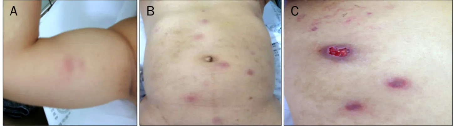
Fig. 1. Multiple tender, raised, annular erythematous lesions on the arms (A) and trunk (B). Round pustular lesions with central necrosis and skin ulcers on the left thigh (C).
Fig. 2. Abdominal computed tomography scan showed a shrunken liver, splenomegaly, and a moderate amount of ascites.
acid and diuretics as prescribed by doctors.
On admission, his vital signs were as follows: blood pres- sure, 100/60 mmHg; pulse rate, 80/minute; respiration rate, 20/minute; and body temperature, 38.5oC. He looked acutely ill but was alert. His heart and lung sounds were normal. He had a moderately distended abdomen, edema- tous lower legs, and swelling of the scrotum. A skin examina- tion revealed multiple tender, erythematous and annular le- sions on both arms and the trunk (Fig. 1A, B) as well as ulcer- ative lesions on the left inguinal region (Fig. 1C).
Peripheral blood tests showed white blood cells, 8,040/
mm3 (neutrophils, 78.2%); hemoglobin, 9.0 g/dL; and plate- lets, 68,000/mm3. Routine chemistry showed total protein, 6.2 g/dL; albumin, 2.5 g/dL; total bilirubin, 2.1 mg/dL; ALP, 95 IU/L; AST, 47 IU/L; ALT, 27 IU/L; and prothrombin time, 20.2 seconds (normal range, 10-14 seconds; INR, 1.78).
HBsAg and HBeAg were positive. The serum HBV DNA titer was 9,737,316 IU/mL. His ESR was 42 mm/hour (normal range, 0-10 mm/hour) and CRP was 0.6 mg/dL (normal range, 0-0.5 mg/dL).
Tumor markers (α-fetoprotein and carcinoembryonic anti- gen) were within normal ranges, and anti-nuclear antibody, anti-mitochondrial antibody, and human immunodeficiency virus screening were negative. An ascites study showed white blood cells, 243/mm3 (polymorphonuclear cells, 70%); pro- tein, 0.5 g/dL; albumin, 0.3 g/dL; and microorganisms were negative in a culture.
Endoscopic findings revealed a well-defined active ulcer at the lesser curvature side of the distal body and duodenal bulb. F1 sized, red color sign (−) esophageal varices, which were classified by the Japanese Society for Portal Hyperten- sion, were noted on the lower one-third of the esophagus.5 A
biopsy specimen from the gastric ulcer site revealed a benign ulcer with Helicobacter pylori.
A chest X-ray finding was normal. Abdominal computed to- mography showed a cirrhotic liver, splenomegaly, and moder- ate amount of ascites (Fig. 2).
Before histological confirmation, he received antibiotic treatment for 3 days to prevent wound infection. A histo- logical examination of tissue obtained from the inguinal re- gion showed dense infiltration of mature neutrophils in the superficial dermis without vascular infiltration (Fig. 3). After histological confirmation, he received 30 mg/day oral pre- dnisolone, and the fever declined and normalized on the fifth day after medication. After 1 week of prednisolone, the cuta- neous lesions had almost completely healed without scars.
Oral prednisolone was tapered gradually during a 2-week period. He did not receive any antiviral agents initially be- cause of economic problems. During regular follow-up of his
Park CW, et al. Sweet’s Syndrome in Liver Cirrhosis by Chronic Hepatitis B 443
Vol. 59 No. 6, June 2012 Fig. 3. Biopsy of a skin lesion showed dense neutrophilic infiltration
into the dermis, without evidence of vasculitis (H&E, ×400).
liver enzymes, the ALT level increased after 2 weeks of prednisolone. So, he took 100 mg lamivudine daily, and his ALT level gradually normalized.
DISCUSSION
SS, also known as acute febrile neutrophilic dermatosis, was first described by Robert Douglas Sweet in 1964.6 SS is characterized by tender, erythematous papules and plaques, fever, and leukocytosis, which respond well to corticosteroid therapy.7 The importance of SS is a marked clinical ex- pressiveness and frequent association with other systemic diseases.
SS occurs more frequently in females and can appear at any age, but peaks in patients in their 50s-70s. SS can be trig- gered by infections, hematological malignancies, other neo- plasms, autoimmune diseases, pregnancy, trauma, and medications.8,9 Immune complex vasculitis, T-cell activation, and altered neutrophil function have been presumed to oc- cur, but there is lacking experimental evidence.2
Diagnostic criteria, which are comprised of two major cri- teria and four minor criteria, were recently modified by von den Driesch in 1994.10 Both major criteria and two of the four minor criteria should be satisfied for a definitive diagnosis of SS. The two major criteria are 1) abrupt onset of painful eryth- ematous plaques or nodules and 2) histopathological evi- dence of a dense neutrophilic infiltrate without evidence of
primary leukocytoclastic vasculitis. The minor criteria are 1) pyrexia (>38oC); 2) an association with an underlying hema- tological or visceral malignancy, inflammatory disease, or pregnancy, or preceded by an upper respiratory or gastro- intestinal infection or vaccination; 3) excellent response to treatment with systemic corticosteroids, potassium iodide, or colchicine; 4) abnormal laboratory values at presentation (three of the following four): ESR >20 mm/hour, positive CRP, >8,000 leukocytes, and >70% neutrophils. The pres- ent case was sufficient to meet these criteria.
SS related skin lesions can be preceded or accompanied by fever and general discomfort11 and should be diagnosed differently from erythema multiforme, erythema elevatum diutinum, erythema nodosum, and pyoderma gangrenosum.
The histological differential diagnosis of SS includes con- ditions microscopically characterized by either neutrophilic dermatoses or neutrophilic panniculitis. Neutrophilic derma- toses include abscesses or cellulitis, intestinal bypass syn- drome, leukocytoclastic vasculitis, pyoderma gangrenosum, and rheumatoid neutrophilic dermatitis. Leukemia cutis can- not occur concurrently with SS, but mimics the dermal changes of SS. In contrast to SS, the dermal infiltrate in pa- tients with leukemia cutis consists of malignant immature leukocytes rather than mature neutrophils. The adipose tis- sue changes that occur with subcutaneous SS are similar to those of other conditions characterized by neutrophillic lobu- lar panniculitis. Similar to pyoderma gangrenosum, SS is known to cause pathergy, in which lesions occur in areas of minor trauma, such as sites of scratches, bites, and ven- ipuncture. The lesions may also be photodistributed or lo- calized to the site of a previous phototoxic reaction.12
In some patients with SS, skin lesions eventually resolve without any therapeutic management. However, the skin le- sions usually persist for weeks to months without treat- ment.13
Systemic steroids are the gold standard therapy for SS.
Dermatosis-associated symptoms improve rapidly after starting steroid therapy, and the cutaneous lesions resolve subsequently. Systemic corticosteroid therapy usually be- gins with 1 mg/kg/day prednisolone as a single oral morning dose.14 However, several studies have suggested starting prednisolone at 30 mg per day, so our patient was also start- ed on 30 mg prednisolone and gradually tapered,4 and his symptoms and skin lesions improved promptly.
444 박창욱 등. 만성 B형간염에 의한 간경변증 환자의 Sweet 증후군
The Korean Journal of Gastroenterology
HBV infection is also an SS trigger factor. HBV is well known to cause several types of skin manifestations such as vasculi- tis, urticaria, and polyarteritis nodosa.15,16 But, the patho- genesis of SS-related hepatitis B is unclear.4
SS pathogenesis is thought to be an abnormal immune re- action triggered by variety of bacterial, viral, or tumorous antigens. Physical condition including generalized edema or deteriorated immunity by hepatic decompensation is thought to provide additional chances for infection or altered T-cell ac- tivation and neutrophil function. Our case was a patient with liver cirrhosis. During the 2 months before he developed SS, he experienced hepatic encephalopathy and consecutive generalized edema. Deteriorated liver status is thought to be a more vulnerable condition than to be infected by a virus or bacteria.
SS skin lesions usually appear similar to other infectious diseases. But, SS skin lesions improve after using steroids not antibiotics.17 Thus, a differential diagnosis of SS should be considered if patients with skin lesions are not effectively controlled by antibiotics. If steroids are not indicated, dap- sone, indomethacin, or potassium iodide can be used to im- prove symptoms.18
Reactivation of HBV replication, which increases HBV DNA and ALT level, has been reported in 20-50% of hepatitis B car- riers undergoing immunosuppressive or cancer chemo- therapy and is more common when chemotherapeutic regi- mens include corticosteroids. Therefore, regular follow-up of serum HBV DNA and ALT level is very important.
According to the practice guidelines of the American Association for the Study of Liver Disease, prophylactic anti- viral therapy should be administered to hepatitis B carriers at the onset of cancer chemotherapy or a finite course of im- munotherapy and maintained for 6 months.19 Our patient al- so experienced an ALT flare up after prednisolone treatment.
Thus, regular monitoring of HBV DNA, ALT level, and prophy- lactic antiviral therapy is very important for hepatitis B car- riers who receive immunosuppressive agents including corticosteroids.
We report a case of SS in a patient with liver cirrhosis caused by chronic hepatitis B that was suspected to be trig- gered by his physical condition including generalized edema and deteriorated immunity by hepatic decompensation.
REFERENCES
1. Ytting H, Vind I, Bang D, Munkholm P. Sweet's syndrome-an ex- traintestinal manifestation in inflammatory bowel disease.
Digestion 2005;72:195-200.
2. Gill HH, Leung AY, Trendell-Smith NJ, Yeung CK, Liang R. Sweet syndrome due to myelodysplastic syndrome: possible therapeu- tic role of intravenous immunoglobulin in addition to standard treatment. Adv Hematol 2010;2010:328316.
3. Cohen PR. Sweet's syndrome--a comprehensive review of an acute febrile neutrophilic dermatosis. Orphanet J Rare Dis 2007;2:34.
4. Tan E, Yosipovitch G, Giam YC, Tan SH. Bullous Sweet's syndrome associated with acute hepatitis B infection: a new association.
Br J Dermatol 2000;143:914-916.
5. Idezuki Y. General rules for recording endoscopic findings of esophagogastric varices (1991). Japanese Society for Portal Hypertension. World J Surg 1995;19:420-422.
6. Sweet RD. An acute febrile neutrophilic dermatosis. Br J Dermatol 1964;76:349-356.
7. Mahajan VK, Sharma NL, Sharma RC. Sweet's syndrome from an Indian perspective: a report of four cases and review of the literature. Int J Dermatol 2006;45:702-708.
8. Cohen PR, Kurzrock R. Sweet's syndrome revisited: a review of disease concepts. Int J Dermatol 2003;42:761-778.
9. Kang HK, Son YM, Na SY, et al. Sweet's syndrome as an initial presentation in a patient with systemic lupus erythematosus.
Korean J Dermatol 2010;48:43-46.
10. von den Driesch P. Sweet's syndrome (acute febrile neutrophilic dermatosis) J Am Acad Dermatol 1994;31:535-556.
11. Cohen PR, Kurzrock R. Sweet's syndrome: a neutrophilic derma- tosis classically associated with acute onset and fever. Clin Dermatol 2000;18:265-282.
12. Fett DL, Gibson LE, Su WP. Sweet's syndrome: systemic signs and symptoms and associated disorders. Mayo Clin Proc 1995;70:234-240.
13. Belhadjali H, Chaabane S, Njim L, Youssef M, Zakhama A, Zili J.
Sweet's syndrome associated with multiple myeloma. Acta Dermatovenerol Alp Panonica Adriat 2008;17:31-33.
14. Cohen PR, Kurzrock R. Sweet's syndrome: a review of current treatment options. Am J Clin Dermatol 2002;3:117-131.
15. Martínez MI, Sánchez JL, López-Malpica F. Peculiar papular skin lesions occurring in hepatitis B carriers. J Am Acad Dermatol 1987;16:31-34.
16. McElgunn PS. Dermatologic manifestations of hepatitis B virus infection. J Am Acad Dermatol 1983;8:539-548.
17. Lee BT, Yoo JH, Seo BS, Kim JM. Sweet's syndrome involving the auricle. Korean J Otorhinolaryngol-Head Neck Surg 2007;50:
818-821.
18. Kim JH, Lim YS, Choi HY, Myung KB. A case of Sweet's syndrome treated with potassium iodide. Korean J Dermatol 1999;37:
1074-1078.
19. Lok AS, McMahon BJ. Chronic hepatitis B: update 2009.
Hepatology 2009;50:661-662.