ISSN 0378-6471 (Print)⋅ISSN 2092-9374 (Online)
https://doi.org/10.3341/jkos.2019.60.9.909
Case Report
후부다형각막이상증의 특징을 보이는 홍채각막내피증후군
Iridocorneal Endothelial Syndrome with Features of Posterior Polymorphous Corneal Dystrophy
나정호⋅이효경
Jeong Ho Na, MD, Hyo Kyung Lee, MD
인제대학교 의과대학 해운대백병원 안과학교실
Department of Ophthalmology, Haeundae Paik Hospital, Inje University College of Medicine, Busan, Korea
Purpose: To report a case of iridocorneal endothelial syndrome, which overlapped with some of the features of posterior poly- morphous corneal dystrophy.
Case summary: A 61-year-old female presented with tearing pain and blurred vision in her left eye, which was aggravated in the morning. The symptom started approximately 1 year prior to her visit. At the initial visit, the visual acuities were 1.0 in both eyes and the intraocular pressures were normal. On slit-lamp examination, a single pair of horizontal parallel lines was observed at the central corneal endothelial layer in the right eye. In contrast, multiple pairs of oblique parallel lines were observed in the left eye.
The lines of the lesions were more prominent and wavier in the left eye than those of the right eye. The overlying cornea was clear, and the corneal thicknesses were in the normal range in both eyes. Using a gonioscopic examination, localized peripheral anterior synechiae were observed only in the left eye. The pupil and iris were normal in both eyes. On specular microscopic ex- amination, the corneal endothelial cell size in the right eye increased and the corneal endothelial density decreased to 668 cells/mm2. In the left eye, multiple abnormal endothelial cells with dark-light reversal were observed. In conclusion, the patient was subsequently diagnosed with iridocorneal syndrome, rather than posterior polymorphous corneal dystrophy.
Conclusions: Posterior polymorphous corneal dystrophy and iridocorneal endothelial syndrome may present with many similarities. Therefore, in cases of uncertain diagnosis, an understanding of the clinical features is important for proper diagnosis.
J Korean Ophthalmol Soc 2019;60(9):909-914
Keywords: Chandler syndrome, ICE cell, Iridocorneal endothelial syndrome, Posterior polymorphous corneal dystrophy, Specular microscopy
■Received: 2019. 3. 14. ■ Revised: 2019. 4. 21.
■Accepted: 2019. 8. 16.
■Address reprint requests to Hyo Kyung Lee, MD
Department of Ophthalmology, Inje University Haeundae Paik Hospital, #875 Haeundae-ro, Haeundae-gu, Busan 48108, Korea Tel: 82-51-797-2310, Fax: 82-51-797-2030
E-mail: [email protected]
*Conflicts of Interest: The authors have no conflicts to disclose.
ⓒ2019 The Korean Ophthalmological Society
This is an Open Access article distributed under the terms of the Creative Commons Attribution Non-Commercial License (http://creativecommons.org/licenses/by-nc/3.0/) which permits unrestricted non-commercial use, distribution, and reproduction in any medium, provided the original work is properly cited.
홍채각막내피증후군(iridocorneal endothelial syndrome;
ICE syndrome)은 비정상 각막내피세포의 증식으로 인해 홍채 실질의 변화, 이차성 폐쇄각녹내장, 각막부전 등을 동
반하는 진행성 질환이다.1 대개 단안을 침범하고 중년 여성 에서 호발하는 것으로 알려져 있으며 유전되지는 않는다. 홍채의 변형에 따라 진행성 홍채 위축(progressive iris atro- phy), Chandler 증후군, 홍채모반 증후군(iris nevus syndrome or Cogan-Reese syndrome)으로 분류된다.2 임상적 특징 및 경면현미경검사에서 관찰되는 특징적인 모습의 ICE 세포 로 인해 대부분의 홍채각막내피증후군은 진단이 어렵지 않 다. 하지만 후부각막의 변화와 함께 각막부종, 폐쇄각녹내 장 등이 동반될 수 있는 질환들, 특히 후부다형각막이상증 (posterior polymorphous corneal dystrophy)과 꼭 감별해야
A B
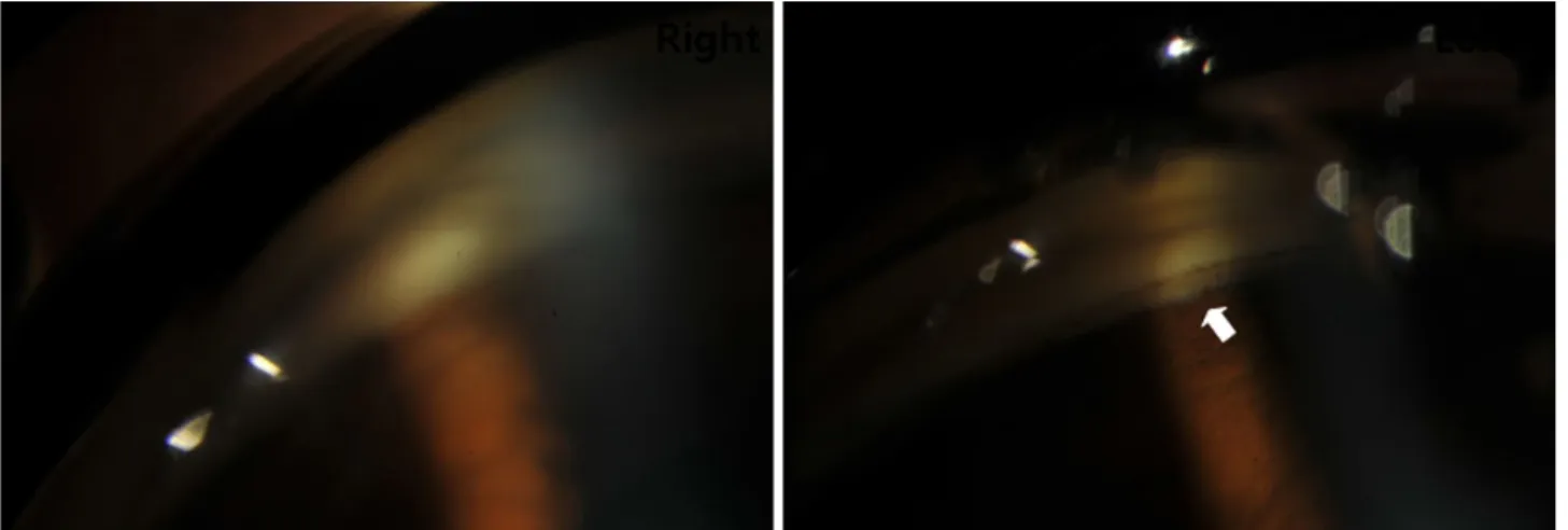
Figure 1. Slit-lamp photography of both eyes at initial examination. (A) In the right eye, a single pair of horizontal parallel lines were
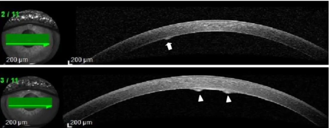
observed at corneal endothelial layer (arrows). (B) In the left eye, the multiple pairs of parallel lines were observed at endothelial layer. The lines were running obliquely through the center of the cornea (arrowheads).Figure 2. Anterior segment optical coherence tomography of both eyes. The lesions were protruded from the endothelial layer. The
lesions were more prominent in the left eye (arrowheads) compared to that of right eye (arrow).한다고 알려져 있다. 후부다형각막이상증은 각막상피세포 의 특징을 가지는 비정상적인 각막내피세포들이 관찰되는 질환으로써 대부분 양안을 침범하는 비진행성의 유전성 질 환으로, 홍채각막내피증후군과는 구분된다. 하지만 두 질환 은 임상적으로 매우 유사한 경우가 있어 일부에서는 두 질 환을 같은 질환군의 변종(variants)으로 여기기도 한다.3,4 본 증례에서는 좌안의 간헐적 시력 저하를 주소로 내원한 61세 여자 환자의 양안에서 홍채내피증후군과 후부다형각막이 상증에 특징적인 소견이 각각 관찰되었기에 문헌 고찰과 함께 보고하는 바이다.
증례보고
61세 여자 환자가 약 1년 전부터 시작된, 아침 기상 시 간헐적으로 발생하는 좌안의 찢어질 듯한 통증 및 시야 흐 림을 주소로 내원하였다. 발생한 증상은 치료용 렌즈를 착
용하면 호전되었지만 렌즈 착용을 중지하면 다시 재발한다 고 하였다. 안구 외상이나 수술의 기왕력은 없었고, 전신 및 안과적 질환의 과거력도 없었다. 출생 당시 난산 혹은 겸자 분만 등의 과거력에 대해서는 부인하였다. 내과적 혹은 안 과적 질환의 가족력도 없었다. 양안 최대교정시력은 양안 모두 1.0이었고, 안압은 우안 15 mmHg, 좌안 14 mmHg였 다. 세극등현미경검사상 각막상피 및 실질은 좌안 중심부 각막의 점상상피미란 외에 특이 소견은 관찰되지 않았다. 양안 모두에서 각막 후면부에 평행한 반투명 선이 관찰되 었으나, 그 양상은 양안에서 차이가 있었다. 우안은 각막내 피세포층에서 선형의 평행한 불투명 선이 각막 중심부를 가로지르며 각막윤부에서 윤부로 이어져 있었다(Fig. 1A).
좌안에서 관찰되는 평행한 선들은 우안보다 두께가 굵고 반투명이었고 더 구불구불한 모습이었다. 각막 후면 중심 부를 사선으로, X자 모양으로 가로지르고 있었다(Fig. 1B). 양 안 모두에서 평행한 선 사이의 각막기질 및 내피는 투명하
Figure 4. Gonioscopy of both eyes. The angle of the right eye was open. In contrast, the focal peripheral anterior synechiae were ob-
served in the left eye (arrow).A B
Figure 3. Specular microscopy of both eyes at initial examination. (A) In the right eye, the endothelial cells became large and the
density of the cells decreased to 668 cells/mm2. (B) In the left eye, the normal hexagonal shape of the endothelial cells disappeared.The multiple abnormal endothelial cells with dark-light reversal were observed (arrows).
였다. 전안부 단층촬영상 양안 모두 병변 부위의 데스메막 은 두꺼워져 있었고 고반사도를 보이며 전방 쪽으로 돌출되 어 있었다(Fig. 2). 샤임플러그 카메라(PentacamTM, OCULUS, Wetzlar, Germany)로 측정한 중심부 각막 두께는 우안 554 μm, 좌안 472 μm로 양안 모두에서 각막 두께는 정상 범위였다.
양안 모두 홍채 및 동공의 이상은 관찰되지 않았다.
환자의 우안에서 관찰되는 병변은 후부다형각막이상증 에서 관찰되는 tram-track line appearance와 유사하였으나, 감별진단을 위하여 경면현미경검사와 전방각경검사를 추 가로 시행하였다. 중심부 경면현미경검사상 우안은 각막내 피세포 밀도가 668 cells/mm2로 감소해 있었고, 평균 세포
의 크기가 증가되어 있었다(Fig. 3A). 좌안의 검사에서 내 피세포의 정상적인 육각형 모양은 소실되어 있었고, 중심 부는 어둡고 가장자리는 밝게 나타나는 명암 반전을 보이 는 세포들이 관찰되었다(Fig. 3B). 시행한 전방각경검사상 개방된 전방각을 보인 우안과는 달리 좌안은 국소적인 주 변부 홍채 앞 유착 소견이 관찰되었다(Fig. 4). 따라서 경면 현미경검사에 따라 환자는 좌안의 재발성 각막미란을 동반 한 홍채각막내피증후군으로 진단하였고, 현재 6개월째 경 과 관찰 중이며 질환의 진행 소견은 관찰되지 않았다.
PPCD ICE syndrome
Laterality Bilateral Unilateral
Inheritance Autosomal dominant Acquired
Sex Male = female Male < female
Progression Non-progressive Progressive
Symptoms Asymptomatic Symptomatic
Onset Childhood to 20s Young to middle-age
Visual impairment Uncommon Common
Endothelial cells Epithelium-like Large, light-dark reversal
Immunostaining Cytokeratin positive Cytokeratin negative
Descemet’s membrane Abnormal posterior nonbanded layer Normal posterior nonbanded layer
Corneal edema Uncommon Common
Glaucoma Uncommon Common
PPCD = posterior polymorphous corneal dystrophy; ICE = iridocorneal endothelial.
Table 1. Comparison of posterior polymorphous corneal dystrophy and iridocorneal endothelial syndrome
고 찰
후부다형각막이상증은 전형적으로 양안을 침범하는 상 염색체 우성 유전의 드문 유전성 각막이상증으로, 대개 천 천히 진행되고 무증상이며 20-30대에 우연히 발견되는 경 우가 많다.5 1916년 Grayson6이 각막 표면에 수포성 병변을 가진 여섯 명의 환자를 보고하며 처음 소개되었고 이후 1974년 Grayson6을 비롯한 다수의 병리학적 연구에서 전자 현미경 소견상 정상적인 단층의 육각형 구조의 각막내피세 포와는 달리 다층의 배열로 현저한 keratin 염색을 보이는 각막상피세포의 특성을 가지는 변형된 내피세포를 관찰하 여 보고하였다. 병리학적으로 데스메막에서 이런 비정상적 인 외피세포상 세포들이 발현하고 증식을 일으켜 정상적인 각막내피세포를 대체하여 각막내피층에 특징적인 변화를 야기한다. 후부다형각막이상증에서 관찰되는 각막 데스메 막 및 내피층의 이상 소견은 임상적으로 수포형태 병변 (vesicle-like lesion), 띠 모양 병변(band lesion), 다형성 혼 탁(polymorphous opacity) 등 세 가지 형태로 분류된다.5 수 포형태 병변은 데스메막층에서 회색의 halo를 가지는 원형 혹은 타원형의 투명한 cyst로, 대부분의 후부다형각막이상 증 환자에서 관찰되므로 질환의 hallmark로 여겨진다. 띠 모양 병변은 대개 각막 중심부 바로 아래에서 물결 모양의 가장자리(scalloped edge)를 보이는 평행한(parallel) 선 모 양으로 관찰되며 끝이 점점 가늘어지지 않으므로, 선천성 녹내장, 외상, 각막수종 등에서 보이는 데스메막 찢어짐 (Haab’s striae)과는 구분된다.7 다형성 혼탁은 가장 드문 형 태로 병변과 인접한 심부 기질층까지 혼탁이 관찰된다.
홍채각막내피증후군은 전형적으로 단안을 침범하는 드 문 후천성 각막질환으로 특징적인 비정상적 각막내피세포 가 증식하여 전방각 및 홍채로 과성장한 후 수축함으로써,
각막내피층의 변화로 인한 각막부전과 함께 홍채의 변형 및 이차성 녹내장을 동반하는 질환이다.2 1979년 Yanoff8에 의해 명명되었으며, 유사한 특징을 가지는 진행성 홍채 위 축(progressive iris atrophy), Chandler 증후군, 홍채모반 증 후군(iris nevus syndrome or Cogan-Reese syndrome)의 세 질환으로 분류된다.9 진행성 홍채 위축은 심한 동공 편위 및 위축, 홍채의 구멍이 특징적이다. Chandler 증후군은 가 장 흔한 형태로 홍채의 이상은 경미하거나 없지만 각막내 피 기능장애로 인한 각막부종이 관찰된다. 홍채모반 증후 군은 홍채의 색소 결절이 특징적이다. 하지만 세 질환은 서 로 독립된 질환이 아니고 임상형이 바뀌거나 공유할 수 있 는 것으로 알려져 있으며, 세 질환 모두에서 경면현미경검 사상 특징적인 ICE 세포를 관찰함으로써 홍채각막증후군 으로 진단할 수 있다.10 ICE 세포는 정상적인 내피세포보다 크기가 크고 육각형 모양이 소실되어 다양한 형태로 관찰 된다. 특히 세포의 가장자리는 밝고 중심부는 어두운 명암 반전(light-dark reversal)이 특징적이다. 대개 중년의 여성에 서 호발하는 것으로 알려져 있는데, 점점 진행하는 질환이 므로 각막내피세포의 정상적인 기능을 상실하여 각막부종 이 발생 가능하며, 비정상 내피세포가 홍채 및 전방각으로 과 증식하여 주변부 홍채 유착과 이차성 녹내장, 동공 편위 등을 유발하여 시력예후는 불량한 경우가 많다.
후부다형각막이상증과 홍채각막내피증후군은 비슷한 임 상양상을 보일 수 있으므로 감별이 필요하다(Table 1). 대 부분의 경우 임상양상 및 경과, 경면현미경 소견에서 감별 이 가능하나 본 증례와 같이 일부 감별이 어려운 경우 공초 점현미경 혹은 전자현미경검사, 면역 염색 등이 도움이 된 다. 공초점현미경검사에서 후부다형각막이상증은 고반사의 핵과 microvilli 등 상피세포의 특징을 가지는 다층의 작은 세포들이 관찰되는 것으로 알려져 있으나 검사 기기의 부
재로 본 증례에서는 검사가 불가능하였다.3,11 각막 조직에 서 전자현미경으로 상피양세포의 유무 혹은 데스메막의 구 조를 확인하거나 면역 염색을 하여 내피세포에서 cytoker- atin의 발현 여부를 보는 것이 감별진단에 도움이 될 것이 나 조직 채취에 어려움이 있으므로 본 증례에서는 확인이 어렵다.4 본 증례에서 관찰되는 우안의 각막내피층의 띠 모 양 병변은 각막 중심부 바로 아래에서 평행한 선이 수평으 로 진행하며 끝이 점점 가늘어지지 않고, 물결 모양의 가장 자리를 가지는 등 후부다형각막이상증에서 관찰되는 띠 모 양 병변과 매우 유사하였다. 전안부 단층촬영을 통해 얻은 각막의 단면 사진에서는 불규칙하게 혼탁한 병변이 각막 후면에서 전방 쪽으로 약간 돌출된 모습으로 관찰되었으며 이는 기존의 보고와 유사한 모습이었다.12 또한 우안의 경 면현미경검사상 각막내피세포의 수가 감소되어 있으며 동 반되는 홍채 및 전방각의 이상 소견은 없었다. 하지만 좌안 의 내피세포층에서 관찰되는 선은 우안에 비해 두께가 굵 고 전방으로 확연히 돌출되어 있었고 사선으로 진행하며 여러 방향에서 관찰되어, 후부다형각막이상증에서 일반적 으로 관찰되는 병변의 모습과는 구분되었다. 각막 단층 사 진에서도 병변의 돌출된 정도가 확연하였고, 나란한 두 선 형 병변의 사이 및 인접한 내피세포층이 불규칙하게 두껍 고 혼탁한 양상으로 관찰되어 우안 병변의 단층촬영 모습 과는 차이가 있었다. 좌안에서도 홍채의 이상 소견은 관찰 되지 않았으나 주변부 홍채 유착이 관찰되었다. 특히 경면 현미경검사상 명암 반전을 보이는 세포가 다수 관찰되어 홍채각막내피증후군, 그중에서도 Chandler 증후군으로 진 단하였다. 따라서 내피세포 층에서 관찰되는 구불구불하고 후면부로 돌출된 평행한 선들은 비정상적 ICE 세포들이 증 식하여 수축함으로써 발생하였을 것으로 추측할 수 있다.
하지만 여전히 우안의 띠 모양 각막 병변에 대해서는 홍채 각막내피증후군에 의해 발생한 것으로 추측하기에는 무리 가 있어, 후부다형각막이상증이 같이 동반되었을 가능성을 배제할 수 없다. 질환의 정확한 감별과 진단을 위해 공초점 현미경검사 등이 도움이 되겠으나 본 기관에서는 시행이 어려워 한계가 있었다. 또한 환자는 안과적 가족력이 없다 고 하였으나, 대부분의 후부다형각막이상증은 무증상이므 로 직계 가족을 동반하여 세극등검사 등을 시행하면 질환 의 감별에 도움이 될 것이다. 검사 장비의 부재 등으로 인 한 감별진단의 제한에도 불구하고 특별한 과거력이 없는 중년 여성에서 단안(좌안)의 시력 저하를 동반한 점, 증상 의 점진적인 진행, 경면현미경검사에서 관찰되는 명암 반 전의 비정상적인 내피세포 등의 소견을 종합하였을 때 본 증례의 좌안은 홍채각막내피증후군에 합당하다고 판단하 였다. 하지만 반대 안(우안)에서 관찰되는 내피세포의 띠
모양 병변과 내피세포 수가 감소된 소견은 후부다형각막이 상증의 동반을 완전히 배제할 수 없을 것으로 생각된다.
현재 환자는 좌안의 반복적인 각막미란과 기상 시 악화 되는 시력 저하를 호소하고 있다. 이는 좌안의 각막내피세 포의 기능이 저하되며 발생하는 것으로 추정된다. 하지만 현재까지 각막부종은 발생하지 않았고, 주변부 홍채유착이 좌안의 전방각에서 관찰되고 있으나 그 정도가 경미하여 안압의 상승은 동반되지 않았다. 따라서 현재는 좌안의 반 복 각막미란에 대해서 보존적인 치료로 경과 관찰을 하고 있다. 하지만 홍채각막내피증후군은 진행하는 질환이므로 추후 좌안의 각막부전이나 이차성 녹내장이 발생할 가능성 이 높을 것으로 사료되어 주기적인 경과 관찰을 환자에게 권유하였고, 각막부전이 발생하는 경우 전층각막이식 혹은 각막내피층이식술이, 이차성 녹내장이 발생하는 경우 녹내 장 약물 치료 혹은 섬유주절제술이나 녹내장 밸브삽입술 등이 필요할 것이다. 우안의 경우 각막내피세포의 수가 저 하되어 있으나 역시 현재까지 각막부종은 발생하지 않았고, 진행 여부에 대해서는 앞으로 경과 관찰이 필요할 것으로 사료된다. 저자들은 반대 안에서 후부다형 각막이상증에서 관찰되는 띠 모양 내피세포 병변을 가지는 홍채각막내피증 후군 증례를 경험하였기에 이를 관계 문헌과 함께 보고하 는 바이다.
REFERENCES
1) Feng B, Tang X, Chen H, et al. Unique variations and character- istics of iridocorneal endothelial syndrome in China: a case series of 58 patients. Int Ophthalmol 2018;38:2117-26.
2) Sacchetti M, Mantelli F, Marenco M, et al. Diagnosis and manage- ment of iridocorneal endothelial syndrome. Biomed Res Int 2015;
2015:763093.
3) Lefebvre V, Sowka JW, Frauens BJ. The clinical spectrum between posterior polymorphous dystrophy and iridocorneal endothelial syndromes. Optometry 2009;80:431-6.
4) Anderson NJ, Badawi DY, Grossniklaus HE, Stulting RD. Posterior polymorphous membranous dystrophy with overlapping features of iridocorneal endothelial syndrome. Arch Ophthalmol 2001;119:
624-5.
5) Kim NH, Kim MS. The clinical features and progression of the disease in posterior polymorphous corneal dystrophy (PPCD). J Korean Ophthalmol Soc 2014;55:368-73.
6) Grayson M. The nature of hereditary deep polymorphous dys- trophy of the cornea: its association with iris and anterior chamber dygenesis. Trans Am Ophthalmol Soc 1974;72:516-59.
7) Cibis GW, Tripathi RC. The differential diagnosis of Descemet's tears (Haab's striae) and posterior polymorpous dystrophy bands.
A clinicopathologic study. Ophthalmology 1982;89:614-20.
8) Yanoff M. Iridocorneal endothelial syndrome: unification of a dis- ease spectrum. Surv Ophthalmol 1979;24:1-2.
9) Wilson MC, Shields MB. A comparison of the clinical variations of
= 국문초록 =
후부다형각막이상증의 특징을 보이는 홍채각막내피증후군
목적: 후부다형각막이상증의 특징이 혼재된 홍채각막내피증후군 1예를 보고하고자 한다.
증례요약: 61세 여자 환자가 1년 전부터 시작된, 아침 기상 시 좌안의 찢어질 듯한 통증과 시야 흐림을 주소로 내원하였다. 초진 시 시력은 양안 1.0이었고 안압은 정상이었다. 세극등검사상 우안 각막 중심부의 내피층에서 수평으로 주행하는 한 쌍의 평행한 선이, 좌안에서는 비스듬하게 주행하는 여러 쌍의 평행한 선이 관찰되었다. 띠 모양 병변은 좌안에서 더 두껍고 구불구불하였다. 내피세포 병변 위의 각막은 깨끗하였고, 각막두께는 모두 정상 범위였다. 전방각경검사상 좌안에서만 국소적인 주변부 홍채 앞 유착이 관찰되 었다. 홍채 및 동공은 양안 모두 정상이었다. 경면현미경검사상 우안은 각막내피세포의 크기가 커져 있었고 밀도가 668 cells/mm2로 감소해 있었으며 좌안은 명암 반전이 뚜렷한 비정상적인 내피세포가 다수 관찰되었다. 결론적으로 본 증례는 후부다형각막이상증보 다는 홍채각막내피증후군에 더 합당할 것으로 판단하였다.
결론: 후부다형각막이상증과 홍채각막내피증후군에서는 다수의 유사한 소견이 관찰될 수 있으므로 적절한 진단을 위하여 임상적 특 징을 이해하는 것이 중요하다.
<대한안과학회지 2019;60(9):909-914>
나정호 / Jeong Ho Na
인제대학교 의과대학 해운대백병원 안과학교실 Department of Ophthalmology,
Haeundae Paik Hospital, Inje University College of Medicine the iridocorneal endothelial syndrome. Arch Ophthalmol 1989;107:
1465-8.
10) Laganowski HC, Kerr Muir MG, Hitchings RA. Glaucoma and the iridocorneal endothelial syndrome. Arch Ophthalmol 1992;110:
346-50.
11) Chiou AG, Kaufman SC, Beuerman RW, et al. Confocal micro-
scopy in the iridocorneal endothelial syndrome. Br J Ophthalmol 1999;83:697-702.
12) Liskova P, Palos M, Hardcastle AJ, Vincent AL. Further genetic and clinical insights of posterior polymorphous corneal dystrophy 3. JAMA Ophthalmol 2013;131:1296-303.