pISSN: 0378-6471 eISSN: 2092-9374
http://dx.doi.org/10.3341/jkos.2014.55.2.182
= 증례보고 =
직난시 및 도난시에 따른 동축 소절개 백내장 수술의 난시와 고위수차의 비교분석
정현진⋅정병주⋅황형빈⋅김현승 가톨릭대학교 의과대학 안과 및 시과학교실
목적: 직난시와 도난시 환자에서 투명각막절개 백내장 수술 이후 발생된 수술유발난시량과 각막전면과 후면의 고위수차 변화량을 비교 하고자 하였다.
대상과 방법: 2.8 mm 투명각막절개 백내장 수술 시에 상측 절개를 시행한 직난시 군 26안과 이측 절개를 시행한 도난시 군 24안을 대상으로 하였다. 수술 전, 수술 후 1주, 1달, 2달에 나안시력, 최대교정시력을 측정하였고, 수술유발난시와 각막 전면과 후면의 고위수 차를 PentacamⓇ으로 측정하여 비교하였다.
결과: 수술유발난시는 직난시 군에서 도난시 군보다 유의하게 높았으나(p<0.05), 난시교정효과는 두 군에서 통계적인 유의성은 없었 다(p>0.05). 각막 고위수차는 술 후 1주째 두 군 모두에서 증가하였으나 두 군간에 유의한 차이가 없었고(p>0.05), 술 후 2개월째 두 군 모두에서 감소되었으나 각막 전면수차에서 총 고위수차(Total Higher Order Aberrations, HOA), 사축트레포일(oblique trefoil), 후면수차에서 총 고위수차(HOA), 수평코마(horizontal coma) 값에서는 유의한 차이를 보였다(p<0.05).
결론: 백내장 수술 시, 직난시 환자에서 상측 절개를 시행하는 것이 도난시 환자에서 이측 절개를 시행하는 것보다 수술유발난시가 더 커졌고, 고위수차 변화도 유의하게 감소하여 술 후 각막난시 교정효과는 더 호전되었다.
<대한안과학회지 2014;55(2):182-189>
■Received: 2013. 9. 7. ■ Revised: 2013. 9. 17.
■Accepted: 2013. 12. 7.
■Address reprint requests to Hyun Seung Kim, MD, PhD Department of Ophthalmology, The Catholic University of Korea Yeouido St. Mary’s Hospital, #10 63-ro, Yeongdeungpo-gu, Seoul 150-713, Korea
Tel: 82-2-3779-1243, Fax: 82-2-761-6869 E-mail: [email protected]
* This study was presented as a poster at the 107th Annual Meeting of the Korean Ophthalmological Society 2012.
백내장 수술 후 시력의 질을 떨어뜨리는 요인으로 난시 와 고위수차 증가의 문제가 제기되어 왔으며, 광학적으로 이를 줄이기 위해 많은 연구들이 이루어지고 있다.1,2
수술유발난시(surgically induced astigmatism, SIA)는 절개창의 폭이 넓을수록, 각막 중심부로부터 가까이에 절개 할수록 값이 커지는 것으로 알려져 있어3최근 수술절개창 의 크기를 최소화하려는 미세절개백내장 수술이 시행되고 있고, 각막곡률이 가파른 축의 방향으로 투명각막절개창을 형성하면 술 전 난시를 교정하는데 효과적이라는 보고들이 있다.4
수차는 렌즈 등의 광학계에서 상을 맺을 때 원거리에서 오는 평행광선은 광학계를 지난 다음 한 점에 맺혀야 하지 만, 한 점에 모이지 않아 빛 번짐이 있거나 일그러져 보이
는 현상을 말하며,5 고위수차는 일반 안경이나 콘택트렌즈로 교정이 불가능한 모든 굴절이상 또는 불규칙난시를 일컫는 용어로 코마(coma), 트레포일(trefoil), 구면수차(spherical aberration) 등 용어와 Zernike계수로 표현된다.6백내장 수 술시 투명각막절개로 인해 각막자체에 광학적인 변화가 생 기면서 안구 내 수정체가 제거되고 기존의 수정체와 다른 생물학적, 광학적인 성질을 가진 인공수정체가 삽입되므로 고위수차의 변화가 동반된다. 이러한 고위수차의 변화는 환 자에게 사물의 실제적인 상과 차이를 느끼게 하여 시력의 질적인 저하를 가져오는 원인이 되는 것이다.7
고위수차 중 코마수차는 인공수정체의 낭내 수직 중심부 이탈이나 기울어짐 및 이중상(double image)과 관련있으며, 구면 수차는 눈부심(glare), 달무리(halo), 대비감도(contrast sensitivity)저하를 가져올 수 있다고 알려졌다.8,9 비구면 인공수정체의 개발을 통해 구면 인공수정체에 비하여 각막 의 구면수차를 상쇄시킴으로써 보다 나은 대비감도를 얻을 수 있게 되었으나,10여전히 구면 수차 외의 여러 고위수차 문제가 남아있어 이를 줄이기 위한 연구가 지속되고 있는 실정이다.
최근 시기능의 정도와 시력의 질을 객관적, 과학적으로 측정하기 위한 수차 측정기에 대한 개발이 활발히 이루어 져, Pentacam® (Oculus, Wetzlar, Germany) 장비로 360도
로 회전하는 Scheimpflug 카메라를 이용하여 측정된 각막 전, 후면의 elevation map을 구하고, 전체 안구 수차 중에서 각막 전면과 후면의 수차를 계측할 수 있게 되었다.11
백내장 수술 후 삽입된 인공수정체의 종류 및 각막 절개 창의 크기에 따른 고위수차의 변화양상에 대한 보고가 활 발한 반면,12 아직까지 국내에서는 투명각막절개창의 위치 에 따른 고위수차에 관한 보고가 없다. 따라서 본 연구에서 는 2.8 mm 가파른 각막곡률축을 통한 동축 투명각막절개 백내장 수술에서 직난시를 가진 환자 군과 도난시를 가진 환자 군을 대상으로 Pentacam®을 이용하여 수술유발난시, 술 전과 술 후 난시변화 및 각막 고위수차가 어떠한 차이가 있는지 알아보고자 하였다.
대상과 방법
2012년 1월부터 2012년 5월까지 본원 안과에서 백내장 으로 수정체유화술 및 후방인공수정체를 삽입한 50명 50 안을 대상으로 의무기록을 기초로 한 후향적인 연구를 시행 하였다. 수술 전 각막질환이나 안구 내 염증, 녹내장, 약시, 시신경 병증 그리고 당뇨병성 망막병증의 병력이 없는 환자 들을 대상으로 하였고 LOCS (Lens Opacities Classification System) III 분류에 의해 핵 경화(Lens Opacities opa- lescence)정도가 5 이상이거나 술 전 전층각막이식술이나 굴절교정수술, 녹내장 수술, 유리체 망막수술 등을 받은 경 우, 술 중 후낭 파열, 인공수정체 이탈, 중심이탈 및 기울임 이 발생한 경우는 연구 대상에서 제외하였다. 대상 환자 중 직난시 군은 26명 26안이었으며, 도난시 군은 24명 24안이 었다. Pentacam® 각막지형도의 simK 값을 기준으로 직난 시 군은 각막곡률 중 가장 가파른 축이 90 ±20°, 도난시 군은 180 ± 20°에 위치한 환자로 정의하였다.
인공수정체의 도수 결정은 IOL master®(Carl Zeiss, Jena, Germany)로 안축장 및 각막굴절력을 측정하여 인공수정체 도수를 구하였고, 목표굴절치는 SRK-II 공식을 이용하여 계산하였다.
백내장 수술은 동일한 술자(HSK)에 의해 0.5% propar- acaine hydrochloride (AlcaineⓇ, Alcon, Puurs, Belgium) 를 이용한 점안마취 후 술 전 pentacam® 각막지형도의 simK 값을 기준으로 각막곡률이 가파른 축에 따라 직난시 인 경우 상측 각막에, 도난시인 경우 이측 각막에 2.8 mm 투명각막절개창을 3 plane으로 만들었다. 점탄물질(Hyal 2000Ⓡ, LG Life Sciences, Korea)을 전방에 주입한 후 앞 낭 집게를 사용하여 5.5 mm 크기로 원형전낭절개술을 시 행하고 평형염액(BSS PlusⓇ, Alcon, Fort Worth, TX, USA) 을사용하여 수력분리술과 수력분층술을 실시한 다음, 초음
파수술기구(Infiniti Vision SystemⓇ, Alcon Laboratories, Inc., Fort Worth, USA)를 이용하여 수정체유화술로 수정 체 핵을 제거하였으며 관류흡입기로 남아있는 수정체 피질 을 제거하였다. 수정체낭내에 비구면 인공수정체인 AcrySof IQ SN60WF (Alcon, Inc., Forth Worth, TX)를 삽입하였 다. 이후 관류흡입기를 통해 잔류 점탄물질을 제거 후, 수술 절개창은 봉합하지 않고 기질 수화로 창상을 폐쇄하였다.
술 후에는 prednisolone acetate eyedrop (PredForteⓇ) 와 Moxyfloxacin eyedrop (VigamoxⓇ)을 하루 4회 점안하 고, 술 후 매주 관찰하면서 술 후 1주일부터 VigamoxⓇ는 하 루 3회 점안 후 술 후 2주 이후 점안을 중단하였고 PredForteⓇ 는 술 후 1주일부터 하루 2회로 감량 후 전방 염증이 없어 지면 점안을 중단하였다.
수술로 인한 난시변화(SIA)는 vector 분석을 이용하여 파악하였으며, 각막 고위수차는 Pentacam® (Occlus, Wetzlar, Germany)으로 각막 중심 직경 6 mm 검사한 후 Zernike 계수로 수직코마 RMS (Vertical Coma Root Mean Square (Z(3 -1)), 수평코마 RMS (Horizontal Coma Root Mean Square(Z(3, 1)), 사축트레포일 RMS (Oblique Trefoil Root Mean Square (Z(3, -3)), 수평트레포일 RMS (Horizontal Trefoil Root Mean Square (Z(3, 3)), 구면수차 RMS (Spherical Aberration Root Mean Square (Z(4, 0)), 총 고위수차 RMS (Total Higher Order (3rd-6th order) aberrations Root Mean Square, HOA RMS)를 각막의 전면과 후면으로 나누 어 술 전, 술 후 1주일, 1개월, 2개월에 조사하였다. 그 외 나안시력, 최대교정시력, 구면렌즈대응치, 각막난시를 계측 하였다.
통계분석은 SPSS 20.0 (SPSS Inc., Chicago, IL, USA) 을 이용하였고, Kolmogorov-Smirnov 검정상 정규성 분포 를 보이지 않아 두 군간 수술유발난시, 난시의 증감 및 각 막 고위수차 비교는 Mann-Whitney 검정을 시행하였으며, 각 군내 술 전과 술 후 각막 고위수차의 변화는 Wilcoxon 부호순위 검정을 시행하였다.
결 과
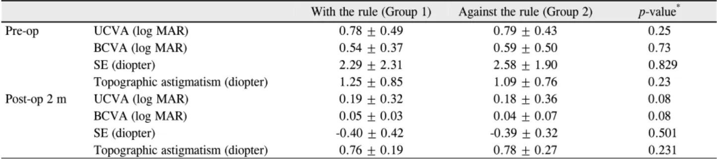
수술 환자는 총 50안으로 직난시(1군) 26안, 도난시(2 군) 24안이었다. 평균 연령은 각각 68.4세, 73.9세로 유의 한 차이는 없었다(p=0.062). 그 외 각막두께, 각막내피세 포 수, 안축장 길이도 유의한 차이는 없었다(Table 1). 술 전 측정한 logMAR 나안 시력과 술 후 logMAR 나안시력 및 logMAR 최대교정시력, 구면렌즈대응치에서 두 군간에 통계적인 차이는 없었으며(Table 2), 각막난시는 수술 전 각각 1.25 ±0.85 D, 1.09 ±0.76 D에서 수술 후 2개월째
Table 1. Patient characteristics
Variables With the rule (Group 1) Against the rule (Group 2) p-value*
No. of eyes 26 24
Laterality (OD:OS) 14 : 12 12 : 12
Sex (M:F) 13 : 13 11 : 13
Age (years) 68.40 ± 6.87 73.90 ± 10.83 0.062
Pachymetry (μm) 563.64 ± 31.19 569.55 ± 33.75 0.558
Endothelial cell count (cell/mm2) 2703 ± 457.13 2825 ± 367.95 0.527
Axial length (mm) 23.95 ± 0.11 23.55 ± 0.53 0.424
Values are presented as mean ± SD.
*By Mann-Whitney U test.
Table 2. Data summary of UCVA, BCVA, SE and topographic astigmatism
With the rule (Group 1) Against the rule (Group 2) p-value*
Pre-op UCVA (log MAR) 0.78 ± 0.49 0.79 ± 0.43 0.25
BCVA (log MAR) 0.54 ± 0.37 0.59 ± 0.50 0.73
SE (diopter) 2.29 ± 2.31 2.58 ± 1.90 0.829
Topographic astigmatism (diopter) 1.25 ± 0.85 1.09 ± 0.76 0.23
Post-op 2 m UCVA (log MAR) 0.19 ± 0.32 0.18 ± 0.36 0.08
BCVA (log MAR) 0.05 ± 0.03 0.04 ± 0.07 0.08
SE (diopter) -0.40 ± 0.42 -0.39 ± 0.32 0.501
Topographic astigmatism (diopter) 0.76 ± 0.19 0.78 ± 0.27 0.231
Values are presented as mean ± SD.
UCVA = uncorrected visual acuity; BCVA = best corrected visual acuity; SE = spherical equivalent; Pre-op = preoperative; Post-op 2 m
= post-operative 2 months.
*By Mann-Whitney U test.
1.4
1.2
1
0.8
0.6
0.4
0.2
0
Topographic astigmatism (D)
1.25 0.85±
1.09 0.76±
0.75 0.19± 0.8 0.33± 0.78 0.27±
0.76 0.19±
0.5 0.13± 0.68 0.21±
Group 1 Group 2
Figure 1. Change of topographic astigmatism. Values are pre-
sented as mean ± SD. Pre-op = preoperative; Post-op 1 w = postoperative 1 week; Post-op 1 m = postoperative 1 month;Post-op 2 m = postoperative 2 months. p-value by Mann-Whitney U test. There is no statistically significant difference between two groups (p > 0.05).
0.6 0.55 0.5 0.45 0.4 0.35 0.3 0.25 0.2
Surgically induced astigmatism (D) * = 0.003p
0.49 0.13±
0.31 0.17± 0.33 0.28±
Group 1 Group 2
* = 0.008p
0.48 0.11±
0.36 0.15±
0.51 0.19± * = 0.004p
Figure 2. Surgically-induced astigmatism calculated by vector
analysis. Values are presented as mean ± SD. Pre-op = pre- operative; Post-op 1 w = postoperative 1 week; Post-op 1 m= postoperative 1 month; Post-op 2 m = postoperative 2 months. p-value by Mann-W hitney U test (*p < 0.05).
각각 0.76 ±0.19 D, 0.78 ±0.27 D를 보여 난시 감소량이 각각 0.48 ±0.52 D, 0.31 ±0.38 D로 직난시 군이 도난시 군에 비해 난시교정효과가 좋았으나 통계적으로 유의하지 는 않았다(p=0.06)(Fig. 1).
수술유발난시는 1군에서 술 후 1주 0.51 ±0.19 D, 1개
월 0.49 ±0.13 D, 2개월 0.48 ±0.11였으며, 2군에서 술 후 1주 0.36 ±0.15 D, 1개월 0.33 ±0.28 D, 2개월 0.31
±0.17 D로 직난시 군이 도난시 군보다 유의하게 높게 측 정되었다(p=0.003, 0.008, 0.004)(Fig. 2).
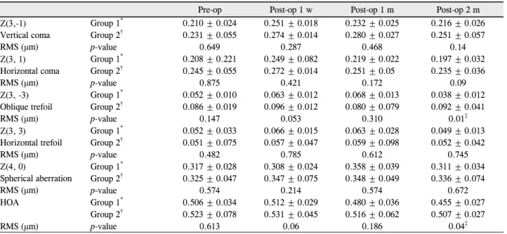
두 군간의 비교에서 각막전면 고위수차는 술 후 1주째 두 군에서 모두 술 전에 비해 증가하였으나 두 군간의 차이가 통
Table 3. Postoperative changes of corneal aberrations on the front side
Pre-op Post-op 1 w Post-op 1 m Post-op 2 m
Z(3,-1) Vertical coma RMS (μm)
Group 1* 0.210 ± 0.024 0.251 ± 0.018 0.232 ± 0.025 0.216 ± 0.026
Group 2† 0.231 ± 0.055 0.274 ± 0.014 0.280 ± 0.027 0.251 ± 0.057
p-value 0.649 0.287 0.468 0.14
Z(3, 1) Horizontal coma RMS (μm)
Group 1* 0.208 ± 0.221 0.249 ± 0.082 0.219 ± 0.022 0.197 ± 0.032
Group 2† 0.245 ± 0.055 0.272 ± 0.014 0.251 ± 0.05 0.235 ± 0.036
p-value 0.875 0.421 0.172 0.09
Z(3, -3) Oblique trefoil RMS (μm)
Group 1* 0.052 ± 0.010 0.063 ± 0.012 0.068 ± 0.013 0.038 ± 0.012
Group 2† 0.086 ± 0.019 0.096 ± 0.012 0.080 ± 0.079 0.092 ± 0.041
p-value 0.147 0.053 0.310 0.01‡
Z(3, 3)
Horizontal trefoil RMS (μm)
Group 1* 0.052 ± 0.033 0.066 ± 0.015 0.063 ± 0.028 0.049 ± 0.013
Group 2† 0.051 ± 0.075 0.057 ± 0.047 0.059 ± 0.098 0.052 ± 0.042
p-value 0.482 0.785 0.612 0.745
Z(4, 0)
Spherical aberration RMS (μm)
Group 1* 0.317 ± 0.028 0.308 ± 0.024 0.358 ± 0.039 0.311 ± 0.034
Group 2† 0.325 ± 0.047 0.347 ± 0.075 0.348 ± 0.049 0.336 ± 0.074
p-value 0.574 0.214 0.574 0.672
HOA Group 1* 0.506 ± 0.034 0.512 ± 0.029 0.480 ± 0.036 0.455 ± 0.027
Group 2† 0.523 ± 0.078 0.531 ± 0.045 0.516 ± 0.062 0.507 ± 0.027
RMS (μm) p-value 0.613 0.06 0.186 0.04‡
Values are presented as mean ± SD.
Z = zernike coefficients; RMS = root mean square; HOA = total higher order aberrations (3rd-6th order); Pre-op = preoperative; Post-op 1 w = postoperative 1 week; Post-op 1 m = postoperative 1 month; Post-op 2 m = postoperative 2 months.
*With the rule; †Against the rule; ‡By Mann-Whitney U test (p < 0.05).
1.4 1.2 1 0.8 0.6 0.4 0.2 0
(m)µ
Oblique trefoil
* = 0.04p * = 0.01p
0.25
0.2
0.15
0.1
0.05
0
Group 1 Group 2
(m)µ
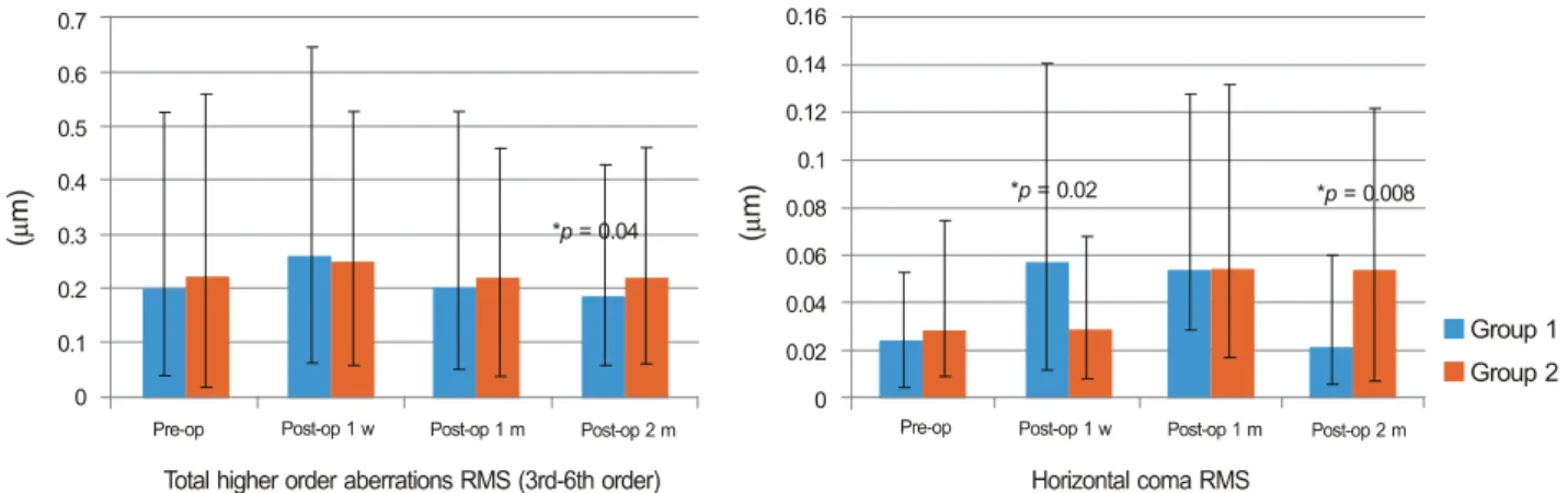
Figure 3. Comparision of corneal total higher order aberrations RMS and oblique trefoil (Z(3,-3)) RMS on front side in two groups.
Z = zernike coefficients; RMS = root mean square; Pre-op = preoperative; Post-op 1 w = postoperative 1 week; Post-op 1 m = postoperative 1 month; Post-op 2 m = postoperative 2 months. p-value by Mann-W hitney U test (*p < 0.05).
계적으로 유의하지는 않았다(p>0.05). 술 후 2개월에는 총 고위수차가 1군에서 0.455 ±0.027 μm, 2군에서 0.507 ± 0.027 μm으로 두 군간에 유의한 차이를 보였고(p=0.04), 고위수차 지표들 중 특히 사축트레포일 수치가 1군에서 0.038 ±0.012 μm, 2군에서 0.092 ±0.041 μm으로 유의 하게 직난시 군에서 도난시 군에 비해 감소됨을 알 수 있었 다(p=0.01)(Table 3, Fig. 3).
직난시 군에서 총 고위수차가 수술 전 0.506 ±0.034 μm 에비해 수술 2개월째 0.455 ±0.027 μm로 유의한 감소를 보였고(p=0.04), 고위수차 지표들 중 사축트레포일 수치 가 수술 전 0.052 ± 0.010 μm, 수술 2개월째 0.038 ±
0.012 μm로 통계적으로 유의하게 감소하였으나(p=0.008), 도 난시 군에서는 고위수차를 나타내는 어떠한 지표도 수술 전과 비 교 시에 수술 후 유의한 변화를 보이지 않았다(Table 3, Fig. 4).
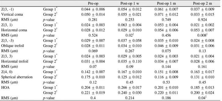
각막 후면의 고위수차는 각막 전면에 비해 전반적으로 모든 지표에서 낮은 값으로 측정되었다. 술 후 1주째 두 군 의 각막 후면 고위수차는 수술 전에 비해 증가하였으나 1군 0.063 ±0.004 μm, 2군 0.029 ±0.010 μm를 보였던 수평 코마 수치만의 유의한 차이를 보였고(p=0.02), 그 외 지표 에서는 두 군간의 차이가 통계적으로 유의하지는 않았다 (p>0.05). 술 후 2개월째 고위수차 지표들 중 총 고위수차 가 1군 0.185 ±0.0131 μm, 2군 0.200 ±0.024 μm로 유
* = 0.008p
* = 0.04p 0.6
0.5 0.4 0.3 0.2 0.1 0
Vertical coma RMS Horizontal coma RMS
Oblique trefoil RMS Horizontal trefoil RMS
Spherical aberration RMS HOA RMS
Group 1
(m)µ
(m)µ
0.6 0.5 0.4 0.3 0.2 0.1 0
Vertical coma RMS Horizontal coma RMS
Oblique trefoil RMS Horizontal trefoil RMS
Spherical aberration RMS HOA RMS
Group 2
Pre-op Post-op 1 w Post-op 1 m Post-op 2 m
Figure 4. Comparision of corneal total higher order aberations on front side in two groups. Z = zernike coefficients; RMS = root
mean square; HOA = total higher order aberrations (3rd-6th order); Pre-op = preoperative; Post-op 1 w = postoperative 1 week;Post-op 1 m = postoperative 1 month; Post-op 2 m = postoperative 2 months. p-value by W ilcoxon rank test (*p < 0.05).
Table 4. Postoperative changes of corneal aberrations on the rear side
Pre-op Post-op 1 w Post-op 1 m Post-op 2 m
Z(3, -1) Vertical coma RMS (μm)
Group 1* 0.044 ± 0.006 0.054 ± 0.012 0.061 ± 0.007 0.037 ± 0.009
Group 2† 0.050 ± 0.014 0.055 ± 0.021 0.071 ± 0.012 0.033 ± 0.015
p-value 0.281 0.253 0.749 0.924
Z(3, 1) Horizontal coma RMS (μm)
Group 1* 0.024 ± 0.003 0.063 ± 0.004 0.053 ± 0.004 0.021 ± 0.002
Group 2† 0.028 ± 0.012 0.029 ± 0.010 0.054 ± 0.006 0.053 ± 0.007
p-value 0.524 0.02‡ 0.456 0.008‡
Z(3, -3) Oblique trefoil RMS (μm)
Group 1* 0.029 ± 0.007 0.037 ± 0.058 0.035 ± 0.010 0.024 ± 0.008
Group 2† 0.028 ± 0.011 0.034 ± 0.010 0.046 ± 0.009 0.031 ± 0.006
p-value 0.069 0.267 0.075 0.13
Z(3, 3)
Horizontal trefoil RMS (μm)
Group 1* 0.024 ± 0.003 0.029 ± 0.005 0.024 ± 0.003 0.021 ± 0.004
Group 2† 0.031 ± 0.004 0.035 ± 0.110 0.034 ± 0.007 0.028 ± 0.006
p-value 0.07 0.09 0.144 0.161
Z(4, 0)
Spherical aberration RMS (μm)
Group 1* 0.142 ± 0.007 0.167 ± 0.010 0.151 ± 0.008 0.163 ± 0.017
Group 2† 0.175 ± 0.010 0.125 ± 0.012 0.116 ± 0.009 0.131 ± 0.010
p-value 0.12 0.45 0.53 0.45
HOA Group 1* 0.204 ± 0.011 0.266 ± 0.017 0.201 ± 0.010 0.185 ± 0.013
Group 2† 0.221 ± 0.019 0.240 ± 0.030 0.220 ± 0.011 0.200 ± 0.024
RMS (μm) p-value 0.4 0.214 0.186 0.04‡
Values are presented as mean ± SD.
Z = zernike coefficient; RMS = root mean square; HOA = total higher order aberrations (3rd-6th order); Pre-op = preoperative; Post-op 1 w
= postoperative 1 week; Post-op 1 m = postoperative 1 month; Post-op 2 m = postoperative 2 months.
*With the rule; †Against the rule; ‡By Mann-Whitney U test (p < 0.05).
의한 차이를 보였으며(p=0.04), 여러 지표들 중 수평코마 에서 1군 0.021 ±0.002 μm, 2군 0.053 ±0.007 μm로 유 의한 차이를 나타내었다(p=0.008) (Table 4, Fig. 5). 하 지만 전면 고위수차와는 달리 각각의 군에서 후면 고위수 차는 술 전과 비교하여 수술 후에 유의한 변화를 보이지 않 았다(Table 4, Fig. 6).
고 찰
백내장 수술 후 안구 수차를 변화시킬 수 있는 요인으로
는 크게 인공수정체의 성질과 수술절개창의 영향 두 가지 를 들 수 있다. 전자에 대해서는 비구면 인공수정체 개발과 발전이 활발히 이루어지고 있으며, 후자에 대한 연구로 Guiaro et al13은 백내장 수술에서 각막절개창은 술 후 각막 의 광학적 성질 및 수차를 변화시키는 중요한 역할을 한다 고 보고하면서 각막난시, 트레포일, 코마 등의 수차는 증가 하나 구면수차는 큰 변화가 없다고 하였다. 또한 국내 연구 에서는 절개창 크기에 따른 각막 전면 고위수차는 변화가 없다는 보고가 있었고,14절개창이 전면보다 후면에서 각막 중심에 더 근접하기 때문에 각막 전면보다 각막 후면에서
(m)µ (m)µ* = 0.04p
0.7 0.6 0.5 0.4 0.3 0.2 0.1 0
0.16 0.14 0.12 0.1 0.08 0.06 0.04 0.02 0
* = 0.02p * = 0.008p
Group 1 Group 2
Figure 5. Comparision of corneal total higher order aberations RMS and Horizontal coma (Z(3, 1)) RMS on rear side in two groups.
Z = zernike coefficients; RMS = root mean square; HOA = total higher order aberrations (3rd-6th order); Pre-op = preoperative;
Post-op 1 w = postoperative 1 week; Post-op 1 m = postoperative 1 month; Post-op 2 m = postoperative 2 months. p-value by Mann-Whitney U test (*p < 0.05).
(m)µ
(m)µ
0.25
0.2
0.15
0.1
0.05
0
Vertical coma RMS
Oblique trefoil RMS Horizontal trefoil RMS
Spherical aberration RMS HOA RMS
Group 1
0.25
0.2
0.15
0.1
0.05
0
Group 2
Pre-op Post-op 1 w Post-op 1 m Post-op 2 m
Horizontal coma RMS
Vertical coma RMS
Oblique trefoil RMS Horizontal trefoil RMS
Spherical aberration RMS HOA RMS Horizontal coma RMS
Figure 6. Comparision of corneal total higher order aberrations on rear side in two groups. Z = zernike coefficients; RMS = root
mean square; HOA = total higher order aberrations (3rd-6th order); Pre-op = preoperative; Post-op 1 w = postoperative 1 week;Post-op 1 m = postoperative 1 month; Post-op 2 m = postoperative 2 month. p-value by Wilcoxon rank test. There is no statistically significant difference among preoperative and postoperative 1 week, postoperative 1 month, postoperative 2 months in two groups, respectively (p > 0.05).
고위수차의 변화가 더 많았다는 연구가 있었다.1각막 중심 에서의 거리가 짧을수록 콜라겐 피브릴들이 큰 힘을 받으 므로, 각막절개 부위에서 각막 중심까지의 거리가 가까울수 록 각막의 형태 변화나 난시유발을 크게 한다는 이론이 여 러 연구의 주장들을 뒷받침하고 있다.15 그러나 본 연구에 서는 상측 절개 직난시 환자에서 이측 절개 도난시 환자에 비해 수술유발난시가 높게 측정되었고, 각막 후면의 고위수 차 값이 전면 고위수차에 비해 낮게 측정되어 절개창과 각 막 중심과의 거리뿐만 아니라 복합적인 인자가 난시유발에 영향을 미치는 것으로 생각된다.
또한 본 연구에서는 각막곡률이 가파른 축에 절개를 시 행한 동축 백내장 수술 후 각막의 고위 수차가 술 후 1주일 직난시, 도난시 모두에서 증가하였으나 통계적으로 유의하
지 않았고, 수술 2개월에는 양군에서 모두 술 전에 비하여 모두 감소하였다. Oh et al16은 3 mm 상측부 공막절개창을 통한 수정체유화술 후 1주째 수술절개창의 각막부종으로 인해 각막 후면에서 더 많은 변화를 일으킨다고 보고하였 는데, 본 연구에서도 수술 후 1주일째에는 각막의 부종이나 절개 창상의 불안정성이 고위수차의 증가에 영향을 미치고, 시간이 지나면서 절개 창상의 양상이 안정화되면서 고위수 차 변화를 가져온 것으로 보인다. 특히 2개월째 각막 전면 에서는 사축트레포일이 감소하였으며 각막 후면에서는 수 평코마수차가 감소하였다. 이런 수술교정효과는 도난시보 다는 직난시 군에서 유의하게 감소하였으며 각막곡률이 가 파른 축 절개가 난시교정효과뿐만 아니라 고위수차를 줄이 는 데도 도움이 되기에, 직난시 군에서 가파른 난시축에 동
축 소절개 백내장 수술이 유용하게 사용될 것으로 생각한다.
하지만 코마수차 및 다른 고위수차 들은 난시나 구면수 차에 비해 개인별로 차이가 큰 편이며 시기능에 미치는 영 향이 정확히 규명되지 않아 이에 대한 원인에 대해서는 더 많은 연구가 필요할 것으로 생각한다. 본 연구의 제한점으 로는 대상환자수가 적고 비교적 추적관찰 기간이 짧았다는 점, 시력의 질에 대한 대비감도 측정과 환자의 주관적 증상 에 대한 문진과 같은 시도가 없어 향 후 보완이 필요하다.
그리고 안구 내부의 수차 변화가 각막의 고위수차와 어떻 게 상보적으로 반응해 안구 전체의 고위수차에 영향을 주 는지 향후 각막의 고위수차와 안구 전체의 고위수차를 모 두 측정해 비교해보는 연구도 임상적인 가치가 있을 것으 로 생각된다.
REFERENCES
1) Moon SJ, Lee DJ, Lee KH. Induced astigmatism and high-order aberrations after 1.8-mm, 2.2-mm and 3.0-mm coaxial phacoemulsi- fication incisions. J Korean Ophthalmol Soc 2011;52:407-13.
2) Artal P, Berrio E, Guirao A, Piers P. Contribution of the cornea and internal surfaces to the change of ocular aberrations with age. J Opt Soc Am A Opt Image Sci Vis 2002;19:137-43.
3) Borasio E, Mehta JS, Maurino V. Surgically induced astigmatism after phacoemulsification in eyes with mild to moderate corneal as- tigmatism: temporal versus on-axis clear corneal incisions. J Cataract Refract Surg 2006;32:565-72.
4) Alió J, Rodríguez-Prats JL, Galal A, Ramzy M. Outcomes of mi- croincision cataract surgery versus coaxial phacoemulsification.
Ophthalmology 2005;112:1997-2003.
5) Artal P, Guirao A, Berrio E, Williams DR. Compensation of cor- neal aberrations by the internal optics in the human eye. J Vis
2001;1:1-8.
6) Artal P, Guirao A. Contributions of the cornea and the lens to the aberrations of the human eye. Opt Lett 1998;23:1713-5.
7) Guirao A, Redondo M, Geraghty E, et al. Corneal optical aberra- tions and retinal image quality in patients in whom monofocal in- traocular lenses were implanted. Arch Ophthalmol 2002;120:
1143-51.
8) Oshika T, Kawana K, Hiraoka T, et al. Ocular higher-order wave- front aberration caused by major tilting of intraocular lens. Am J Ophthalmol 2005;140:744-6.
9) Fujikado T, Kuroda T, Maeda N, et al. Light scattering and optical aberrations as objective parameters to predict visual deterioration in eyes with cataracts. J Cataract Refract Surg 2004;30:1198-208.
10) Lee SY, Chung JL, Hong JP, et al. Comparative study of two asphe- ric, aberration-free intraocular lenses in cataract surgery. J Korean Ophthalmol Soc 2009;50:1520-6.
11) Holladay JT, Hill WE, Steinmueller A. Corneal power measure- ments using scheimpflug imaging in eyes with prior corneal re- fractive surgery. J Refract Surg 2009;25:862-8.
12) Yao K, Tang X, Ye P. Corneal astigmatism, high order aberrations, and optical quality after cataract surgery: microincision versus small incision. J Refract Surg 2006;22(9 Suppl):S1079-82.
13) Guirao A, Tejedor J, Artal P. Corneal aberrations before and after small-incision cataract surgery. Invest Ophthalmol Vis Sci 2004;
45:4312-9.
14) Hwang SJ, Choi SK, Oh SH, et al. Surgically induced astigmatism and corneal higher order aberrations in microcoaxial and conven- tional cataract surgery. J Korean Ophthalmol Soc 2008;49:1597- 602.
15) Meek KM, Newton RH. Organization of collagen fibrils in the cor- neal stroma in relation to mechanical properties and surgical practice. J Refract Surg 1999;15:695-9.
16) Oh HC, Lee DJ, Park WC. Changes of the corneal aberration fol- lowing cataract surgery. J Korean Ophthalmol Soc 2009;50:518- 22.
=ABSTRACT=
Comparison of Higher Order Aberrations and Astigmatism after On-Axis Small Incision Cataract Surgery
Hyun Jin Jeong, MD, Byung Ju Jung, MD, Hyung Bin Hwang, MD, Hyun Seung Kim, MD, PhD
Department of Ophthalmology and Visual Science, The Catholic University of Korea College of Medicine, Seoul, Korea
Purpose: To assess the changes of corneal astigmatism and higher order aberrations (HOAs) of the anterior and posterior corneal surface after cataract surgery with on-axis clear corneal incision in eyes with-the-rule (WTR) astigmatism and against-the-rule (ATR) astigmatism.
Methods: This study included 50 patients who underwent phacoemulsification and IOL insertion through a 2.8-mm on-axis clear corneal incision. The eyes were divided into two groups: (1) 26 eyes with WTR astigmatism with a superior incision and (2) 24 eyes with ATR astigmatism with a temporal incision. During the follow-up period, visual acuity was measured, and the surgically induced astigmatism (SIA) and HOAs of the anterior and posterior corneal surface were measured with Pentacam® (Occlus, Wetzlar, Germany) preoperatively and 1 week, 1 month, and 2 months postoperatively.
Results: There were no significant differences in UCVA and BCVA between the two groups. HOAs increased in both groups 1 week after surgery, but no significant differences were found between the groups (p > 0.05). Surgically induced astigmatism was larger in the WTR group than in the ATR group (p < 0.05). At postoperative 2 months, there were sig- nificant differences in HOAs between the two groups, and there were statistically significant differences in HOAs, oblique trefoil at front side, and in HOAs, horizontal coma at rear side (p < 0.05).
Conclusions: In conclusion, superior incision in eyes with WTR astigmatism resulted in higher SIA compared to temporal incision in eyes with ATR astigmatism. Moreover, HOAs was significantly decreased in eyes with WTR astigmatism with superior incision. Thus, superior incision could be more effective in reducing corneal astigmatism in eyes with WTR astigmatism.
J Korean Ophthalmol Soc 2014;55(2):182-189
Key Words: Against-The-Rule (ATR), Higher Order Aberrations (HOAs), On-axis Incision, Surgically Induced Astigmatism (SIA), With-The-Rule (WTR)
Address reprint requests to Hyun Seung Kim, MD, PhD
Department of Ophthalmology, The Catholic University of Korea Yeouido St. Mary’s Hospital
#10 63-ro, Yeongdeungpo-gu, Seoul 150-713, Korea
Tel: 82-2-3779-1243, Fax: 82-2-761-6869, E-mail: [email protected]