Yonsei Med J http://www.eymj.org Volume 52 Number 2 March 2011 322
Original Article
DOI 10.3349/ymj.2011.52.2.322pISSN: 0513-5796, eISSN: 1976-2437 Yonsei Med J 52(2):322-325, 2011
Central Corneal Thickness and Corneal Endothelial Cell Changes Caused by Contact Lens Use in Diabetic Patients
Hyun Sung Leem,
1Koon Ja Lee,
1and Ki Cheul Shin
21Department of Optometry, Eulji University, Seongnam;
2Department of Ophthalmology, College of Medicine, Konkuk University, Seoul, Korea.
Received: April 30, 2010 Revised: June 10, 2010 Accepted: June 11, 2010
Corresponding author: Dr. Ki Cheul Shin, Department of Ophthalmology, College of Medicine, Konkuk University, 4-12 Hwayang-dong, Gwangjin-gu, Seoul 143-729, Korea.
Tel: 82-2-2030-7655, Fax: 82-2-2030-5273 E-mail: [email protected]
∙ The authors have no financial conflicts of interest.
© Copyright:
Yonsei University College of Medicine 2011 This is an Open Access article distributed under the terms of the Creative Commons Attribution Non- Commercial License (http://creativecommons.org/
licenses/by-nc/3.0) which permits unrestricted non- commercial use, distribution, and reproduction in any medium, provided the original work is properly cited.
Purpose: To analyze the effects of soft contact lenses on central corneal thickness and morphologic characteristics of the corneal endothelium in diabetic patients.
Materials and Methods: Ultrasound pachymetry and noncontact specular micros- copy were performed on 26 diabetic patients who regularly use soft contact lenses (group 1), 27 diabetic patients who do not use soft contact lenses (group 2) and 30 normal subjects (group 3). We compared the values in each group using the Mann- Whitney test. Results: The central cornea was found to be thicker in diabetic pa- tients, both those who use and do not use contact lenses, than in the normal control group. The central corneal thickness was significantly higher in group 1 (564.73 ± 35.41 µm) and group 2 (555.76 ± 45.96 µm) than in the control group (534.05 ± 27.02 µm), but there was no statistically significant difference between groups 1 and 2. Endothelial cell density was significantly different between the groups, and was smallest in the group of diabetic patients using contact lenses. The coefficient of variation of cell size was significantly higher and the percentage of hexagonal cells was significantly lower in contact lens using diabetic patients than in non- contact lens using diabetic patients and in the control group. Conclusion: Central corneal thickness and endothelial cell density is more affected by diabetes melli- tus, and corneal endothelial cell morphology is more affected by contact lens use, when compared with normal subjects.
Key Words: Central corneal thickness, morphology of corneal endothelial cell, diabetics using contact lenses, pachymetry, specular microscopy
INTRODUCTION
Corneal endothelial cells are arranged in a single layer and extremely stable. They have a metabolism-favorable regular hexagonal structure, thus controlling water balance and maintaining corneal transparency. Human corneal endothelial cells do not regenerate after injury but heal through their hyperplasia and mobilization. In severe cases, corneal edema and opacity occur and subsequently lead to vision loss. The corneal endothelium is affected by various factors including age, dura- tion of contact lens use, diabetes mellitus and so on.1
Previous studies have demonstrated histopathologic changes of the corneal en-
Corneal Changes by Contact Lens in Diabetics
Yonsei Med J http://www.eymj.org Volume 52 Number 2 March 2011 323
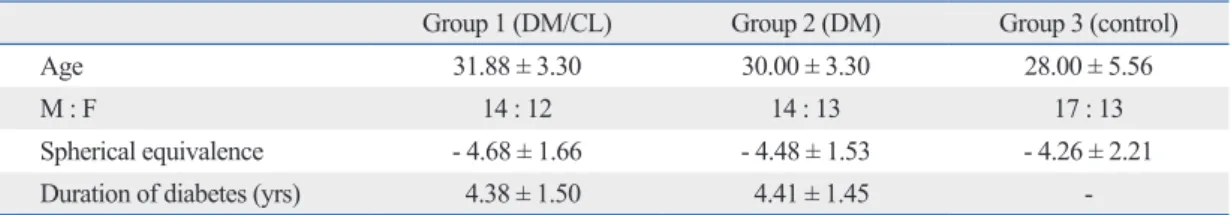
lenses [group 2 (DM), n = 54]. The control subjects [group 3 (control), n = 60] were free of diabetes mellitus and did not wear soft contact lenses. The mean age was 31.88 ± 3.30 years in group 1, 30.00 ± 3.30 years in group 2, 28.00 ± 5.56 years in group 3. There were no statistical differences in age, the difference of sex and the refractive errors among the groups (Table 1).
Both eyes were examined at the same time. In all groups, a complete medical history was taken and a slit-lamp ex- amination and indirect ophthalmoscopic examination were performed. The thickness of the cornea (µm), endothelial cell density (cells/mm2), CV of cell size (SD/area) and per- centage of hexagonal cells (%) were measured using a non- contact specular microscope (Noncon Robo-CA Konan SP- 9000p, Tokyo, Japan) and an objective refraction test was performed using ARK8800 (Topcon, Tokyo, Japan). None of the patients used topical ocular medications, and any pa- tients having a history of ocular diseases, previous ophthal- mic intervention or systemic disease besides diabetes melli- tus were excluded from study.
Corneal endothelial cells were examined by a single exam- iner using a non-contact specular microscope. After photo- graphing the center of the cornea, the number of endothelial cells was calculated using the ‘dot’ method. The data ob- tained were analyzed in terms of endothelial cell density, CV of cell size and hexagonality. We compared the values of cor- neal factors such as corneal thickness and endothelial mor- phology among the groups by using the Mann-Whitney test.
RESULTS
The duration of diabetics was 4.38 ± 1.50 years in group 1 and 4.41 ± 1.45 years in group 2 (p > 0.05). The duration of contact lens use was 5.27 ± 1.76 years in group 1.
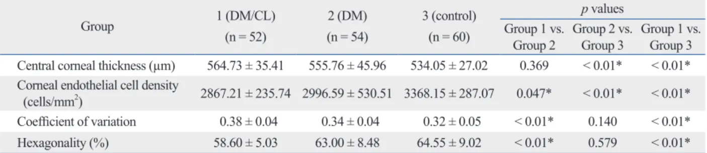
Central corneal thickness was significantly greater in group 1 (564.73 ± 35.41 µm) and group 2 (555.76 ± 45.96 µm) than in the control group (534.05 ± 27.02 µm), but there was no statistically significant difference between groups 1 and 2.
dothelium in contact lens wearers, such as corneal swell- ing.2-4 It is well known that long-term contact lens use causes changes in keratometry, corneal topography and morphology of endothelial cells. Although the exact mech- anisms for such changes have not yet been completely elu- cidated, chronic hypoxia has been reported to be the main cause.5,6 Since Connor and Zagrod7 reported in 1986 that polymegathism was increased in long-term contact lens wearers, numerous studies on corneal endothelial damage induced by contact lens use have been conducted. Previous studies have indicated that cell size increases while the per- centage of hexagonal endothelial cells and endothelial cell density decrease.8,9
Investigation of the relationship between the corneal en- dothelium and diabetes mellitus has so far been conducted using human and animal models. It has been reported that cell size and coefficients of variation (CV) of cell size in patients with a 10-year history of diabetes mellitus are dif- ferent from those in normal subjects and that the thickness of the cornea correlates significantly with the duration of diabetes mellitus.10,11 However, there have been few studies of the changes in thickness and morphology of the corneal endothelium in contact lens-wearing, non contact lens- wearing diabetic patients and normal control.
Therefore, this study was carried out to investigate the morphological characteristics of corneal endothelial cells and corneal thickness in contact lens-wearing diabetic pa- tients and to compare these variables with non contact lens- wearing diabetic patients and age-matched control group.
MATERIALS AND METHODS
This study included patients aged 15 to 39 years who visit- ed the Department of Ophthalmology, Konkuk University Hospital from August 2006 to August 2007. The patients were divided into 2 categories: those who were diagnosed with diabetes mellitus and continued to wear soft contact lenses [group 1 (DM/CL), n = 52] and those who were diag- nosed with diabetes mellitus and did not wear soft contact
Table 1. Patient Characteristics
Group 1 (DM/CL) Group 2 (DM) Group 3 (control)
Age 31.88 ± 3.30 30.00 ± 3.30 28.00 ± 5.56
M : F 14 : 12 14 : 13 17 : 13
Spherical equivalence - 4.68 ± 1.66 - 4.48 ± 1.53 - 4.26 ± 2.21
Duration of diabetes (yrs) 4.38 ± 1.50 4.41 ± 1.45 -
Hyun Sung Leem, et al.
Yonsei Med J http://www.eymj.org Volume 52 Number 2 March 2011 324
contact lens users and that the proportion of hexagonal cells and corneal endothelilal cell density in those using soft con- tact lens for more than six years were significantly lower than in the control group.
Many authors have studied the effects of diabetes and contact lens wearing on corneal endothelial cells and cen- tral corneal thickness, but only one study has been per- formed on the effects of contact lens wearing in diabetic subjects. O’Donnell and Efron27 reported that the mor- phometry of corneal endothelial cells and the central corne- al thickness values in diabetic patients who wear soft con- tact lens were not appreciably different from those found in lens-wearing control subjects. They compared diabetic pa- tients who were soft contact lens to a lens-wearing control group, rather than a normal control group.
In our study, we compared cornea characteristics of dia- betic patients who wear contact lenses to those of diabetic patients who do not wear contact lenses and those of normal control subjects without contact lens. The central corneal thickness was not significantly different between diabetic patients with or without contact lenses but was significantly greater than that of normal control group. Endothelial cell density was significantly lower in diabetic patients with contact lenses than in diabetic patients without contact lens or control group. Our results showed that central corneal thickness and corneal endothelial cell density are more af- fected by diabetes than contact lens use.
On the other hand, the CV of cell size was significantly higher and the percentage of hexagonal cells was signifi- cantly lower in diabetic patients who wore contact lenses as compared with both diabetic patients who did not wear contact lenses and the normal control group. These results imply that morphologic characteristics of corneal endotheli- al cell were more affected by contact lens than diabetes.
Recently, as the incidence of diabetes has been increasing among young adults,28 there has been a coinciding increase Endothelial cell density was significantly less in group 1
(2867.21 ± 235.74 cells/mm2) than in groups 2 (2996.59 ± 530.51 cells/mm2) and 3 (3368.15 ± 287.07 cells/mm2).
The CV of cell size variation was 0.38 ± 0.04 in group 1, 0.34 ± 0.04 in group 2, and 0.32 ± 0.05 in group 3; the per- centage of hexagonal cells was 58.60 ± 5.03 (%) in group 1, 63.00 ± 8.48 (%) in group 2, and 64.55 ± 9.02 (%) in group 3. The CV of cell size was significantly higher and percentage of hexagonal cells was significantly lower in group 1 as compared to groups 2 and 3 (Table 2).
DISCUSSION
Diabetes mellitus affects structural and functional changes in corneal endothelial cells and their thickness.12-17 Many studies have suggested that diabetic patients have corneal abnormalities such as higher autofluorescence, lower corne- al sensitivity, greater corneal thickness, less corneal endothe- lial cell density and increased endothelial permeability. The central cornea of diabetic patients is generally thicker than that of normal persons, and lower corneal endothelial cell density, lower hexagonality and higher CV of cell size have been reported in the cases of diabetes. Such results have also been revealed in experimental studies on diabetic mice or dogs.18,19 It is thought that diabetes reduces the activity of Na + -K + ATPase of the corneal endothelium and this causes the morphological and functional changes of diabet- ic cornea.20
Contact lenses also affect corneal endothelial cells by in- ducing chronic hypoxia, which causes lactate accumula- tion, elevated carbon dioxide levels and decrease of pH.21,22 The results are increases in polymegathism and pleomor- phism and a decrease in corneal endothelial cell density.23-25 Lee, et al.26 reported that the CV of cell size in the soft con- tact lens use group was significantly greater than in the non-
Table 2. Central Corneal Thickness and Morphologic Characteristics of Endothelial Cell by Group
Group 1 (DM/CL)
(n = 52)
2 (DM) (n = 54)
3 (control) (n = 60)
p values Group 1 vs.
Group 2 Group 2 vs.
Group 3 Group 1 vs.
Group 3 Central corneal thickness (µm) 564.73 ± 35.41 555.76 ± 45.96 534.05 ± 27.02 0.369 < 0.01* < 0.01*
Corneal endothelial cell density
(cells/mm2) 2867.21 ± 235.74 2996.59 ± 530.51 3368.15 ± 287.07 0.047* < 0.01* < 0.01*
Coefficient of variation 0.38 ± 0.04 0.34 ± 0.04 0.32 ± 0.05 < 0.01* 0.140 < 0.01*
Hexagonality (%) 58.60 ± 5.03 63.00 ± 8.48 64.55 ± 9.02 < 0.01* 0.579 < 0.01*
Mann-Whitney Test.
*Statistically significant difference.
Corneal Changes by Contact Lens in Diabetics
Yonsei Med J http://www.eymj.org Volume 52 Number 2 March 2011 325 tion control in diabetes mellitus. Invest Ophthalmol Vis Sci 1995;
36:586-95.
15. Larsson LI, Bourne WM, Pach JM, Brubaker RF. Structure and function of the corneal endothelium in diabetes mellitus type I and type II. Arch Ophthalmol 1996;114:9-14.
16. Busted N, Olsen T, Schmitz O. Clinical observations on the corne- al thickness and the corneal endothelium in diabetes mellitus. Br J Ophthalmol 1981;65:687-90.
17. Ziadi M, Moiroux P, d’Athis P, Bron A, Brun JM, Creuzot-Garch- er C. Assessment of induced corneal hypoxia in diabetic patients.
Cornea 2002;21:453-7.
18. Meyer LA, Ubels JL, Edelhauser HF. Corneal endothelial mor- phology in the rat. Effects of aging, diabetes, and topical aldose reductase inhibitor treatment. Invest Ophthalmol Vis Sci 1988;29:
940-8.
19. Yee RW, Matsuda M, Kern TS, Engerman RL, Edelhauser HF.
Corneal endothelial changes in diabetic dogs. Curr Eye Res 1985;
4:759-66.
20. Herse PR. Corneal hydration control in normal and alloxan-induced diabetic rabbits. Invest Ophthalmol Vis Sci 1990;31:2205-13.
21. Carlson KH, Bourne WM. Endothelial morphologic features and function after long-term extended wear of contact lenses. Arch Ophthalmol 1988;106:1677-9.
22. Connor CG, Zagrod ME. Contact lens-induced corneal endothelial polymegathism: functional significance and possible mechanisms.
Am J Optom Physiol Opt 1986;63:539-44.
23. Hirst LW, Auer C, Cohn J, Tseng SC, Khodadoust AA. Specular microscopy of hard contact lens wearers. Ophthalmology 1984;
91:1147-53.
24. Holden BA, Sweeney DF, Vannas A, Nilsson KT, Efron N. Effects of long-term extended contact lens wear on the human cornea. In- vest Ophthalmol Vis Sci 1985;26:1489-501.
25. Mac Rae SM, Matsuda M, Shellans S, Rich LF. The effects of hard and soft contact lenses on the corneal endothelium. Am J Ophthalmol 1986;102:50-7.
26. Lee JS, Park WS, Lee SH, Oum BS, Cho BM. A comparative study of corneal endothelial changes induced by different dura- tions of soft contact lens wear. Graefes Arch Clin Exp Ophthalmol 2001;239:1-4.
27. O’Donnell C, Efron N. Corneal endothelial cell morphometry and corneal thickness in diabetic contact lens wearers. Optom Vis Sci 2004;81:858-62.
28. Zimmet P, Alberti KG, Shaw J. Global and societal implications of the diabetes epidemic. Nature 2001;414:782-7.
in contact lens use in diabetic patients. These patients should be cautioned that not only diabetes but also their contact lens use can affect the corneal endothelial cells, and should have their corneal endothelial cells examined regularly.
REFERENCES
AC
1. Sheng H, Bullimore MA. Factors affecting corneal endothelial morphology. Cornea 2007;26:520-5.
2. Holden BA, Mertz GW, McNally JJ. Corneal swelling response to contact lenses worn under extended wear conditions. Invest Oph- thalmol Vis Sci 1983;24:218-26.
3. Kanai A, Kaufman HE. Electron microscopic studies of swollen corneal stroma. Ann Ophthalmol 1973;5:178-90.
4. Bergmanson JP, Chu LW. Corneal response to rigid contact lens wear. Br J Ophthalmol 1982;66:667-75.
5. Bruce AS, Brennan NA. Corneal pathophysiology with contact lens wear. Surv Ophthalmol 1990;35:25-58.
6. Liesegang TJ. Physiologic changes of the cornea with contact lens wear. CLAO J 2002;28:12-27.
7. Connor CG, Zagrod ME. Contact lens-induced corneal endothelial polymegathism: functional significance and possible mechanisms.
Am J Optom Physiol Opt 1986;63:539-44.
8. Holden BA, Sweeney DF, Vannas A, Nilsson KT, Efron N. Effects of long-term extended contact lens wear on the human cornea. In- vest Ophthalmol Vis Sci 1985;26:1489-501.
9. Setälä K, Vasara K, Vesti E, Ruusuvaara P. Effects of long-term contact lens wear on the corneal endothelium. Acta Ophthalmol Scand 1998;76:299-303.
10. Larsson LI, Bourne WM, Pach JM, Brubaker RF. Structure and function of the corneal endothelium in diabetes mellitus type I and type II. Arch Ophthalmol 1996;114:9-14.
11. Lee JS, Oum BS, Choi HY, Lee JE, Cho BM. Differences in cor- neal thickness and corneal endothelium related to duration in dia- betes. Eye (Lond) 2006;20:315-8.
12. Schultz RO, Matsuda M, Yee RW, Edelhauser HF, Schultz KJ.
Corneal endothelial changes in type I and type II diabetes mellitus.
Am J Ophthalmol 1984;98:401-10.
13. Sánchez-Thorin JC. The cornea in diabetes mellitus. Int Ophthal- mol Clin 1998;38:19-36.
14. Weston BC, Bourne WM, Polse KA, Hodge DO. Corneal hydra-