ABSTRACT
Purpose: The aim of this study was to evaluate clinical factors affecting the longevity of fixed retainers and the influence of fixed retainers on periodontal health in periodontitis patients.
Methods: In total, 52 patients with at least 2 years of follow-up after periodontal and orthodontic treatment were included in this study. After scaling and root planing,
orthodontic treatment with fixed appliances or clear aligners was performed. Fixed retainers with twist-flex stainless steel wires were bonded to the palatal or lingual sides of anterior teeth. Changes in clinical parameters, including the plaque index, gingival index, calculus index (CI), probing pocket depth, and radiographic bone levels, were evaluated before bonding of fixed retainers and at a 12-month follow-up. Cumulative survival rates (CSRs) for retainer failure were evaluated according to sex, site, CI, stage of periodontitis, and the severity of the irregularity with the log-rank test and hazard ratios (HRs).
Results: Twelve months after bonding of fixed retainers, improvements were observed in all clinical parameters except CI and radiographic bone gain. The overall CSR of the retainers with a CI <1 at the 12-month follow-up after bonding of fixed retainers was significantly higher than that of the retainers with a CI ≥1 at the 12-month follow-up (log-rank test;
P<0.001). Patients with stage III (grade B or C) periodontitis had a higher multivariate HR for retainer failure (5.4; 95% confidence interval, 1.22–23.91; P=0.026) than patients with stage I (grade A or B) periodontitis.
Conclusions: Although fixed retainers were bonded in periodontitis patients, periodontal health was well maintained if supportive periodontal treatment with repeated oral hygiene education was provided. Nonetheless, fixed retainer failure occurred more frequently in patients who had stage III (grade B or C) periodontitis or a CI ≥1 at 12-month follow-up after bonding of fixed retainers.
Keywords: Dental calculus; Oral hygiene; Orthodontics; Orthodontic appliances;
Orthodontic retainers; Periodontitis
Research Article
Received: May 11, 2020 Revised: Jan 15, 2021 Accepted: Mar 22, 2021
*Correspondence:
Chang-Joo Park
Division of Oral & Maxillofacial Surgery, Department of Dentistry, Hanyang University College of Medicine, 222 Wangsimni-ro, Seongdong-gu, Seoul 04763, Korea.
E-mail: fastchang@hanyang.ac.kr Tel: +82-2-2290-8671
Fax: +82-2-2290-8673
Copyright © 2021. Korean Academy of Periodontology
This is an Open Access article distributed under the terms of the Creative Commons Attribution Non-Commercial License (https://
creativecommons.org/licenses/by-nc/4.0/).
ORCID iDs Ji-Young Han
https://orcid.org/0000-0002-2364-8366 Seo Hee Park
https://orcid.org/0000-0003-2680-4966 Joohyung Kim
https://orcid.org/0000-0001-5472-7721 Kyung-Gyun Hwang
https://orcid.org/0000-0002-8713-660X Chang-Joo Park
https://orcid.org/0000-0002-6895-9854 Funding
This research was supported by a grant of the Korea Health Technology R&D Project through the Korea Health Industry Development Institute, funded by the Ministry of Health
& Welfare, Republic of Korea (grant No.
HI20C0013).
Ji-Young Han 1, Seo Hee Park 2, Joohyung Kim 3, Kyung-Gyun Hwang 4, Chang-Joo Park 4,*
1Division of Periodontology, Department of Dentistry, Hanyang University College of Medicine, Seoul, Korea
2Division of Periodontology, Department of Dentistry, Hanyang University Medical Center, Seoul, Korea
3Division of Orthodontics, Department of Dentistry, Hanyang University Medical Center, Seoul, Korea
4 Division of Oral & Maxillofacial Surgery, Department of Dentistry, Hanyang University College of Medicine, Seoul, Korea
Clinical factors affecting the longevity of fixed retainers and the influence of fixed retainers on periodontal health in periodontitis patients:
a retrospective study
Periodontal Science
Author Contributions
Conceptualization: Ji-Young Han; Formal analysis: Chang-Joo Park, Joohyung Kim, Kyung-Gyun Hwang; Investigation: Seo Hee Park; Methodology: Joohyung Kim, Ji-Young Han; Project administration: Ji-Young Han;
Writing - original draft: Ji-Young Han; Writing - review & editing: Ji-Young Han, Kyung-Gyun Hwang, Chang-Joo Park.
Conflict of Interest
No potential conflict of interest relevant to this article was reported.
INTRODUCTION
Crowded or malposed teeth sometimes make it difficult for periodontitis patients to perform proper oral hygiene. Orthodontic treatment can successfully resolve pathologically migrated or malaligned teeth to facilitate proper oral hygiene [1]. However, teeth have a tendency to relapse to their initial positions because of tension in the periodontal ligament and supra- alveolar gingival fibers [2,3]. Unwanted tooth movement after orthodontic treatment can also occur as a result of normal age-related changes, and is also possible in patients who have not received orthodontic treatment [3]. This deterioration in the alignment of teeth is reported to be due to changes in soft tissue pressure and skeletal structure around the teeth [3]. Therefore, orthodontic retention is mandatory for maintaining the corrected position of teeth following orthodontic treatment and minimizing age-related changes [4]. Retention after orthodontic treatment is particularly important in periodontitis patients.
No consensus exists regarding the ideal duration of retention [3,5,6]. However, the first 8 months post-treatment, when remodeling of the elastic fibers around the neck of the teeth, the dento-gingival fibers, and interdental fibers occurs, have been reported to be a critical period for orthodontic retention [2,7]. Several types of retainers have been used and a flexible multistranded wire bonded to each anterior tooth is currently considered the gold standard [8-10]. However, orthodontic retainers installed to maintain the corrected tooth position after orthodontic treatment may affect the periodontal tissue. Some authors have reported that orthodontic retainers did not cause clinically significant damage to periodontal health if proper oral hygiene and professional plaque and calculus removal were provided [11,12]. However, a recent systematic review reported that patients with long-term retention exhibited higher calculus accumulation, greater gingival recession, and increased probing depths [7].
Moreover, the failure of fixed retainers may result in a relapse of orthodontically treated teeth.
Therefore, it is necessary to elucidate the factors influencing retainer failure. To the authors' knowledge, the prolonged effects of fixed retainers on periodontal health and factors influencing retainer failure in periodontitis patients have not been adequately addressed.
Therefore, it is important to clarify the relationship of fixed retainers with periodontal health and their influence on oral hygiene in periodontitis patients.
The aim of this study was to evaluate clinical factors affecting the longevity of fixed retainers and the influence of fixed retainers on periodontal health in periodontitis patients after periodontal and orthodontic treatment.
MATERIALS AND METHODS
Subject sample
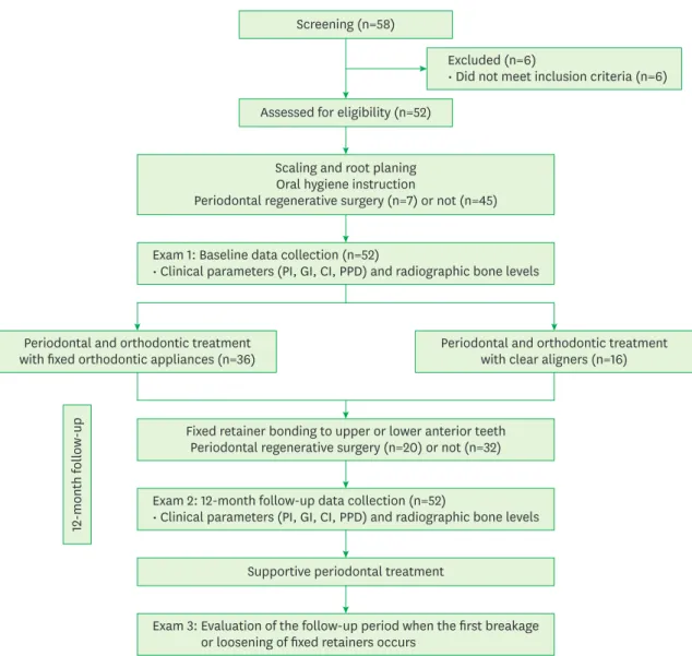
In total, 58 patients who underwent periodontal and orthodontic treatment at the Department of Periodontology, Hanyang University Medical Center, between March 2005 and August 2019 were screened for eligibility for this study (Figure 1).
The inclusion criteria were as follows: 1) Patients who were more than 20 years old; 2) Patients who had no relevant systemic conditions or diseases; 3) Patients who underwent periodontal and orthodontic treatment and had fixed orthodontic retainers in the anterior
area; 4) Patients who had more than 1 to 2 mm of interdental clinical attachment loss (≥
stage I) [13]; 5) Patients who were compliant with supportive periodontal treatment after periodontal and orthodontic treatment.
The exclusion criteria were as follows: 1) Patients who had periodontal and orthodontic treatment for prosthodontic treatment in the posterior area; 2) Patients who had an active infection or disease affecting bone metabolism and wound healing; 3) Patients with regular use of steroids or other medications affecting bone turnover; 4) Patients who were pregnant.
This study was approved by the Institutional Review Board of Hanyang University Hospital (No. 2019-10-037). The STrengthening the Reporting of OBservational studies in Epidemiology guidelines were observed in the preparation of the manuscript.
Excluded (n=6)
• Did not meet inclusion criteria (n=6) Screening (n=58)
Assessed for eligibility (n=52)
Scaling and root planing Oral hygiene instruction
Periodontal regenerative surgery (n=7) or not (n=45)
Fixed retainer bonding to upper or lower anterior teeth Periodontal regenerative surgery (n=20) or not (n=32)
Supportive periodontal treatment Periodontal and orthodontic treatment
with fixed orthodontic appliances (n=36)
12-month follow-up
Periodontal and orthodontic treatment with clear aligners (n=16)
Exam 2: 12-month follow-up data collection (n=52)
• Clinical parameters (PI, GI, CI, PPD) and radiographic bone levels
Exam 3: Evaluation of the follow-up period when the first breakage or loosening of fixed retainers occurs
Exam 1: Baseline data collection (n=52)
• Clinical parameters (PI, GI, CI, PPD) and radiographic bone levels
Figure 1. Flow chart of the participants in this study.
PI: plaque index, GI: gingival index, CI: calculus index, PPD: probing pocket depth.
Periodontal and orthodontic treatment
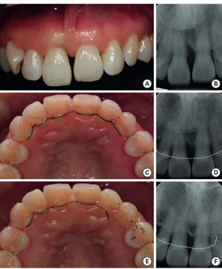
All periodontal treatment and orthodontic treatment were performed by one periodontist (H.J.). After scaling and root planing with repeated oral hygiene education, the patients were re-evaluated. If a patient's oral hygiene was not satisfactory (plaque index [PI] >2), oral hygiene education with intraoral tooth brushing instructions [14] and supportive periodontal treatment were repeated prior to orthodontic treatment. After periodontal treatment with or without regenerative surgical procedures, patients underwent orthodontic treatment either with fixed appliances or with clear aligners according to the decision-making process [15]. From the treatment planning stage, all cases were discussed with orthodontists at the Department of Orthodontics with careful consideration given to the patients' primary needs. Study casts were created and duplicated for orthodontic analysis and diagnosis. Initial records including intraoral photographs and radiographs were collected prior to orthodontic treatment (Figure 2A and B).
For periodontal and orthodontic treatment with fixed appliances, brackets were bonded from canine to canine in the maxilla or the mandible. For the correction of crowded anterior
A B
C D
E F
Figure 2. Maxillary fixed retainer. (A) A clinical photograph before periodontal and orthodontic treatment. (B) A periapical radiograph before periodontal and orthodontic treatment shows a vertical bone defect on the mesial side of the maxillary right central incisor. (C) After periodontal and orthodontic treatment, a fixed retainer with a twist-flex stainless steel wire was bonded to the palatal side of the maxillary anterior teeth. (D) A periapical radiograph after periodontal and orthodontic treatment. (E) Breakage of the fixed retainer occurred on the maxillary left canine at the 5-year follow-up after bonding of fixed retainers. (F) A periapical radiograph shows the detached wire of the fixed retainer.
teeth, progressive interproximal stripping was performed with topical fluoride application to prevent proximal caries within the limits of enamel thickness. The first arch wire placed was a 0.014 inch Ni-Ti wire for leveling. After preliminary alignment, a series of stiffer wires were placed at 3-week intervals. Orthodontic treatment using a clear aligner was performed with a series of clear aligners. Patients were asked to wear the clear aligners full-time except while eating, drinking, and brushing and to change the clear aligners every 2 weeks. Throughout the orthodontic treatment, all patients received professional tooth cleaning with oral hygiene education at every visit.
After periodontal and orthodontic treatment, impressions for retainers were taken and models for the fabrication of fixed retainers were made. Fixed retainers with 0.0175-inch twist-flex stainless steel wire (Tri-flex™, RMO®, Denver, CO, USA) were bonded using flowable resin composite (Filtek™ Supreme Ultra Flowable Restorative, 3M™ ESPN™, St.
Paul, MN, USA) to the palatal or lingual sides of the 6 anterior teeth to prevent relapse. After bonding of fixed retainers, all patients were followed up for supportive periodontal therapy involving oral hygiene education and tri-monthly visits.
Clinical measurements
Data from patients' clinical records were utilized for clinical evaluation. The following clinical parameters before orthodontic treatment (baseline) and at the 12-month follow- up after bonding of fixed retainers were used for the evaluation of clinical measurements.
The PI [16] and gingival index (GI) [17] were evaluated at 4 sites (mesiobuccal, midbuccal, distobuccal, and lingual); the calculus index (CI) [18] was assessed at 4 sites (mesiobuccal, midbuccal, distobuccal, and lingual); and the probing pocket depth (PPD) was measured at 6 sites per tooth (mesiobuccal, mid-buccal, distobuccal, mesiolingual, mid-lingual, and distolingual) as the distance from the free gingival margin to the base of the probable pocket using a periodontal probe (PCP-UNC 15, Hu-Friedy Mfg. Co., Chicago, IL, USA) rounded off to the nearest millimeter following probing with a pressure of approximately 0.25 N.
Patients were classified as having stage I (grade A or B), stage II (grade A or B), stage III (grade B or C), and stage IV (grade B or C) periodontitis based on the severity and complexity of management (evidence or risk of rapid progression) [13]. The severity of the irregularities was classified according to the amount of malalignment [19].
Radiographic measurements
Panoramic and periapical radiographs taken before orthodontic treatment and at the 12-month follow-up after bonding of fixed retainers were used for radiographic assessments.
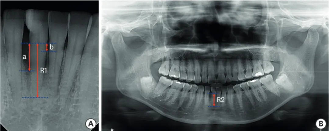
A digital radiography system (CS9300 Select, Carestream Dental LLC., Atlanta, GA, USA) was used for radiographic evaluation. Panoramic and periapical radiographs were imported into Analysis Toolkit (Adobe Photoshop CS6, Adobe Systems Inc., San Jose, CA, USA). Radiographic alveolar crest levels were assessed at the mesial and distal sites using baseline and 12-month follow-up periapical radiographs. To standardize the radiographic measurements, periapical radiographs taken with XCP-DS FIT® Universal sensor holders (Dentsply International Inc., York, PA, USA) were used [20]. One blinded examiner (P.S.) measured the distance between the cemento-enamel junction and the most coronal level of the alveolar bone for all sets of periapical radiographs twice (Figure 3A). The vertical distortion of the periapical radiographs at baseline and 12-month follow-up after bonding of fixed retainers was estimated using panoramic radiographs (Figure 3B). Anatomically non-variable distances from the cemento-enamel junction to the root apex on panoramic views were used for estimating radiographic bone level on periapical radiographs [20]. All
the measurements were repeated after an interval of at least 2 weeks. The mean of the 2 measurements was used as the radiographic bone level value. To test the reproducibility of the radiographic measurements, intra-class correlation coefficients (ICCs) were used.
Failure of retainers
Clinical records were used to evaluate retainer failure. The retainer was considered as a failure if any of the following was demonstrated: breakage at the wire-composite interface, breakage at the adhesive-enamel interface, a stress fracture of the wire, and partial or complete loosening of the fixed retainer from the teeth [21-23]. The point during follow-up when the first breakage or loosening of the fixed retainer occurred was recorded. The interval time between bonding (Figure 2C and D) and retainer failure (Figure 2E and F) was measured in months.
Statistical analysis
All statistical analyses were performed using SPSS (version 21, IBM Corp., Armonk, NY, USA). Descriptive statistics were performed using mean±standard deviation values for quantitative variables. After normality testing using the Shapiro-Wilk test, the differences of clinical parameters between baseline and the 12-month follow-up visit after bonding of fixed retainers were analyzed using the Wilcoxon signed rank test. The differences of clinical parameters and radiographic bone levels according to sex and site were analyzed using the Mann-Whitney U test. The cumulative survival rates (CSRs) of the fixed retainers were estimated using Kaplan-Meier analysis. The log-rank test was used to identify significant differences in the survival functions between the groups. Univariate and multivariate Cox proportional hazard regression models were used to analyze the hazard ratios (HRs) for failure of the fixed retainers according to clinical factors. A P value <0.05 was considered to indicate statistical significance.
RESULTS
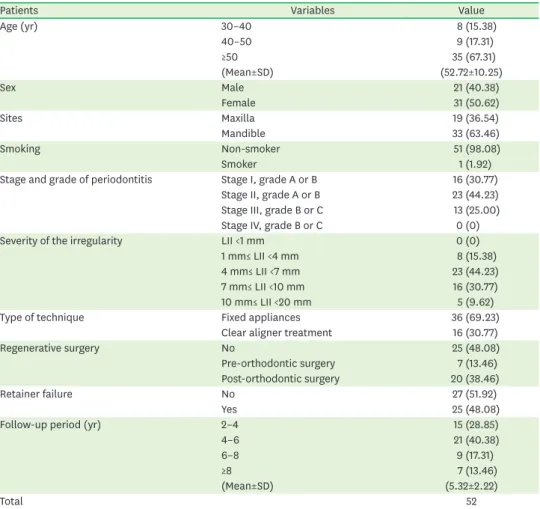
In total, 52 patients (21 male and 31 female) with a mean age of 52.72±10.25 years (range, 35–74 years) were included in this study (Table 1). Nineteen (36.54%) and 33 (63.46%)
A B
a b
R1
R2
Figure 3. Radiographic measurements. (A) The distances between the cemento-enamel junction and the most coronal level of the alveolar bone were measured at distal (a) and mesial (b) sites on all sets of periapical radiographs. The distances from the cemento-enamel junction to the root apex (R1) were measured on the periapical radiographs. (B) The distances from the cemento-enamel junction to the root apex (R2) were measured on the panoramic radiographs. The BL was calculated using the following formula: BL =𝑎𝑎𝑎𝑎 + 𝑏𝑏𝑏𝑏
2 × 𝑅𝑅𝑅𝑅2 𝑅𝑅𝑅𝑅1. BL: radiographic bone level.
patients underwent periodontal and orthodontic treatment in the maxilla and the mandible, respectively. They were all non-smokers (n=51) except for 1 patient. The mean follow-up period was 5.32±2.22 years (range, 2.08–9.83 years).
Clinical outcomes
At 12-month follow-up after bonding of fixed retainers, the clinical parameters (including PI, GI, and PPD) improved significantly compared to the baseline values (Table 2).
Before periodontal and orthodontic treatment, the mean values of the PI in the maxilla and mandible were 1.39±0.62 and 1.83±0.59, respectively (Table 3). The mean values of the PI in the maxilla and mandible at the 12-month follow-up after bonding of fixed retainers were Table 1. Demographic characteristics of the patients
Patients Variables Value
Age (yr) 30–40 8 (15.38)
40–50 9 (17.31)
≥50 35 (67.31)
(Mean±SD) (52.72±10.25)
Sex Male 21 (40.38)
Female 31 (50.62)
Sites Maxilla 19 (36.54)
Mandible 33 (63.46)
Smoking Non-smoker 51 (98.08)
Smoker 1 (1.92)
Stage and grade of periodontitis Stage I, grade A or B 16 (30.77)
Stage II, grade A or B 23 (44.23)
Stage III, grade B or C 13 (25.00)
Stage IV, grade B or C 0 (0)
Severity of the irregularity LII <1 mm 0 (0)
1 mm≤ LII <4 mm 8 (15.38)
4 mm≤ LII <7 mm 23 (44.23)
7 mm≤ LII <10 mm 16 (30.77)
10 mm≤ LII <20 mm 5 (9.62)
Type of technique Fixed appliances 36 (69.23)
Clear aligner treatment 16 (30.77)
Regenerative surgery No 25 (48.08)
Pre-orthodontic surgery 7 (13.46)
Post-orthodontic surgery 20 (38.46)
Retainer failure No 27 (51.92)
Yes 25 (48.08)
Follow-up period (yr) 2–4 15 (28.85)
4–6 21 (40.38)
6–8 9 (17.31)
≥8 7 (13.46)
(Mean±SD) (5.32±2.22)
Total 52
Values are presented as number (%).
SD: standard deviation, LII: Little's irregularity index.
Table 2. Changes of clinical parameters and BLs
Variable Baseline 12-month follow-up Difference P value
PI 1.67±0.64 0.88±0.53 0.79±0.58 <0.001
GI 0.85±0.49 0.60±0.27 0.25±0.29 <0.001
CI 0.86±0.35 0.82±0.40 0.07±0.32 0.354
PPD (mm) 2.72±0.71 2.43±0.68 0.29±0.34 <0.001
BL (mm) 3.64±1.13 3.31±0.91 0.33±0.53 <0.001
Data are shown as mean±standard deviation.
PI: plaque index, GI: gingival index, CI: calculus index, PPD: probing pocket depth, BL: radiographic bone level.
0.82±0.56 and 0.92±0.51, respectively. The CI of the mandible (0.92±0.38) was significantly greater than that of the maxilla (0.63±0.39) at the 12-month follow-up after bonding of fixed retainers (P=0.014) (Figure 4).
There were no significant differences according to sex in clinical parameters, including PI, GI, and CI, at the baseline and 12-month follow-up after bonding of fixed retainers (Table 4).
Radiographic evaluation
The overall radiographic bone levels significantly improved at the 12-month follow-up after bonding of fixed retainers (P<0.001) (Table 2). At 12-month follow-up after bonding of fixed retainers, the radiographic bone levels in the maxilla and mandible were 3.33±1.29 mm and 3.29±0.62 mm, respectively (Table 3). There was no significant difference between the maxilla and mandible. In addition, there were no significant differences in radiographic bone levels between male and female at baseline and the 12-month follow-up after bonding of fixed retainers (Table 4). The ICC of the radiographic measurements at baseline was 0.972. At the 12-month follow-up after bonding of fixed retainers, the ICC of the radiographic bone level was 0.945. High intraexaminer reliability was found for the radiographic measurements.
Failure of retainers
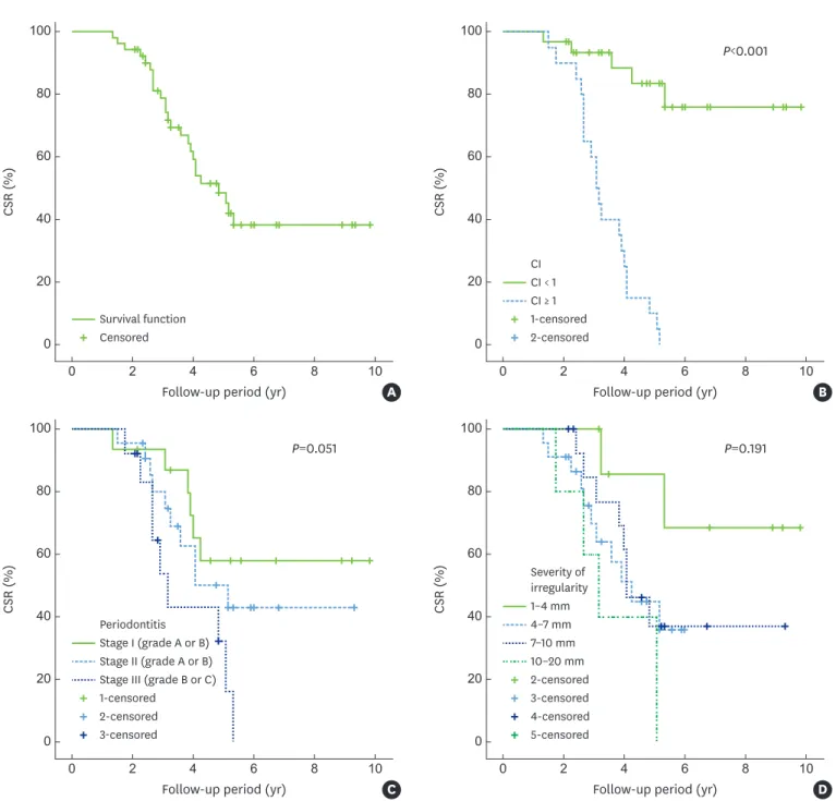
Failure of fixed retainers occurred in 48.08% (n=25) of patients (Table 1). Figure 5 shows Kaplan-Meier survival probability plots. The median survival time of the fixed retainers was 4.83 years. The median survival time of the group with a CI ≥1 at the 12-month follow-up after bonding of fixed retainers was 3.08 years. The CSR of the retainers with a CI <1 at the 12-month follow-up after bonding of fixed retainers was significantly higher than that of the retainers with a CI ≥1 at the 12-month follow-up after bonding of fixed retainers (log-rank test; P<0.001). The median survival time of retainers in patients with stage III (grade B or C) periodontitis (3.17 years) was shorter than that of patients with stage I (grade A or B) periodontitis (9.83 years) (log-rank test; P=0.051).
Univariate and multivariate Cox regression models are shown in Table 5. Patients with fixed retainers in the maxilla had a significantly lower multivariate HR for failure of retainers (0.22;
95% confidence interval, 0.08–0.65; P=0.006) compared to patients with fixed retainers in Table 3. Comparison of clinical parameters and BLs according to site
Variable Site Baseline 12-month follow-up
PI Mx (n=19) 1.39±0.62 0.82±0.56
Mn (n=33) 1.83±0.59 0.92±0.51
P value 0.087 0.812
GI Mx (n=19) 0.90±0.61 0.62±0.34
Mn (n=33) 0.83±0.43 0.59±0.23
P value 0.495 0.53
CI Mx (n=19) 0.74±0.39 0.63±0.39
Mn (n=33) 0.92±0.31 0.92±0.38
P value 0.046 0.014
PPD (mm) Mx (n=19) 2.89±0.74 2.58±0.68
Mn (n=33) 2.62±0.69 2.34±0.68
P value 0.389 0.548
BL (mm) Mx (n=19) 3.63±1.45 3.33±1.29
Mn (n=33) 3.64±0.92 3.29±0.62
P value 0.539 0.279
Data are shown as mean±standard deviation.
PI: plaque index, GI: gingival index, CI: calculus index. PPD: probing pocket depth, BL: radiographic bone level, Mx: maxilla, Mn: mandible.
A B
C D
E F
Figure 4. Mandibular fixed retainer. (A) A clinical photograph before periodontal and orthodontic treatment. (B) A periapical radiograph when the mandibular right central incisor was avulsed with trauma. (C) The mandibular right central incisor, which had been avulsed with trauma, was splinted to the adjacent teeth using adhesive resin and wire. (D) A periapical radiograph before periodontal and orthodontic treatment. (E) Calculus deposition (white arrows) and partially detached fixed retainer (a black arrow) are shown on the lingual side of the mandibular anterior teeth. (F) A periapical radiograph at the 12-month follow-up after bonding of fixed retainers.
Table 4. Comparison of clinical parameters and BLs according to sex
Variable Sex Baseline 12-month follow-up
PI Male (n=21) 1.83±0.75 1.02±0.66
Female (n=31) 1.56±0.53 0.79±0.39
P value 0.251 0.238
GI Male (n=21) 0.76±0.33 0.56±0.19
Female (n=31) 0.91±0.58 0.63±0.31
P value 0.779 0.822
CI Male (n=21) 0.82±0.42 0.74±0.42
Female (n=31) 0.88±0.29 0.87±0.39
P value 0.415 0.326
PPD (mm) Male (n=21) 3.01±0.75 2.57±0.71
Female (n=31) 2.52±0.63 2.33±0.66
P value 0.038 0.318
BL (mm) Male (n=21) 3.93±1.39 3.51±1.08
Female (n=31) 3.44±0.87 3.17±0.77
P value 0.211 0.259
Data are shown as mean±standard deviation.
PI: plaque index, GI: gingival index, CI: calculus index, PPD: probing pocket depth, BL: radiographic bone level.
the mandible. The multivariate HR for failure of retainers with a CI ≥1 at the 12-month follow- up after bonding of fixed retainers was 20.34 (95% confidence interval, 5.63–73.52; P<0.001).
Patients with stage III (grade B or C) periodontitis had a higher multivariate HR for retainer failure (5.4; 95% confidence interval, 1.22–23.91; P=0.026) compared to patients with stage I (grade A or B) periodontitis.
A 0
2
0 4 6 8 10
20 60 100
CSR (%) 40
80
Follow-up period (yr) Survival function
Censored
B 0
2
0 4 6 8 10
20 60 100
CSR (%) 40
80
Follow-up period (yr) 1-censored
2-censored CI < 1 CI ≥ 1
P<0.001
C 0
2
0 4 6 8 10
20 60 100
CSR (%) 40
80
Follow-up period (yr) D
0
2
0 4 6 8 10
20 60 100
CSR (%) 40
80
Follow-up period (yr) 2-censored
3-censored 7–10 mm 10–20 mm 1–4 mm 4–7 mm
4-censored 5-censored Stage III (grade B or C)
Stage II (grade A or B) Stage I (grade A or B)
1-censored 2-censored 3-censored
P=0.191 P=0.051
Severity of irregularity Periodontitis
CI
Figure 5. Kaplan-Meier survival curves. (A) The overall median survival time of the fixed retainers was 4.83 years. (B) The CSR of the fixed retainers in patients who had a calculus index <1 at the 12-month follow-up after bonding of fixed retainers was significantly higher than that of fixed retainers in patients who had a calculus index ≥1 at the 12-month follow-up after bonding of fixed retainers (log-rank test; P<0.001). (C) The median survival time of fixed retainers in patients with stage I (grade A or B) periodontitis was 9.83 years. Patients with stage III (grade B or C) periodontitis had shorter median survival time of fixed retainers (3.17 years) (log-rank test; P=0.051). (D) The median survival time of fixed retainers in patients with severe irregularities (10–20 mm according to Little's irregularity index) was 3.17 years.
CSR: cumulative survival rate, CI: calculus index.
DISCUSSION
This study was conducted to investigate clinical factors affecting the longevity of fixed retainers and the influence of fixed retainers on periodontal health in periodontitis patients.
The overall CSR of the fixed retainers in patients who had a CI <1 at the 12-month follow-up after bonding of fixed retainers was higher than in patients who had a CI ≥1 at the 12-month follow-up after bonding of fixed retainers. Patients who had a CI ≥1 at the 12-month follow-up after bonding of fixed retainers exhibited a significantly higher multivariate HR for retainer failure. In addition, patients with stage III (grade B or C) periodontitis had a higher multivariate HR for retainer failure compared to patients with stage I (grade A or B) periodontitis.
Retention after orthodontic treatment is challenging and especially critical during the first 8 months post-treatment, when remodeling of the supra-alveolar fibers occurs [2]. Sharpe et al. [24] reported that orthodontic patients with previously treated severe periodontal disease or crestal bone loss had an increased risk of deterioration of tooth alignment after orthodontic treatment. Currently, invisible retainers are generally used and lifelong retention is prescribed instead of retention for a limited time [25]. Therefore, we used flexible
multistranded wire retainers for retention in this study.
Many studies have demonstrated the influence of orthodontic retainers on periodontal tissue [11,12,26-32]. Zachrisson [11] emphasized the importance of appropriate oral hygiene for periodontal health in patients who had fixed retainers. Fixed lingual retainers may lead to plaque accumulation and calculus deposition on teeth because of the difficulty in practicing proper oral hygiene [32]. However, Artun et al. [27] reported that the accumulation of plaque and calculus around fixed retainers did not seem to cause apparent damage to the adjacent hard and soft tissues. They also noted that there were no signs of carious white spot lesions and suggested that the accessibility for the free flow of saliva may be a major factor precluding enamel decalcification [27]. Heier et al. [12] reported that slightly more plaque and calculus accumulation were observed on the lingual surfaces in the fixed retainer group. However, they suggested that periodontal health should not be compromised by the Table 5. Cox proportional HRs of retainer failure according to sex, site, calculus index, stage (grade) of periodontitis, and severity of the irregularity
Variable Univariate HR 95% confidence interval P value Multivariate HR 95% confidence interval P value Sex
Male 1.42 0.63–3.18 0.401 3.39 1.72–10.75 0.038
Female 1 1
Site
Maxilla 0.89 0.39–2.01 0.775 0.22 0.08–0.65 0.006
Mandible 1 1
CI (at 12-month follow-up)
CI <1 1 1
CI ≥1 13.36 4.40–40.52 <0.001 20.34 5.63–73.52 <0.001
Periodontitis
Stage I, grade A or B 1 0.067 1 0.058
Stage II, grade A or B 1.49 0.54–4.12 0.436 0.89 0.29–2.80 0.848
Stage III, grade B or C 3.25 1.14–9.24 0.027 5.4 1.22–23.91 0.026
Severity of the irregularity
1 mm≤ LII <4 mm 1 0.244 1 0.144
4 mm≤ LII <7 mm 3.52 0.77–16.05 0.103 1.58 0.29–8.39 0.59
7 mm≤ LII <10 mm 2.95 0.62–13.98 0.174 0.57 0.10–3.22 0.522
10 mm≤ LII <20 mm 5.8 1.04–32.39 0.045 0.27 0.03–2.58 0.252
HR: hazard ratio, CI: calculus index, LII: Little's irregularity index.
presence of bonded lingual wires if professional plaque and calculus removal accompanied by a session on motivation and oral hygiene instruction is repeated every 6 months. Storey et al. [30] also showed that there were no clinically significant changes in periodontal health after 12 months of retention using bonded fixed retainers. In the present study, there were no significant differences in PI and GI according to sex and sites. This finding is similar to that of a previous study, which reported 20-year outcomes of fixed retainers and found that the mean GI in the upper and lower anterior teeth was 0.50 and 0.58, respectively [31]. At the 12-month follow-up after bonding of fixed retainers, even though the fixed retainers were bonded, clinical parameters including PI, GI, and PPD improved in the present study.
Eroglu et al. [32] reported that oral hygiene improved after orthodontic treatment with fixed appliances regardless of retainer type. They also showed that fixed and removable orthodontic retainers did not differ in terms of salivary Streptococcus mutans and Lactobacillus casei levels and periodontal status.
However, the current study showed that the CI of the lower anterior teeth was significantly greater than that of the upper anterior teeth after the bonding of fixed retainers. During the follow-up period after orthodontic treatment, general dentists and dental hygienists found it difficult to clean around fixed retainers, contributing to the risk of developing periodontal issues. Moreover, the lingual surfaces of mandibular anterior teeth are one of the most common locations for the development of supragingival calculus [33]. Therefore, the meticulous removal of plaque and calculus sometimes results in the breakage of fixed retainers. Juloski et al. [34] also reported that the long-term presence of fixed lingual retainers increased the accumulation of calculus. They also emphasized that professional plaque and calculus removal and good oral hygiene could improve periodontal health.
Multistranded bonded orthodontic retainers have been reported to exhibit risks of failure ranging from 12% [28] to 46.4% [35]. The present study showed that the CSR of fixed retainers in patients who had a CI <1 at the 12-month follow-up after bonding of fixed retainers was significantly higher than that of fixed retainers in patients who had a CI ≥1 at the 12-month follow-up after bonding of fixed retainers (P<0.001). Pandis et al. [35]
compared the survival rates of mandibular lingual retainers with either chemically cured or light-cured adhesives after orthodontic treatment. They concluded that there was no significant difference between the 2 groups. Gökçe and Kaya [36] reported that the periodontal outcomes and survival rates of mandibular fixed retainers were not affected by bonding technique or wire thickness. They also showed that the survival rates were 85%
for 0.0215-inch and 90% for 0.0175-inch retainer wires at the 6-month follow-up visit. The survival rate of fixed retainers in their study was higher than in the present study. It seems that the mean follow-up period of the present study (5.32±2.22 years) was longer than the follow-up in their study. Scheibe and Ruf [37] suggested that retainer failures were related to operator experience and that operators with less experience may have a higher failure rate of retainers. In the present study, all the fixed retainers were bonded with light-cured adhesive by 1 experienced operator, resulting in no bias based on operator experience.
Salehi et al. [22] showed that the most frequent type of failure in multi-stranded retainers was retainer loosening, which was observed in 84.48% of cases in the maxilla and 96.42% of cases in the mandible, even though there were no significant differences between the survival times of the maxillary and mandibular retainers. However, Tacken et al. [28] reported that failure of retainers occurred more frequently in the maxilla than in the mandible. In addition, Radlanski and Zain [38] explained that the frequent failure of maxillary retainers is due to
the occlusal contacts of opposing teeth in the area of the bonded composite of the retainers.
Bearn also suggested that this may reflect the role of occlusal factors [8]. In contrast, the present study showed that the multivariate HR of the maxilla for retainer failure was 0.22 (95% confidence interval, 0.08–0.65; P=0.006) after adjustment for confounding factors.
Moreover, patients with a CI ≥1 at the 12-month follow-up after bonding of fixed retainers had a higher HR for the failure of fixed retainers. It is suggested that more force may be applied to the retainer wire and bonded resin during the meticulous removal of calculus and stain around the fixed retainers and teeth. This may result in the detachment of the bonding sites, with the retainer sometimes remaining in situ. During professional tooth cleaning, the operator can find detached retainers that were unrecognizable to the patient. In this study, we included periodontitis patients who were compliant with the supportive periodontal treatment after periodontal and orthodontic treatment. If patients had not been followed up regularly, the breakage of the fixed retainers could not have been detected and might have resulted in relapse of the corrected alignment of the teeth. Shaughnessy et al. [39] reported that a fixed retainer made with dead soft wire was the least likely to create torque problems, but was the most likely to break, and that a fixed retainer made with a flexible twist wire can produce inadvertent tooth movement. They also emphasized the need for regular observation of patients wearing fixed retainers because patients might not notice partial debonding.
No significant differences were found in the multivariate HRs for the failure of fixed retainers according to the severity of the irregularity. However, patients with stage III (grade B or C) periodontitis had a higher multivariate HR for failure of retainers (5.4; 95% confidence interval, 1.22–23.91; P=0.026) compared to patients with stage I (grade A or B) periodontitis.
Artun and Urbye [40] reported that 10 failures of fixed retainers were recorded in 9 of 19 patients with advanced loss of marginal periodontium after periodontal and orthodontic treatment. They also suggested that relapse within the retained segment was associated with failures of the retainer.
There are several limitations in this study. One is the inherent bias of retrospective studies.
We included patients who were compliant with the supportive periodontal treatment.
Therefore, clinical parameters including PI, GI, and PPD improved even though the fixed retainers had been bonded to the palatal and lingual sides of the upper and lower anterior teeth. Second, we did not evaluate the formation of caries after the bonding of fixed retainers. Nonetheless, this study has several strengths. First, previous studies investigating the influence of fixed retainers on periodontal health mostly included young patients without periodontitis. On the contrary, the present study included patients who already had periodontitis. Therefore, the results of this study on the influence of fixed retainers on periodontal health are particularly meaningful. Second, all periodontal, orthodontic, and supportive periodontal treatment was performed by 1 periodontist with the same protocol for oral hygiene instructions. Third, the long-term survival rate of fixed retainers could be evaluated because all the patients who were included in this study were compliant with the supportive periodontal treatment.
Although the fixed retainers were bonded to the palatal or lingual sides of the upper or lower anterior teeth, clinical parameters including PI, GI, and PPD improved after periodontal and orthodontic treatment as long as repeated oral hygiene education and professional tooth cleaning were provided. At the 12-month follow-up after bonding of fixed retainers, the CI of the lower anterior teeth was significantly higher than that of the upper anterior teeth. Moreover, the failure of fixed retainers occurred more frequently in patients who had
stage III (grade B or C) periodontitis or a CI ≥1 at the 12-month follow-up after bonding of fixed retainers. However, the results of this study should be interpreted with caution. Future studies directly comparing fixed retainers in periodontitis patients with those in healthy patients with a larger sample size are needed.
ACKNOWLEDGEMENTS
We would like to thank Prof. En-Woo Nam of the Biostatistical Consulting and Research Lab, Hanyang University for assistance with statistical analysis and Seong-Jin Ahn of Hanyang Medical Center for radiographic assistance.
REFERENCES
1. Hirschfeld J, Reichardt E, Sharma P, Hilber A, Meyer-Marcotty P, Stellzig-Eisenhauer A, et al. Interest in orthodontic tooth alignment in adult patients affected by periodontitis: a questionnaire-based cross- sectional pilot study. J Periodontol 2019;90:957-65.
PUBMED | CROSSREF
2. Reitan K. Clinical and histologic observations on tooth movement during and after orthodontic treatment. Am J Orthod 1967;53:721-45.
PUBMED | CROSSREF
3. Johnston CD, Littlewood SJ. Retention in orthodontics. Br Dent J 2015;218:119-22.
PUBMED | CROSSREF
4. Horowitz SL, Hixon EH. Physiologic recovery following orthodontic treatment. Am J Orthod 1969;55:1-4.
PUBMED | CROSSREF
5. Littlewood SJ, Kandasamy S, Huang G. Retention and relapse in clinical practice. Aust Dent J 2017;62 Suppl 1:51-7.
PUBMED | CROSSREF
6. Little RM, Riedel RA, Artun J. An evaluation of changes in mandibular anterior alignment from 10 to 20 years postretention. Am J Orthod Dentofacial Orthop 1988;93:423-8.
PUBMED | CROSSREF
7. Arn ML, Dritsas K, Pandis N, Kloukos D. The effects of fixed orthodontic retainers on periodontal health:
a systematic review. Am J Orthod Dentofacial Orthop 2020;157:156-164.e17.
PUBMED | CROSSREF
8. Bearn DR. Bonded orthodontic retainers: a review. Am J Orthod Dentofacial Orthop 1995;108:207-13.
PUBMED | CROSSREF
9. Iliadi A, Kloukos D, Gkantidis N, Katsaros C, Pandis N. Failure of fixed orthodontic retainers: a systematic review. J Dent 2015;43:876-96.
PUBMED | CROSSREF
10. Wouters C, Lamberts TA, Kuijpers-Jagtman AM, Renkema AM. Development of a clinical practice guideline for orthodontic retention. Orthod Craniofac Res 2019;22:69-80.
PUBMED | CROSSREF
11. Zachrisson BU. Clinical experience with direct-bonded orthodontic retainers. Am J Orthod 1977;71:440-8.
PUBMED | CROSSREF
12. Heier EE, De Smit AA, Wijgaerts IA, Adriaens PA. Periodontal implications of bonded versus removable retainers. Am J Orthod Dentofacial Orthop 1997;112:607-16.
PUBMED | CROSSREF
13. Papapanou PN, Sanz M, Buduneli N, Dietrich T, Feres M, Fine DH, et al. Periodontitis: consensus report of workgroup 2 of the 2017 world workshop on the classification of periodontal and peri-implant diseases and conditions. J Periodontol 2018;89 Suppl 1:S173-82.
PUBMED | CROSSREF
14. Park SH, Cho SH, Han JY. Effective professional intraoral tooth brushing instruction using the modified plaque score: a randomized clinical trial. J Periodontal Implant Sci 2018;48:22-33.
PUBMED | CROSSREF
15. Han JY. A comparative study of combined periodontal and orthodontic treatment with fixed appliances and clear aligners in patients with periodontitis. J Periodontal Implant Sci 2015;45:193-204.
PUBMED | CROSSREF
16. Silness J, Löe H. Periodontal disease in pregnancy. ii. correlation between oral hygiene and periodontal condition. Acta Odontol Scand 1964;22:121-35.
PUBMED | CROSSREF
17. Löe H, Silness J. Periodontal disease in pregnancy. I. Prevalence and severity. Acta Odontol Scand 1963;21:533-51.
PUBMED | CROSSREF
18. Greene JC, Vermillion JR. The simplified oral hygiene index. J Am Dent Assoc 1964;68:7-13.
PUBMED | CROSSREF
19. Little RM. The irregularity index: a quantitative score of mandibular anterior alignment. Am J Orthod 1975;68:554-63.
PUBMED | CROSSREF
20. Huh KH, Lee SS, Jeon IS, Yi WJ, Heo MS, Choi SC. Quantitative analysis of errors in alveolar crest level caused by discrepant projection geometry in digital subtraction radiography: an in vivo study. Oral Surg Oral Med Oral Pathol Oral Radiol Endod 2005;100:750-5.
PUBMED | CROSSREF
21. Ardeshna AP. Clinical evaluation of fiber-reinforced-plastic bonded orthodontic retainers. Am J Orthod Dentofacial Orthop 2011;139:761-7.
PUBMED | CROSSREF
22. Salehi P, Zarif Najafi H, Roeinpeikar SM. Comparison of survival time between two types of orthodontic fixed retainer: a prospective randomized clinical trial. Prog Orthod 2013;14:25.
PUBMED | CROSSREF
23. Labunet AV, Badea M. In vivo orthodontic retainer survival - a review. Clujul Med 2015;88:298-303.
PUBMED
24. Sharpe W, Reed B, Subtelny JD, Polson A. Orthodontic relapse, apical root resorption, and crestal alveolar bone levels. Am J Orthod Dentofacial Orthop 1987;91:252-8.
PUBMED | CROSSREF
25. Padmos JAD, Fudalej PS, Renkema AM. Epidemiologic study of orthodontic retention procedures. Am J Orthod Dentofacial Orthop 2018;153:496-504.
PUBMED | CROSSREF
26. Artun J. Caries and periodontal reactions associated with long-term use of different types of bonded lingual retainers. Am J Orthod 1984;86:112-8.
PUBMED | CROSSREF
27. Artun J, Spadafora AT, Shapiro PA. A 3-year follow-up study of various types of orthodontic canine-to- canine retainers. Eur J Orthod 1997;19:501-9.
PUBMED | CROSSREF
28. Tacken MP, Cosyn J, De Wilde P, Aerts J, Govaerts E, Vannet BV. Glass fibre reinforced versus multistranded bonded orthodontic retainers: a 2 year prospective multi-centre study. Eur J Orthod 2010;32:117-23.
PUBMED | CROSSREF
29. Bazargani F, Jacobson S, Lennartsson B. A comparative evaluation of lingual retainer failure bonded with or without liquid resin. Angle Orthod 2012;82:84-7.
PUBMED | CROSSREF
30. Storey M, Forde K, Littlewood SJ, Scott P, Luther F, Kang J. Bonded versus vacuum-formed retainers:
a randomized controlled trial. Part 2: periodontal health outcomes after 12 months. Eur J Orthod 2018;40:399-408.
PUBMED | CROSSREF
31. Booth FA, Edelman JM, Proffit WR. Twenty-year follow-up of patients with permanently bonded mandibular canine-to-canine retainers. Am J Orthod Dentofacial Orthop 2008;133:70-6.
PUBMED | CROSSREF
32. Eroglu AK, Baka ZM, Arslan U. Comparative evaluation of salivary microbial levels and periodontal status of patients wearing fixed and removable orthodontic retainers. Am J Orthod Dentofacial Orthop 2019;156:186-92.
PUBMED | CROSSREF
33. Corbett TL, Dawes C. A comparison of the site-specificity of supragingival and subgingival calculus deposition. J Periodontol 1998;69:1-8.
PUBMED | CROSSREF
34. Juloski J, Glisic B, Vandevska-Radunovic V. Long-term influence of fixed lingual retainers on the
development of gingival recession: a retrospective, longitudinal cohort study. Angle Orthod 2017;87:658-64.
PUBMED | CROSSREF
35. Pandis N, Fleming PS, Kloukos D, Polychronopoulou A, Katsaros C, Eliades T. Survival of bonded lingual retainers with chemical or photo polymerization over a 2-year period: a single-center, randomized controlled clinical trial. Am J Orthod Dentofacial Orthop 2013;144:169-75.
PUBMED | CROSSREF
36. Gökçe B, Kaya B. Periodontal effects and survival rates of different mandibular retainers: comparison of bonding technique and wire thickness. Eur J Orthod 2019;41:591-600.
PUBMED | CROSSREF
37. Scheibe K, Ruf S. Lower bonded retainers: survival and failure rates particularly considering operator experience. J Orofac Orthop 2010;71:300-7.
PUBMED | CROSSREF
38. Radlanski RJ, Zain ND. Stability of the bonded lingual wire retainer-a study of the initial bond strength. J Orofac Orthop 2004;65:321-35.
PUBMED | CROSSREF
39. Shaughnessy TG, Proffit WR, Samara SA. Inadvertent tooth movement with fixed lingual retainers. Am J Orthod Dentofacial Orthop 2016;149:277-86.
PUBMED | CROSSREF
40. Artun J, Urbye KS. The effect of orthodontic treatment on periodontal bone support in patients with advanced loss of marginal periodontium. Am J Orthod Dentofacial Orthop 1988;93:143-8.
PUBMED | CROSSREF