http://dx.doi.org/10.4174/astr.2014.86.3.115 Annals of Surgical Treatment and Research
The pattern and significance of the calcifications of papillary thyroid microcarcinoma presented in
preoperative neck ultrasonography
Eun Mee Oh, Yoo Seung Chung, Won Jong Song, Young Don Lee
Department of Thyroid and Endocrine Surgery, Gachon University Gil Medical Center, Incheon, Korea
INTRODUCTION
The prevalence of papillary thyroid microcarcinoma (PTMC) has increased recently, due in part to the increased use of neck ultrasonography (NUS) and NUS-guided fine needle aspiration cytology [1]. NUS features that suggest malignancy in a thyroid nodule include microcalcifications, the absence of “halo” sign, marked hypoechogenicity, extrathyroidal extension, an irregular or microlobulated margin, and a heterogeneous echo structure
[2]. Calcifications on thyroid ultrasonography can be classified as micro- or macrocalcifications, and microcalcification considered to be the most specific sonographic indicator in the diagnosis of papillary thyroid carcinoma (PTC) [3].
PTCs are the most common type of thyroid cancer, often forming concentric calcified foci, called psammoma bodies, which are strongly diagnostic for PTC [4]. On NUS, psammoma bodies appear as fine, scattered, and punctate bright echoes, indicative of microcalcifications. Other types of calcification,
Received July 24, 2013, Revised December 24, 2013, Accepted December 30, 2013
Corresponding Author: Young Don Lee
Department of Thyroid and Endocrine Surgery, Gachon University Gil Medical Center, 21 Namdong-daero 774beon-gil, Namdong-gu, Incheon 405-760, Korea
Tel: +82-32-460-8419, Fax: +82-32-461-3214 E-mail: peacemk@gilhospital.com
Copyright ⓒ 2014, the Korean Surgical Society
cc Annals of Surgical Treatment and Research is an Open Access Journal. All articles are distributed under the terms of the Creative Commons Attribution Non- Commercial License (http://creativecommons.org/licenses/by-nc/3.0/) which permits unrestricted non-commercial use, distribution, and reproduction in any medium, provided the original work is properly cited.
Purpose: To analyze the incidence and patterns of calcification of papillary thyroid microcarcinoma (PTMC) on neck ultrasonography (NUS) and assess the clinical implications of calcification, especially for neck node metastasis.
Methods: The clinical data of 379 patients with PTMC who underwent thyroidectomy between January and December 2011 were retrospectively analyzed. PTMC lesions were classified into four subgroups according to their calcification patterns on preoperative NUS: microcalcification, macrocalcification, rim calcification, and noncalcification. The clinicopathologic characteristics were compared between the patients with and without calcification, and among the four subgroups.
Results: Calcifications were detected on NUS in 203 patients (53.5%) and central neck node metastasis was observed in 119 patients (31.3%). Calcification was associated with larger tumor size (0.68 cm vs. 0.54 cm), higher rate of lymph node metastasis (38.6% vs. 23.2%) and higher lymph node ratio (0.11 vs. 0.06) compared to noncalcification (All P < 0.05). In addition, the extent of calcification correlated with lesion size (0.67 cm vs. 0.69 cm vs. 0.85 cm). Further, the likelihood of lymph node metastasis also correlated with the extent of calcification in the order of non-, micro- and macrocalcification (23.3%, 36.8%, and 44.1%, respectively). The calcification rate was higher in patients with lymph node metastasis than those without it (65.5% vs.47.7%) (All P < 0.05).
Conclusion: PTMC patients positive for calcification on NUS had a higher rate of lymph node metastasis, and a higher lymph node ratio compared to noncalcification patients. Calcification patterns should be assessed carefully in patients with PTMC by preoperative NUS.
[Ann Surg Treat Res 2014;86(3):115-121]
Key Words: Papillary thyroid microcarcinoma, Calcification, Ultrasonography
including coarse, macro, eggshell and rim calcifications, were formerly thought to be more common in benign thyroid tumors than in malignant thyroid tumors. However, these patterns have also been observed in malignant lesions, although microcalcifications remain the most frequent type in thyroid malignancies [5].
The mechanism of formation of microcalcifications in PTC was thought to involve a poor blood supply to the nipple, leading to calcification necrosis [6]. More recently, however, osteopontin (OPN) became known as the cause of formation of microcalcifications [7]. Moreover, OPN expression was found to be significantly related to lymph node metastasis [8,9].
Thus, to date, the molecular mechanism responsible for calcification in PTC has not been determined, nor has the clinical significance of calcification in thyroid malignancy, including PTMCs. This study was therefore designed to analyze the incidence and patterns of PTMC calcification on NUS and to assess the clinical implications of PTMC calcification.
METHODS
The NUS findings and clinical data of 379 patients with PTMC who underwent thyroidectomy between January and December 2011 were retrospectively reviewed. All preoperative NUS examinations were performed by one endocrine surgeon and several thyroid radiologists at Gachon University Gil Medical Center. Electronic patient records were reviewed, and NUS findings were reviewed and analyzed in detail by a single endocrine surgeon, who paid close attention to the presence and patterns of calcifications.
PTMC calcifications were classified by size and pattern as, namely, microcalcification (defined as punctate echogenic foci ≤1 mm with or without posterior shadowing), macro
calcifications (defined as punctate echogenic foci >1 mm in size), or rim calcifications (defined as nodules with peripheral curvilinear or eggshell calcifications) [10]. Of the 379 patients, 136 (35.9%) had microcalcifications, 59 (15.6%)
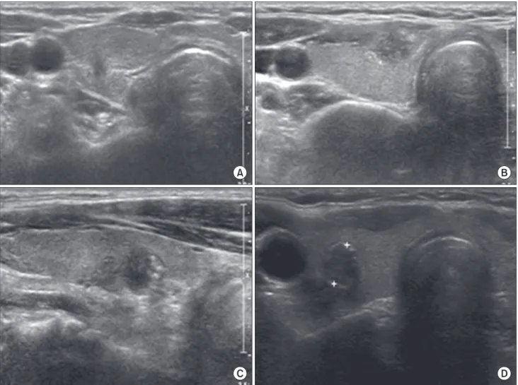
Fig. 1. Papillary thyroid microcarcinoma calcification patterns on neck neck ultrasonography. (A) Noncalcification: taller than wide, illdefined hypoechoic nodule. (B) Microcalcification: several calcifications measuring less than 1 mm. (C) Macrocalcification: one calcification larger than 1 mm. (D) Rim calcification: ringshaped calcification on the periphery of the nodule.
had macrocalcification, 8 (2.1%) had rim calcifications, and remaining 176 patients (46.4%) did not have any calcifications (Fig. 1).
A total thyroidectomy with bilateral central neck compar- tment dissection was performed on 198 of the 379 patients, and 181 underwent unilateral lobectomy including the isthmus with unilateral central neck node dissection. Moreover, 56 patients (14.7%) who had suspicious lymph node enlargement on NUS underwent therapeutic central lymph node dissection (CND), with the remaining 323 (85.2%) undergoing prophylactic CND.
The study protocol was approved by our Institutional Review Board, which waived the requirement for informed consent due to the retrospective nature of this study.
The chi-square and independent t-tests were used to com- pare clinicopathologic data between the 203 patients with calcification and the 176 without calcification. Data in the four patient subgroups were compared using the analysis of variance and Kruskal-Wallis H test. All statistical analyses were performed using the IBM SPSS ver. 19.0 (IBM Co., Armonk, NY, USA). A P-value of <0.05 was considered statistically significant.
RESULTS
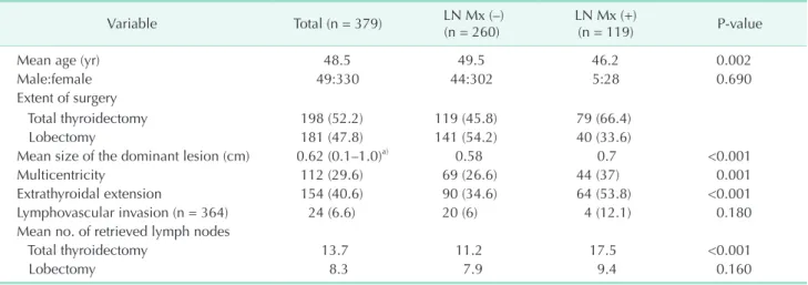
The mean age of the 379 patients was 48.5 years (range, 20- 77 years), and the male to female ratio was 1:7 (49:330). One hundred nineteen patients (31.3%) had neck node metastasis, including 112 (94.1%) with central and 7 (5.9%) with central and lateral neck node metastasis. Patients with lymph node metastasis (LN+) were younger than those without lymph node metastasis (LN-). Lesion size (0.7 cm vs. 0.58 cm, P <
0.001), frequency of extrathyroidal extension (53.8% vs. 34.6%,
P < 0.001) and multicentricity (37% vs. 26.6%, P = 0.001) were significantly higher in the LN+ than in the LN- group. In addition, the average number of retrieved central neck lymph nodes was significantly higher in LN+ than in LN- patients who underwent total thyroidectomy (17.5 vs. 11.2, P < 0.001), but not in patients who underwent lobectomy (Table 1).
Among the 119 patients with lymph node metastasis, 112 (94.1%) had only central neck node metastasis, whereas 7 (5.9%) had both lateral and central neck node metastasis. The mean number of metastatic nodes was 2.4 in patients who underwent lobectomy and 3.7 in patients who underwent total thyroidectomy (Table 2).
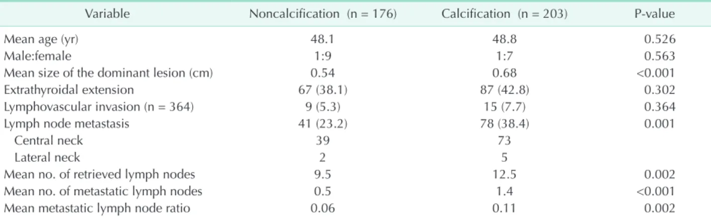
Calcifications on preoperative NUS were detected in 203 of the 379 patients (53.5%). The mean size of the dominant lesion was slightly larger in patients with calcification than without calcification (0.68 cm vs. 0.54 cm, P < 0.001). The percentage of
Table 2. Clinicopathologic characteristics of the 119 papil
lary thyroid microcarcinoma patients with cervical lymph node metastasis
Variable Value
Extent of surgery
Total thyroidectomy 79 (66.4)
Lobectomy 40 (33.6)
Lymph node metastasis
Central neck 112 (94.1)
Central neck and lateral neck 7 (5.9) Mean no. of metastatic lymph nodes
Total thyroidectomy 3.7
Lobectomy 2.4
Values are presented as number (%) unless otherwise indicated.
Table 1. Clinicopathologic characteristics of papillary thyroid microcarcinoma patients with and without lymph node metastasis
Variable Total (n = 379) LN Mx (–)
(n = 260) LN Mx (+)
(n = 119) Pvalue
Mean age (yr) 48.5 49.5 46.2 0.002
Male:female 49:330 44:302 5:28 0.690
Extent of surgery
Total thyroidectomy 198 (52.2) 119 (45.8) 79 (66.4)
Lobectomy 181 (47.8) 141 (54.2) 40 (33.6)
Mean size of the dominant lesion (cm) 0.62 (0.1–1.0)a) 0.58 0.7 <0.001
Multicentricity 112 (29.6) 69 (26.6) 44 (37) 0.001
Extrathyroidal extension 154 (40.6) 90 (34.6) 64 (53.8) <0.001
Lymphovascular invasion (n = 364) 24 (6.6) 20 (6) 4 (12.1) 0.180
Mean no. of retrieved lymph nodes
Total thyroidectomy 13.7 11.2 17.5 <0.001
Lobectomy 8.3 7.9 9.4 0.160
Values are presented as number (%) unless otherwise indicated.
LN, lymph node; Mx, metastasis.
a)Range.
patients with lymph node metastasis (38.4% vs. 23.2%, P = 0.001), the mean number of retrieved nodes (12.5 vs. 9.5, P = 0.002), the mean number of metastatic nodes (1.4 vs. 0.5, P
< 0.001), and the lymph node ratio (0.11 vs. 0.06, P = 0.002) were significantly higher in patients with calcification than without calcification (Table 3). Further analysis of the patients with calcification showed a correlation between older age and larger calcification size. Mean lesion size (0.67 cm vs. 0.69 cm vs. 0.85 cm, P < 0.001) increased significantly with increasing degree of calcification, in the order of microcalcification, macro- calcification and rim calcification. The rate of lymph node metastasis (23.3% vs. 36.8% vs. 44.1%, P = 0.013) increased significantly with increasing extent of calcification, in the order of non-calcification, micro-, macrocalcification except
rim calcification (25%) (Table 4). The overall calcification rate was significantly higher in patients with node metastasis than without node metastasis (65.5% vs. 47.7% P = 0.001).
The analysis of patients with node metastasis showed that the number of retrieved neck nodes was significantly higher in the group with calcification than without calcification (15.9 vs. 12.7, P = 0.001), but there were no differences in the mean number of metastatic lymph nodes and lymph node ratio among the 4 groups (Table 5).
DISCUSSION
Microcalcification on NUS is the most specific indicator in the sonographic diagnosis of PTC [3]. The incidence of calcification
Table 5. Relationship between lymph node status and calcification patterns in the 119 papillary thyroid microcarcinoma patients with lymph node metastasis
Variable Noncalcification
(n = 41)
Calcification (n = 78)
Pvalue All Micro (n = 50) Macro (n = 26) Rim (n = 2)
Mean no. of retrieved lymph nodes 12.7 15.9 16.1 15.7 13.0 0.001
Mean no. of metastatic lymph nodes 2.2 3.8 3.9 3.5 3.0 0.119
Mean metastatic lymph node ratio 0.25 0.30 0.31 0.29 0.24 0.194
Table 3. Clinicopathologic characteristics of papillary thyroid microcarcinoma patients with and without calcification
Variable Noncalcification (n = 176) Calcification (n = 203) Pvalue
Mean age (yr) 48.1 48.8 0.526
Male:female 1:9 1:7 0.563
Mean size of the dominant lesion (cm) 0.54 0.68 <0.001
Extrathyroidal extension 67 (38.1) 87 (42.8) 0.302
Lymphovascular invasion (n = 364) 9 (5.3) 15 (7.7) 0.364
Lymph node metastasis 41 (23.2) 78 (38.4) 0.001
Central neck 39 73
Lateral neck 2 5
Mean no. of retrieved lymph nodes 9.5 12.5 0.002
Mean no. of metastatic lymph nodes 0.5 1.4 <0.001
Mean metastatic lymph node ratio 0.06 0.11 0.002
Values are presented as number (%) unless otherwise indicated.
Table 4. Clinicopathologic characteristics of patients classified by calcification pattern (n = 379)
Variable Noncalcification
(n = 176)
Calcifications (n = 203)
Pvalue Micro (n = 136) Macro (n = 59) Rim (n = 8)
Mean age (yr) 48.1 47.9 49.8 56.8 0.038
Male:female 1:9 1:6 1:6 1:3 0.747
Mean size of the dominant lesion (cm) 0.54 0.67 0.69 0.85 <0.001
Extrathyroidal extension 67 (38.1) 56 (41.2) 29 (49.2) 2 (25.0) 0.379
Lymphovascular invasion (n = 364) 9 (5.4) 8 (6.2) 7 (12.1) 0 (0) 0.279
Lymph node metastasis 41 (23.3) 50 (36.8) 26 (44.1) 2 (25.0) 0.008
Values are presented as number (%) unless otherwise indicated.
in thyroid malignancy has been reported in several studies. One study found that 62% of patients with microcalcifications had malignant lesions, whereas 38% were diagnosed with benign thyroid pathology [5]. Another study reported that calcifications were present in 58.1% of thyroid malignancies, with 44.2%
having microcalcifications, 9.7% having macrocalcifications and 4.2% having rim calcifications [10]. A further study reported that the presence of microcalcifications in a predominantly solid nodule increased a patient's cancer risk about 3-fold [11].
Microcalcifications in thyroid lesions have been shown to have a high predictive value (42%–94%), but a low sensitivity rate (26%–59%) in the diagnosis of malignancy [3,12-14].
Most previous studies regarding calcifications in thyroid cancer were limited to nodules larger than 1 cm. A study com- paring the sensitivity and specificity of microcalcification in large and small nodules, with a cutoff size of 1 cm, discovered that microcalcification was more diagnostic for thyroid cancer in nodules larger than 1 cm (51.4% sensitivity and 91.6% speci- ficity) than in nodules smaller than 1 cm (36.6% sensitivity and 87.9% specificity) [10]. These findings suggested that the frequency of microcalcification was lower in PTMCs than in PTCs and furthermore, microcalcification was not a major predictor of malignancy in nodules ≤1 cm [10]. Therefore, we attempted to find not only the incidence and the pattern of calcification, but also clinical significance of calcification in PTMC through this study.
In our study, in which all patients had PTMC, the incidence of calcification was 53.5% (203/379), whereas the incidence of microcalcification was 35.9% (136/379). Similar results have been reported in other studies. A study showed that 58.3% of patients with PTMC (14/24) had calcified nodules and 20.8%
(5/24) had fine stippled psammomatous calcifications [14]. And another study reported microcalcifications in 68 of 127 (53.5%) patients with PTMC [15].
Other types of calcifications, including macrocalcifications and eggshell or rim calcifications, were thought to be more common in benign lesions than in malignant ones. Benign nodules usually have coarse calcifications, particularly with long disease duration [16]. However, calcification patterns other than microcalcifications have also been observed in malignant lesions. For example, one study reported that macrocalcifications were equally distributed between benign and malignant lesions, with 66.7% of patients having both micro- and macrocalcifications diagnosed with cancer [5].
Although a specific type of rim calcification has been reported as indicative of malignancy [17], the relationship between rim calcification and malignancy has not yet been determined [11].
Of our 379 patients with PTMC, 8 (2.1%) had rim calcification in this study.
Histopathologically, thyroid calcifications can be classified as psammoma bodies and dystrophic calcifications, or as
psammoma bodies, stromal calcifications and bone formation [18]. Psammoma bodies are laminated, basophilic, spherical accretions and are characteristic of papillary carcinomas, although they are occasionally observed in benign thyroid lesions [6,19,20].
The mechanism responsible for the formation of calcifications has not yet been fully elucidated. The rapid proliferation of cancer cells may lead to cancer tissue hyperplasia and hyperplasia mixed with necrosis, resulting in the deposit of calcium and calcifications. Therefore, psammoma bodies in PTC are found in association with tumor cells within lymphatic spaces or within the tumor stroma [6,14,21]. Alternatively, psammoma bodies may be formed by intracellular calcifications in viable cells, such as the nidus. Calcifications in PTC may not necessarily develop in nonviable and dying cells [7].
Little is known about the molecular interactions that result in psammoma bodies, stromal calcification or bone formation in the thyroid, except for the involvement of OPN, which is a member of the bone morphogenetic protein family [22]. OPN has been reported with roles related to initiation, progression and transplantation of malignant tumor in breast tumor and PTC [8,23-27]. OPN has not been detected in normal thyroid tissue [9] and further, OPN mRNA-expressing cells have been observed around psammoma bodies [8]. Moreover, OPN upregulation correlates with aggressive clinicopathological features of PTC. Hence, the presence of lymph node metastases and tumor size both positively correlated with OPN positivity [28].
Discovering that OPN expression correlates with both the formation of microcalcifications and adverse prognostic fac- tors in PTC suggests that calcifications in PTMC may also be associated with adverse prognostic factors, including tumor size and lymph node metastasis. The presence of psammoma bodies was found to correlate significantly with gross lymph node metastasis, persistent disease on follow-up examination, higher incidence of pulmonary metastasis and poorer disease- free survival, suggesting that the presence of psammoma bodies may be a useful predictor of outcome in patients with PTC [18,29].
To date, however, few reports have assessed the clinical significance of calcifications in PTC, particularly in PTMC.
Therefore, the present study focused primarily on the significance of calcifications in PTMC, discovering that patients with calcifications have a significantly higher number of metastatic neck nodes (1.4 vs. 0.5, P < 0.001) and a higher lymph node ratio (0.11 vs. 0.06, P = 0.002) compared to patients without calcifications. In addition, an analysis of patients with node metastasis demonstrated that the number of metastatic neck nodes (3.8 vs. 2.2, P = 0.119) and the lymph node ratio (0.3 vs. 0.25, P = 0.194) was higher in patients with calcification than without calcification even though there was no statistical
REFERENCES
significance. These results suggest that the presence of calcification on NUS may be predictive of neck lymph node metastasis in PTMC patients. Furthermore, the analysis of patients with a calcification group according to calcification patterns indicated a correlation between larger calcification size and higher rate of lymph node metastasis in the order of non- calcification, microcalcification, and macrocalcification except rim calcification (23.3% vs. 36.8% vs. 44.1%, P = 0.008). The reason for the discrepancy in the rim calcification may result from too small number of patients (only eight), and suggesting the need to assess a larger number of patients in order to determine the characteristics of lesions with rim calcification.
The most important limitation of this study is that we were not able to confirm the direct relationship between the role of OPN and histologic aggressiveness of PTMC, particularly with regard to neck node metastasis. Further study will be required
to make such confirmations.
In conclusion, patients with PTMC having calcifications on NUS had larger tumor size, a higher rate of lymph node metastasis, and a higher lymph node ratio compared to patients without calcification. Further, the rate of lymph node metastasis increased in the order of non-, micro-, and macrocalcification.
Calcification patterns should be carefully assessed by preoperative NUS in patients with PTMC. In particular, those having calcifications should undergo a thorough central neck node dissection.
CONFLICTS OF INTEREST
No potential conflict of interest relevant to this article was reported.
1. Ito Y, Uruno T, Nakano K, Takamura Y, Miya A, Kobayashi K, et al. An observation trial without surgical treatment in pa- tients with papillary microcarcinoma of the thyroid. Thyroid 2003;13:381-7.
2. Koike E, Noguchi S, Yamashita H, Murakami T, Ohshima A, Kawamoto H, et al. Ultrasonographic characteristics of thyroid nodules: prediction of mali- gnancy. Arch Surg 2001;136:334-7.
3. Khoo ML, Asa SL, Witterick IJ, Freeman JL. Thyroid calcification and its associ- ation with thyroid carcinoma. Head Neck 2002;24:651-5.
4. Takashima S, Fukuda H, Nomura N, Kishimoto H, Kim T, Kobayashi T. Thyroid nodules: re-evaluation with ultrasound. J Clin Ultrasound 1995;23:179-84.
5. Seiberling KA, Dutra JC, Grant T, Ba- jra movic S. Role of intrathyroidal calci- fications detected on ultrasound as a ma rker of malignancy. Laryngoscope 2004;114:1753-7.
6. Johannessen JV, Sobrinho-Simoes M.
The origin and significance of thyroid psammoma bodies. Lab Invest 1980;43:
287-96.
7. Das DK, Sheikh ZA, George SS, Al-Ba- quer T, Francis IM. Papillary thyroid carcinoma: evidence for intracytoplasmic
formation of precursor substance for calcification and its release from well- preserved neoplastic cells. Diagn Cyto- pathol 2008;36:809-12.
8. Tunio GM, Hirota S, Nomura S, Kitamura Y. Possible relation of osteopontin to development of psammoma bodies in human papillary thyroid cancer. Arch Pathol Lab Med 1998;122:1087-90.
9. Sun Y, Fang S, Dong H, Zhao C, Yang Z, Li P, et al. Correlation between osteopontin messenger RNA expression and micro- calcification shown on sonography in papillary thyroid carcinoma. J Ultrasound Med 2011;30:765-71.
10. Moon WJ, Jung SL, Lee JH, Na DG, Baek JH, Lee YH, et al. Benign and malignant thyroid nodules: US differentiation-- multicenter retrospective study. Radiology 2008;247:762-70.
11. Frates MC, Benson CB, Charboneau JW, Cibas ES, Clark OH, Coleman BG, et al. Management of thyroid nodules detected at US: Society of Radiologists in Ultrasound consensus conference statement. Radiology 2005;237:794-800.
12. Papini E, Guglielmi R, Bianchini A, Cre- scenzi A, Taccogna S, Nardi F, et al. Risk of malignancy in nonpalpable thyroid nodules: predictive value of ultrasound
and color-Doppler features. J Clin Endocrinol Metab 2002;87:1941-6.
13. Kim EK, Park CS, Chung WY, Oh KK, Kim DI, Lee JT, et al. New sonographic criteria for recommending fine-needle aspiration biopsy of nonpalpable solid nodules of the thyroid. AJR Am J Roentgenol 2002;178:687-91.
14. Wang N, Xu Y, Ge C, Guo R, Guo K. Asso- ciation of sonographically detected calci- fication with thyroid carcinoma. Head Neck 2006;28:1077-83.
15. Wang Y, Li L, Wang YX, Feng XL, Zhao F, Zou SM, et al. Ultrasound findings of papillary thyroid microcarcinoma: a review of 113 consecutive cases with histopathologic correlation. Ultrasound Med Biol 2012;38:1681-8.
16. Kuma K, Matsuzuka F, Kobayashi A, Hirai K, Morita S, Miyauchi A, et al. Outcome of long standing solitary thyroid nodules.
World J Surg 1992;16:583-7.
17. Kwak MS, Baek JH, Kim YS, Jeong HJ.
Patterns and significance of peripheral cal ci fications of thyroid tumors seen on ultrasound. J Korean Radiol Soc 2005;
53:401-5.
18. Bai Y, Zhou G, Nakamura M, Ozaki T, Mori I, Taniguchi E, et al. Survival impact of psammoma body, stromal calcification,
and bone formation in papillary thyroid carcinoma. Mod Pathol 2009;22:887-94.
19. Triggiani V, Guastamacchia E, Licchelli B, Tafaro E. Microcalcifications and psam- moma bodies in thyroid tumors. Thyroid 2008;18:1017-8.
20. Ellison E, Lapuerta P, Martin SE. Psa- mmoma bodies in fine-needle aspirates of the thyroid: predictive value for papillary carcinoma. Cancer 1998;84:169-75.
21. Klinck GH, Winship T. Psammoma bodies and thyroid cancer. Cancer 1959;12:656-62.
22. Hopkins DR, Keles S, Greenspan DS. The bone morphogenetic protein 1/Tolloid- like metalloproteinases. Matrix Biol 2007;26:508-23.
23. Senger DR, Wirth DF, Hynes RO. Trans- formed mammalian cells secrete spe- cific proteins and phosphoproteins. Cell
1979;16:885-93.
24. Oldberg A, Franzen A, Heinegard D. Clo- ning and sequence analysis of rat bone sialoprotein (osteopontin) cDNA reveals an Arg-Gly-Asp cell-binding sequence.
Proc Natl Acad Sci U S A 1986;83:8819-23.
25. Sharp JA, Sung V, Slavin J, Thompson EW, Henderson MA. Tumor cells are the source of osteopontin and bone sialoprotein expression in human breast cancer. Lab Invest 1999;79:869-77.
26. Hirota S, Ito A, Nagoshi J, Takeda M, Kurata A, Takatsuka Y, et al. Expression of bone matrix protein messenger ribonucleic acids in human breast cancers. Possi ble involvement of osteopontin in develop- ment of calcifying foci. Lab Invest 1995;
72:64-9.
27. Bellahcene A, Castronovo V. Expression
of bone matrix proteins in human bre ast cancer: potential roles in mic- rocalcification formation and in the genesis of bone metastases. Bull Cancer 1997;84:17-24.
28. Guarino V, Faviana P, Salvatore G, Castellone MD, Cirafici AM, De Falco V, et al. Osteopontin is overexpressed in human papillary thyroid carcinomas and enhances thyroid carcinoma cell invasiveness. J Clin Endocrinol Metab 2005;90:5270-8.
29. Carc ang iu ML , Zampi G, Pupi A , Castagnoli A, Rosai J. Papillary carcinoma of the thyroid. A clinicopathologic study of 241 cases treated at the University of Florence, Italy. Cancer 1985;55:805-28.