Background: The purpose of this study was to investigate the anti-inflammatory response of lipopolysaccharide (LPS) activated macrophages (RAW 264.7 murine cell line) to JCE003 which is an extract including Eucommia ulmoides, Juglans regia, Eleutherococcus senticosus, and Zingiber officinale.
Methods: An MTT [3-(4,5-dimethylthiazole-2-yl)-2,5-diphenyltetrazolium bromide] assay was performed to analyze the survival rate of RAW 264.7 cells. The production of nitric oxide and pro- inflammatory cytokines (IFN-γ, TNF-α, IL-1β, IL-6) in LPS-induced RAW 264.7 cells was measured by enzyme-linked immunosorbent assay. mRNA expression levels of pro-inflammatory cytokines (IFN-γ, TNF-α, IL-1β, and IL-6) were analyzed by quantitative polymerase chain reaction analysis.
Results: Exposure of LPS-activated RAW 264.7 cells to JCE003 was not cytotoxic up to 400 μg/mL, but cell survival was statistically significantly decreased at 800 μg/mL (p < 0.001). Nitric oxide production was not markedly lowered in LPS-activated RAW 264.7 cells by exposure to JCE003 (10, 50, 100, 200, 400, 800 μl/mL) compared with the Control group. In addition, JCE003 reduced the production of TNF-α in LPS-induced RAW 264.7 cells at 400 μg/mL (p < 0.05), but IFN-γ and TNF-α mRNA expression in LPS-induced RAW 264.7 cells was decreased at 100, 200, and 400 μg/mL JCE003 (p < 0.01).
Conclusion: These results suggest that JCE003 inhibited the expression and production of pro-inflammatory cytokines in LPS-activated RAW 264.7 cells. The findings of this study provide basic data for the development of new Korean medicine anti-inflammatory drugs.
©2019 Korean Acupuncture & Moxibustion Medicine Society. This is an open access article under the CC BY- NC-ND license (http://creativecommons.org/licenses/by-nc-nd/4.0/).
Article history:
Submitted: October 11, 2019 Revised: October 30, 2019 Accepted: November 8, 2019 Keywords:
inflammation, mRNA, RAW 264.7 cell line
https://doi.org/10.13045/jar.2019.00276 pISSN 2586-288X eISSN 2586-2898
J Acupunct Res 2019;36(4):256-263
Original Article
Anti-inflammatory Effects of Complex Extract including Eucommia ulmoides in LPS-induced RAW 264.7 Cells
Hwa Yeon Ryu 1, 2, Hyun Lee 1, 2, Hae Jin Kong 1, 2, Jae Hui Kang 1, 2, *
1 Department of Acupuncture and Moxibustion Medicine, College of Korean Medicine, Daejeon University, Cheonan, Korea 2 Department of Acupuncture and Moxibustion Medicine, Cheonan Korean Medicine Hospital of Daejeon University, Cheonan, Korea
ABSTRACT
Journal of Acupuncture Research
Journal homepage: http://www.e-jar.org
Introduction
There is an increase in the numbers and types of inflammatory diseases due to a diverse range of factors such as the modern diet, environmental pollution, and stress [1]. The, inflammatory response initially reacts to injuries or stimuli such as endotoxins, tissue damage, and bacterial infection [2]. However, if inflammation is persistent, it may lead to dysfunctions such as fever, edema, pain, and mucosal damage, which can lead to more severe conditions such as arthritis or cancer [3].
Macrophages differentiate from blood mononuclear cells to have a protective role in the inflammation cascade. They produce proinflammatory cytokines such as interferon-gamma (IFN-γ), tumor necrosis factor-alpha (TNF-α), interleukin 6 (IL- 6), and interleukin-1β (IL-1β), and produce nitric oxide (NO)
and prostaglandin E
2(PGE
2), which induce an inflammatory response and promote the migration of immune cells to the site of inflammation [4]. In particular, TNF-α induces the production of other inflammatory mediators such as IL-6, IL-8, and various proteolytic enzymes which heighten inflammation, promote synovial cell proliferation, and cause joint damage in arthritis [5].
Thus, influencing the level of production of NO and the expression of pro-inflammatory cytokines, including TNF-α, is a key factor in modulating the inflammatory response.
Lipopolysaccharide (LPS), is a component of the outer membrane of gram-negative bacteria, and stimulates toll- like receptor 4 (TLR-4) on macrophages to induce activation of mitogen-activated protein kinases (MAPKs) [6]. Therefore, treatment of murine RAW 264.7 macrophages with LPS, activates the TLR-4 signaling process, resulting in the production of an
*Corresponding author.
Department of Acupuncture and Moxibustion Medicine, Cheonan Korean Medicine Hospital of Daejeon University, Cheonan, Korea E-mail: [email protected]
ORCID: Hwa Yeon Ryu https://orcid.org/0000-0002-9468-1227, Jae Hui Kang https://orcid.org/ 0000-0003-4812-0557
©2019 Korean Acupuncture & Moxibustion Medicine Society. This is an open access article under the CC BY-NC-ND license (http://creativecommons.org/licenses/by-nc- nd/4.0/).
039
inflammatory mediators. LPS-induced RAW 264.7 cells are used for anti-inflammatory substance screening because they are easy to grow in culture and provide a similar inflammatory response as in vivo macrophages [7].
The extract JCE003 has been proposed to have anti- inflammatory properties via dampening of the immune response.
It is a complex extract including Eucommia ulmoides, Juglans regia, Eleutherococcus senticosus, and Zingiber officinale. Cheong Ah- hwan is a prescription for the treatment of lower back pain and arthritis in Korean medicine consists of Juglans regia, Eucommia ulmoides, Psoralea corylifolia, and Zingiber officinale. It is widely used as a treatment for arthritis in Korea. It was first described in
<Tae-pyeong-hye-min-hwa-je-gug-bang>, and later in <Dong- ui-bo-gam> and <bang-yag-hab-pyeon>. Psoralea corylifolia has been widely used for bone diseases such as osteoporosis, but Psoralea corylifolia has toxic components, and the seeds of Psoralea corylifolia are hepatotoxic when ingested at high doses (≥ 10 times the usual dose) [8]. In this study, Eleutherococcus senticosus was used instead of Psoralea corylifolia, because it is non-toxic with multiple purposes for treatment of the musculoskeletal system. In Korean medicine, Juglans regia has been reported to have antioxidant effects and analgesic effects [9]. Eucommia ulmoides is used to treat lower back, and knee pain in Korean medicine, strengthening muscles and bones, and stabilizing the fetus [10]. According to recent studies, Eucommia ulmoides treats lower back pain, and confers cardiovascular and nervous system protection, and has antioxidant and anti-inflammatory effects [11- 13]. In particular, in the OVX mouse model, Eucommia ulmoides effectively inhibited osteoprotegerin-induced bone loss and bone tissue changes [14]. In addition, it exhibits a remarkable anti- inflammatory effect, inhibits NO production, and the production of TNF-α, PGE-2 and IL-6 [15]. Eleutherococcus senticosus is also effective for lower back pain, and is known to be effective in neuralgia, arthralgia, and healing bruises in Korean medicine [16].
In particular, the roots and stem parts of Eleutherococcus senticosus have been reported to contain a large amount of eleutheroside E and eleutheroside B, which are known to be physiologically active ingredients [17]. Zingiber officinale promotes the activation of macrophages, enhancing the immune function of the body [18].
The components of Zingiber officinale are known to have anti- inflammatory and antioxidant effects [19].
The purpose of this study was to analyze the anti-inflammatory effects of LPS-induced RAW 264.7 cells when exposed to a complex extract of Juglans regia, Eucommia ulmoides, Eleutherococcus senticosus, and Zingiber officinale (JCE003).
Materials and Methods Complex extract JCE003
The Juglans regia, Eucommia ulmoides, Eleutherococcus senticosus and Zingiber officinale used in this study to produce JCE003 were selected from Onggi Hanbukkuk (Daegu, Korea) and tested for their sensory qualities. After mixing 100 g of Juglans regia, 100 g of Eucommia ulmoides, 100 g of Eleutherococcus senticosus and 50 g of Zingiber officinale with 3,500 mL of 30% ethanol, the resulting mixture was extracted with a hot water extractor for 2 hours, and then concentrated with a vacuum extractor. The yield was 8.65%.
The concentrated mixture was diluted in distilled water and frozen (-80°C).
Cells
RAW 264.7 cells were ordered from the American Type Culture
Collection (ATCC) (Manassas, VA, USA). A preparation of 10%
fetal bovine serum (FBS), 100 U/mL penicillin, and 100 μg/mL streptomycin in a Dulbecco’s modified Eagle’s medium (DMEM) was added to the cells, and incubated at 37°C, in a 5% CO
2incubator.
Reagents and instruments
DMEM and FBS were purchased from Hyclone (Logan, UT, USA) and hydrogen peroxide (H
2O
2), dimethylsulfoxide (DMSO), 3-(4,5-dimethyl thiazole-2- yl)-2,5-diphenyl tetrazolium bromide (MTT), 2ʹ,7ʹ-dichlorofluorescein diacetate (DCFH-DA) and trypsin-EDTA were purchased from Sigma-Aldrich Co. (St. Louis, MO, USA).
Sigma Aldrich (St. Louis, Mo., USA) was the main provider of reagents in this study. The reagents used were diethyl pyrocarbonate (DEPC), chloroform, isopropanol, Tris-HCl, KCl, MgCl
2, phosphate buffered saline (PBS) and streptomycin. The IL-6 ELISA kit was purchased from R&D Systems (MN, USA), the TNF-α ELISA kit was purchased from AbCam (Cambridge, UK), the FBS was purchased from Gibco (Paisley, UK), and the RNase inhibitor and M-MLV RT were purchased from Invitrogen (CA, USA).
In this study, a hot-water extractor (DWT-1800T, Daewoong, Seoul, Korea), a rotary vacuum evaporator (BUCHI B-480, Flawil, Switzerland) a freeze dryer (EYELA FDU-540, ), a CO
2incubator (HERAcell 150, Thermo Electron Corp., Waltham, MA, USA), a centrifuge (Hanil scientific INC., Gimpo, Korea), a Real-Time quantitative PCR (Applied Biosystems, Molecular Devices, CA, USA), and a ELISA plate reader (Molecular Devices, CA, USA) were used.
Cytotoxicity analysis
The cell viability following exposure to JCE003, was measured using an MTT assay. First, RAW 264.7 cells (5 × 10
4cells/well) were seeded in a 96-well plate using DMEM medium, and the cells were cultured at 37°C, 5% CO
2for 24 hours. The cell supernatant was removed, and the adherent cells washed twice with PBS, and then incubated with DMEM at 37°C, 5% CO
2until the DMEM culture medium was replaced with 20 μL of 5 mg/mL MTT solution and 180 μL of PBS. The cells were incubated at 37°C, 5% CO
2for 4 hours. After which the supernatant was removed, and 200 μL of DMSO was added to each well to dissolve the generated formazan.
The plate was read at 550 nm using an automated ELISA plate reader.
Cell viability (%) was expressed using the following equation.
Cell survival rate (%) = absorbance of the sample treated group / absorbance of the Control group × 100
NO analysis
The RAW 264.7 cells were cultured for 18 hours before being treated with different concentrations (10, 50, 100, 200, 400, 800 μl/
mL) of JCE003 and 500 ng/mL of LCE for 24 hours. The amount of NO produced (in the form of NO
2-), was measured using Griess reagent in the cell culture. 100 μL of the cell culture supernatant and 100 μL of the Griess reagent [1% (w/v) sulfanilamide, 0.1% (w/
v) naphthylethylenediamine in 2.5% (v/v) phosphoric acid] were
mixed in a 96-well plate and left for 10 minutes. The absorbance
was measured at 540 nm. NO concentration was determined using
sodium nitrite (NaNO
2) as a standard solution.
Cytokine analysis
RAW 264.7 cells were cultured in RPMI (Rosewell Park Memorial Institute) 1640 medium supplemented with 10% FBS and 1% penicillin/streptomycin for 24 hours at 37°C, 5% CO2, and treated with different concentrations (10, 50, 100, 200, 400, 800 μl/mL) of JCE003. After the 24 hour culture, IFN-γ, TNF-α, IL-1β, and IL-6 cytokines were measured by enzyme-linked immunosorbent assay (ELISA). Thereafter, 100 μL of 2,2-azinobis (3-ethylbenzothiazoline-6-sulfonic acid) (ABTS) substrate solution was added to each well to induce color development for 10 minutes. The levels of IFN-γ, TNF-α, IL-1β, and IL-6 were analyzed at 450 nm using an ELISA plate reader.
Gene analysis
RNA extraction
RAW 264.7 cells were seeded at 1 × 10
6cells/mL in a 24- well plate. Cells without LPS treatment, cells treated with LPS (500 ng/mL), cells treated with LPS (500 ng/mL) and different concentrations (10, 50, 100, 200, 400, 800 μl/mL) of JCE003, were cultured for 24 hours. The cells were removed from the 24-well plate washed and treated with 1 mL of Trizol (Ambion, USA) reagent, and 100 μL of chloroform in an Eppendorf tube. The cells were incubated for 17 minutes on ice, and centrifuged at 13,000 rpm for 15 minutes, the supernatant was transferred to Eppendorf tubes. There was 1.1 mL of isopropanol added to the cells and incubated on ice for 10 minutes, and centrifuged at 13,000 rpm for 15 minutes. The supernatant was discarded, the cells washed with 80% ethanol (Sigma, USA), centrifuged at 13,000 rpm for 10 minutes, and the supernatant removed. When the supernatant became transparent it was diluted with diethyl pyrocarbonate (DEPC).
RNA extraction
The reverse transcription (RT) reaction was performed by denaturing 3 μg of prepared total RNA at 75°C for 5 minutes, adding 2.5 μL of 10 mM dNTPs mix, 1 μL of random sequence hexanucleotides (25 rM/25 μL) and 1 μL RNase inhibitor (20 U/
μL), 1 μL 100 mM DTT, and 4.5 μL 5× RT buffer (250 mM Tris- HCl, pH 8.3, 375 mM KCl, 15 mM MgCl
2) as RNA inhibitor. Then, 1 μL of M-MLV RT (200 U/μL) was added, and DEPC-treated distilled water was made up to a final volume of 20 μL. This 20 μL reaction mixture was mixed well, centrifuged at 2,000 rpm for 5 seconds, and incubated for 60 minutes in a 37°C constant- temperature water bath. The first strand cDNA was synthesized and allowed to stand at 95°C for 5 minutes. After inactivation, the synthesized cDNA was used for real-time quantitative polymerase chain reaction (qPCR).
cDNA qPCR
qPCR was performed using an Applied Biosystems 7500 Fast qPCR system (Applied Biosystems, USA) The nucleotide sequences of the probes and oligonucleotides used are shown in Table 1.
Mouse GAPDH probe set; Endogenous Control (VIC / MGB Probe, Probe limited) from Applied Biosystems (4352339E) The Taqman PCR Master Mix (ABI) was used for gene expression, and G3PDH was used as the internal standard. The final concentration of the primer was 200 nM.
The relative quantitative (RQ) of the target group was measured by qPCR. Pre-denaturation was performed at 50°C for 2 minutes and 94°C for 10 minutes. There were 40 cycles of 0.15 minutes at 95 °C and 1 minute at 60°C.
y = x(1+e) n, x: starting quantity (y: yield, n: number of cycles, e:
efficiency)
Control and experiment group settings
In the cytotoxicity evaluation, cells not treated with JCE003 were set as a Control group and designated as the Normal group.
Experimental groups were treated with JCE003 (10, 50, 100, 200, 400, 800 μg/mL) in a total of 6 groups, and each group was designated as JCE10, JCE50, JCE100, JCE200, JCE400, and JCE800, respectively.
In order to evaluate the production of NO, cytokine, and cytokine mRNA in the JCE003 group, a LPS Control group (500 ng/mL) was set up. There were 6 experimental groups and the group not treated with JCE003 and LPS, was designated as Normal.
The remaining 5 groups were treated with 500 ng/mL of LPS and JCE003, and each group was designated as JCE10, JCE50, JCE100, JCE200, and JCE400 according to the concentrations of JCE003 (10, 50, 100, 200 and 400 μg).
Statistical analysis
All values were expressed as a mean ± SD and the one-way analysis of variance (ANOVA) was performed using SPSS (22.0 for Windows program). Post-test was used with Dunnett’s test, Dunnett T3, and a p < 0.05 was considered statistically significant.
Results
Cytotoxicity Assessment
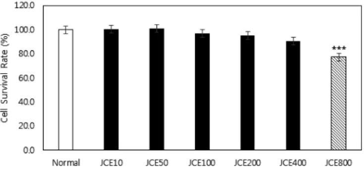
Cell viability was measured using an MTT assay and showed cell viability was 77.32% at 800 μg/mL JCE003, 90.77% at 400 μg/mL, 95.29% at 200 μg/mL, 96.81% at 100 μg/mL, 101.14% at 50 and 100.61% at 10 μg/mL.
Cell viability at JCE003 800 μg/mL was statistically significantly
Target
gene Primer Sequences
IFN-γ
Forward 5’-GGATGTAACGACAGCCCTCT-3’
Reverse 5’-GTGTTCCTTGTTGCCGTAAG-3’,
TNF-α
Forward 5′-GCT GCT GTG CTG TGT AGG AG -3′
Reverse 5′-CAG ATG CCT CGG TTT TGT TA-3′
IL-1 β
Forward 5′-TCCTACCCCAACTTCCAATGCTC-3′
Reverse 5′-TTGGATGGTCTTGGTCCTTAGCC-3′
IL-6
Forward 5’-CAGTTGCAATGCCATCCACA-3’
Reverse 5’-AGCCACATCCGAGGCCTTT-3’,
GAPDH-
VIC Probe 5’-CATGTTCCAGTATGACTCCACTCACG-3
qPCR, real-time quantitative polymerase chain reaction; IFN-γ , interferon-gamma;
TNF-α, tumor necrosis factor-alpha; IL-1 β, interleukin-1β; IL-6, interleukin 6; GAPDH, Human Glyceraldehyde 3-phosphate dehydrogenase.
Table 1. Primer Sequence for qPCR Analysis.
lower than that of the Control group (p < 0.001; Fig.1).
Assessment of inhibition of NO production
Production of NO is shown in Table 2. The production of NO for JCE10, JCE50, JCE100, JCE200, and JCE400 groups was 60.88, 51.99, 50.25, 44.7, and 40.09 μM, respectively. The differences were not significant (Fig. 2).
Cytokine production analysis
Cytokine (IFN-γ, TNF-α, IL-1β, and IL-6) production in the Normal, Control, JCE10, JCE50, JCE100, JCE200, and JCE400 groups was analyzed by ELISA (Table 3). The differences of production of IFN-γ, IL-1β and IL-6 in JCE10, JCE50, JCE100, JCE200, and JCE400 groups were not significant. In JCE400 group, TNF-α production was statistically significantly lower than that of the Control group (p < 0.05).
IFN-γ
IFN-γ production in JCE10, JCE50, JCE100, JCE200, and JCE400 groups was 29.15, 33.19, 26.86, 27.65, and 25.05 pg/mL, respectively. The differences were not significant (Fig. 3A).
TNF-α
TNF-α production in JCE10, JCE50, JCE100, and JCE200 groups was 453.84, 439.03, 412.19, and 398.9 rg/mL, respectively.
The differences were not significant. In contrast, the amount of TNF-α produced in JCE400 group was 382.98 rg/mL, which was statistically significantly lower than that of the Control group (p <
0.05; Fig. 3B).
IL-1β
IL-1β production in JCE10, JCE50, JCE100, JCE200, and JCE400 groups was 54.48, 49.41, 52.94, 48.67, and 48.22 rg/mL, respectively. The differences were not significant (Fig. 3C).
IL-6 IL-6 production in the JCE10, JCE50, JCE100, JCE200, and JCE400 groups was 333.56, 327.34, 340.7, 321.63, and 305.69 rg/
mL, respectively. The differences were not significant (Fig. 3D).
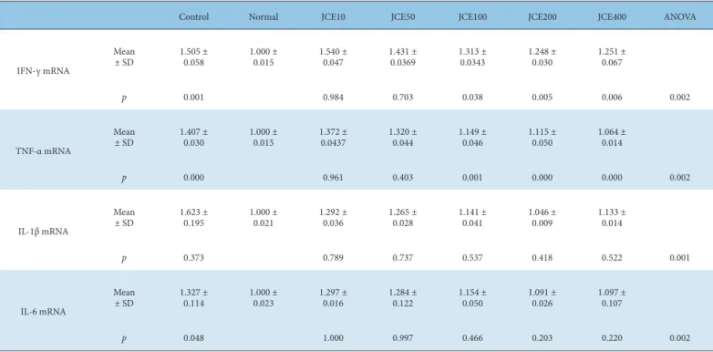
Analysis of cytokine mRNA expression
The expression levels of cytokine (IFN-γ, TNF-α, IL-1β, and IL- 6) in the Normal, Control, and JCE10, JCE50, JCE100, JCE200, and JCE400 groups were analyzed by qPCR and the data are shown in Table 4. The expression level of IL-1β and IL-6 mRNA in JCE10,
Fig. 1. The cytotoxicity effect of JCE003 on of RAW264.7 cells.
Normal group was not treated with JCE003. Cells in experimental groups were incubated in the presence of JCE003 (10, 50, 100, 200, 400 and 800 μg/mL) for 24 h. Cell cytotoxicity was determined by MTT assay. Cell cytotoxicity (% of control) = (sample OD / control O) × 100.
*p < 0.001 vs normal by Dunnett’s test
MTT, 3-(4,5-dimethyl thiazole-2- yl)-2,5-diphenyl tetrazolium bromide; OD, optical density.
Fig. 2. Inhibitory effect of JCE003 on the production of NO in LPS-treated RAW264.7 cells.
The Control group was only treated with LPS 500 ng/mL. The Normal group was not treated with JCE003 and LPS. Cells in experimental groups were incubated in the presence of LPS (500 μg/mL) alone or in combination with JCE003 (10, 50, 100, 200 and 400 μg/mL) for 24 h. The culture media of the treated cells was used to measure NO level.
*p < 0.001 vs normal by Dunnett’s test NO, nitric oxide; LPS, lipopolysaccharide.
Control Normal JCE10 JCE50 JCE100 JCE200 JCE400 ANOVA
(μM)NO
Mean ± SD 51.16 ± 7.530 3.83 ± 3.236 60.88 ± 13.898 51.988 ± 17.111 50.247 ± 8.870 44.697 ± 2.509 40.093 ± 2.603
p < 0.001 0.635 1.000 1.000 0.893 #0.518 0.045
Data are presented as mean ± SD (n = 3). ANOVA was performed after regularity test using SPSS (22.0 for Windows program). Dunnett’s test was used when the equality of variances was tested, and Dunnett’s T3 was used if the test was not performed.
*p-value vs normal by Dunnett’s test.
†p-value vs control by Dunnett’s test.
NO, nitric oxide; ANOVA, one-way analysis of variance.
Table 2. Analysis of Inhibition of NO Production.
Fig. 3. Inhibitory effect of JCE003 on the production of cytokines [(A) IFN-γ, (B) TNF-α, (C) IL-1β, (D) IL-6] in LPS treated RAW264.7 cells.
Control group was only treated with LPS 500 ng/mL. Normal group was not treated with JCE003 and LPS. Cells in experimental groups were incubated in the presence of LPS (500 μg/mL) in combination with JCE003 (10, 50, 100, 200 and 400 μg/mL) for 24 h. The culture media of the treated cells were used to measure IL-6 level.
*p < 0.001 vs normal by Dunnett’s test.
†p < 0.05 vs control by Dunnett’s test.
IFN-γ , interferon-gamma; TNF-α, tumor necrosis factor-alpha; IL-1 β, interleukin-1β; IL-6, interleukin 6; LPS, Lipopolysaccharide.
(A)
(C)
(B)
(D)
Control Normal JCE10 JCE50 JCE100 JCE200 JCE400 ANOVA
IFN-γ (pg/mL)
Mean
± SD 30.281 ±
4.986 4.207 ±
3.322 29.151 ±
5.889 33.191 ±
4.831 26.860 ±
3.885 27.651 ±
2.743 25.050 ±
3.638
p 0.000 0.998 0.845 0.744 0.891 0.375 0.012
TNF-α (pg/mL)
Mean
± SD 470.669 ±
38.578 14.268 ± 2.168 453.836 ±
32.670 439.026 ±
50.709 412.189 ±
32.927 398.904 ±
29.234 382.983 ±
31.346
p 0.000 0.960 0.653 0.143 0.056 0.017 0.000
IL-1β (pg/mL)
Mean
± SD 55.370 ±
6.951 6.410 ±
3.964 54.480 ±
5.755 49.410 ±
6.181 52.938 ±
1.169 48.670 ±
10.397 48.218 ±
8.594
p 0.000 1.000 0.702 0.991 0.604 0.546 0.046
(pg/mL)IL-6
Mean
± SD 351.773 ±
31.725 5.796 ±
2.335 333.556 ±
1.893 327.341 ±
10.216 340.697 ±
23.656 321.632 ±
19.688 305.686 ±
12.439
p 0.003 0.657 0.533 1.000 0.613 0.137 0.000
Data are presented as mean ± SD (n = 3). ANOVA was performed after regularity test using SPSS (22.0 for Windows program). Dunnett’s test was used when the equality of variances was tested, and Dunnett’s T3 was used if the test was not performed.
*p-value vs normal by Dunnett’s test.
†p-value vs normal by Dunnett T3.
‡p-value vs control by Dunnett’s test.
§p-value vs control by Dunnett T3.
ELISA, enzyme-linked immunosorbent assay; IFN-γ , interferon-gamma; TNF-α, tumor necrosis factor-alpha; IL-1 β, interleukin-1β; IL-6, interleukin 6; ANOVA, one-way analysis of variance.
Table 3. Analysis of Cytokine Production by ELISA.
Control Normal JCE10 JCE50 JCE100 JCE200 JCE400 ANOVA
IFN-γ mRNA
Mean
± SD 1.505 ±
0.058 1.000 ±
0.015 1.540 ±
0.047 1.431 ±
0.0369 1.313 ±
0.0343 1.248 ±
0.030 1.251 ±
0.067
p 0.001 0.984 0.703 0.038 0.005 0.006 0.002
TNF-α mRNA
Mean
± SD 1.407 ±
0.030 1.000 ±
0.015 1.372 ±
0.0437 1.320 ±
0.044 1.149 ±
0.046 1.115 ±
0.050 1.064 ±
0.014
p 0.000 0.961 0.403 0.001 0.000 0.000 0.002
IL-1β mRNA
Mean
± SD 1.623 ±
0.195 1.000 ±
0.021 1.292 ±
0.036 1.265 ±
0.028 1.141 ±
0.041 1.046 ±
0.009 1.133 ±
0.014
p 0.373 0.789 0.737 0.537 0.418 0.522 0.001
IL-6 mRNA
Mean
± SD 1.327 ±
0.114 1.000 ±
0.023 1.297 ±
0.016 1.284 ±
0.122 1.154 ±
0.050 1.091 ±
0.026 1.097 ±
0.107
p 0.048 1.000 0.997 0.466 0.203 0.220 0.002
Data are presented as mean ± SD (n = 3). ANOVA was performed after regularity test using SPSS (22.0 for Windows program). Dunnett’s test was used when the equality of variances was tested, and Dunnett’s T3 was used if the test was not performed.
*p-value vs normal by Dunnett’s test.
†p-value vs normal by Dunnett T3.
‡p-value vs control by Dunnett’s test.
§p-value vs control by Dunnett T3.
qPCR, real-time quantitative polymerase chain reaction; IFN-γ , interferon-gamma; TNF-α, tumor necrosis factor-alpha; IL-1 β, interleukin-1β; IL-6, interleukin 6; ANOVA, one-way analysis of variance.
Table 4. Analysis of Cytokine mRNA Expression Level by qPCR.
JCE50, JCE100, JCE200, and JCE400 groups were not significantly different from the Control group. The expression level of IFN-γ and TNF-α mRNA were statistically significantly lower than in the Control group in the JCE100, JCE200 and JCE400.
IFN-γ mRNA
IFN-γ mRNA expression in the JCE100 group (1.31 ± 0.03) was statistically significantly lower than that of the Control group (p <
0.05). In addition, IFN-γ mRNA expression in JCE200 and JCE400 groups were 1.25 ± 0.03 and 1.25 ± 0.07, respectively, which was also statistically significantly lower than in the Control group (p <
0.01; Fig. 4A). The expression levels of TNF-α mRNA in JCE10 and JCE50 groups were 1.37 ± 0.04 and 1.32 ± 0.04, respectively, which were not significantly difference.
TNF-α mRNA
The expression levels of TNF-α mRNA in JCE100, JCE200, and JCE400 groups were 1.15 ± 0.05, 1.11 ± 0.05 and 1.06 ± 0.01, respectively, which was significantly lower than that of the Control group (p < 0.01; Fig. 4B). The differences in expression levels of TNF-α mRNA in JCE10 and JCE50 groups were 1.37 ± 0.04 and 1.32 ± 0.04, respectively, which were not significant.
IL-1β mRNA
The expression level of IL-1β mRNA in JCE10, JCE50, JCE100, JCE200, and JCE400 groups were 1.29 ± 0.04, 1.26 ± 0.03, 1.14 ± 0.04, 1.05 ± 0.01, and 1.13 ± 0.01, respectively, but not significantly
different from the Control group (Fig. 4C).
IL-6 mRNA
The expression levels of IL-6 mRNA in JCE10, JCE50, JCE100, JCE200, and JCE400 groups were 1.3 ± 0.01, 1.28 ± 0.12, 1.15 ± 0.05, 1.09 ± 0.03, and 1.1 ± 0.11, respectively, but the differences were not significant (Fig. 4D).
Discussion
The objective of this study was to investigate the anti- inflammatory effect of JCE003 which included Eucommia ulmoides, in LPS-activated RAW 264.7 cells.
An inflammatory reaction is the body’s first immune response against injury and invasion of external foreign bodies [20].
Typically, during inflammatory reactions, any foreign bodies at the site of a wound are recognized and the immune system is activated by cytokines, which are inflammatory mediators.
When appropriate, the immune response is dampened and pro-
inflammatory cytokines are inhibited by anti-inflammatory
cytokines where there has been injury and the tissue is regenerated
[21]. The inflammatory response is essential in maintaining
homeostasis in the body. When there is a failure to regulate
the immune system, hyper-secreted inflammatory mediators
cause dysfunction and necrosis of cells and tissues. Maintaining
the survival of activated immune cells through the lack of
immunoregulation could lead to conditions such as inflammatory
bowel disease or Type-2 diabetes [22-24].
Macrophages are immune cells that exist in all tissues and organs and when they recognize foreign substances or wounds they release inflammatory mediators enabling an acute inflammatory reaction, and the migration of more macrophages and other inflammatory cells into the area [25]. During the inflammatory reaction, macrophages produce NO and PGE
2, promoting the migration of immune cells to the site of inflammation [26]. Macrophages also provide an immediate defense against pathogens (prior to the chemotaxis of leukocytes). Stimulation with LPS encountered on the outer membrane of gram-negative bacteria initiates the production of inflammatory mediators such as interleukins and TNF-α [27,28].
In arthritis patients, pro-inflammatory cytokines such as TNF-α and IL-1β are expressed primarily by macrophages in the synovial fluid, and exhibit various pathological actions. TNF-α induces the expression of cytokines such as IL-1β and IL-6 inducing immune cell infiltration into inflammatory sites, and increases matrix metalloproteinases (MMPs) in neutrophils, synovial cells and chondrocytes [29]. In addition, the activity of IL-1β is similar to the range of TNF-α activity, and plays an important role in the inflammatory response, and adhesion of leukocytes to endothelial cells.
The concentration of IL-1β in the blood is also associated with joint destruction [30]. IL-6 is involved in a variety of acute inflammatory diseases, which promote T-cell activity, and rheumatoid factor production in rheumatoid arthritis patients [31,32].
Anti-inflammatory drugs are largely classified into steroidal
Fig. 4. The expression of cytokine [(A) IFN-γ, (B) TNF-α, (C) IL-1β, (D) IL-6] mRNA by LPS activated RAW264.7 cells following treatment with JCE003.
Control group was only treated with LPS 500 ng/mL. Normal group was not treated with JCE003 and LPS. Cells in experimental groups were incubated in the presence of LPS (500 μg/mL) in combination with JCE003 (10, 50, 100, 200 and 400 μg/mL) for 24 h. The mRNA expression was evaluated by qPCR analysis.
*p < 0.05 vs normal by Dunnett’s test.
†p < 0.01 vs normal by Dunnett’s test.
‡p < 0.05 vs control by Dunnett’s test.
§p < 0.01 vs control by Dunnett’s test.
IFN-γ , interferon-gamma; TNF-α, tumor necrosis factor-alpha; IL-1 β, interleukin-1β; IL-6, interleukin 6; LPS, Lipopolysaccharide; qPCR, real-time quantitative polymerase chain reaction.
and nonsteroidal types, all of which exert their effect through the inhibition of the synthesis of prostaglandin (PG), a major mediator of the inflammatory response. However, these anti-inflammatory drugs raise some safety concerns due side effects affecting the kidney, heart, and gastrointestinal system [33]. Therefore, active research into anti-inflammatory drugs that are natural and minimize side effects, is beneficial [34].
In conclusion, JCE003 significantly inhibited the production of TNF-α during cytokine production in LPS-activated RAW 264.7 macrophage cells, and significantly decreased IFN-γ and TNF-α mRNA expression. The production of TNF-α decreased at a concentration of 400 μg/mL JCE003, and the expression of IFN-γ and TNF-α mRNA decreased above a concentration of 100 μg/
mL JCE003, suggesting that JCE003 is more effective at inhibiting the expression of cytokine mRNA than inhibiting the production of cytokines. In particular, inhibition of the gene expression and production of TNF-α, which plays a significant role in the inflammatory response of arthritis, may be related to the anti- inflammatory effects of JCE003.
Conclusion
JCE003 showed no cytotoxicity to LPS-activated RAW 264.7 macrophage cells, up to the concentration of 400 μg/mL. Toxicity was observed at a concentration of 800 μg/mL, where cell survival rate was statistically significantly decreased (p < 0.001). NO production was not significantly different in the experimental group compared to the Control group. The production of TNF-α was statistically significantly decreased at a concentration of 400
(A)(C)
(B)
(D)
μg/mL JCE003 (p < 0.05). The expression of IFN-γ and TNF-α mRNA was statistically significantly decreased at concentrations
≥ 100 μg/mL JCE003 (p < 0.01). These results suggest that the complex extract JCE003 (including Eucommia ulmoides) may be effective at dampening the inflammatory immune response.
However, future studies are required to substantiate these findings.
Conflicts of Interest
The authors have no conflicts of interest to declare.
References
[1] Vlassara H. Advanced glycation in health and disease: role of the modern environment. Ann N Y Acad Sci 2005;1043:452-460.
[2] Lee HJ, Hyun EA, Yoon WJ, Kim BH, Rhee MH, Kang HK et al. In vitro anti-inflammatory and anti-oxidative effects of Cinnamomum camphora extracts. J Ethnopharmacol 2006;103:208-216.
[3] Ljung T, Lundberg S, Varsanyi M, Johansson C, Schmidt PT, Herulf M et al. Rectal nitric oxide as biomarker in the treatment of inflammatory bowel disease: responders versus nonresponders. World J Gastroenterol 2006;12:3386-3392.
[4] Zhang G, Ghosh S. Molecular mechanisms of NF-kappaB activation induced by bacterial lipopolysaccharide through Toll-like receptors. J Endotoxin Res 2000;6:453-457.
[5] Smolen JS, Aletaha D, Koeller M, Weisman MH, Emery P. New therapies for treatment of rheumatoid arthritis. Lancet 2007;370:1861-1874.
[6] Akira S, Takeda K. Toll-like receptor signalling. Nat Rev Immunol 2004;4:499-511.
[7] Kang CH, Choi YH, Choi IW, Lee JD, Kim GY. Inhibition of Lipopolysaccharide-Induced iNOS , COX- 2 , and TNF- α Expression by Aqueous Extract of Orixa Japonica in RAW 264.7 Cells via Suppression of NF- κ B Activity. Trop J Pharm Res 2011;10:161-168.
[8] Nam SW, Baek JT, Lee DS, Kang SB, Ahn BM, Chung KW. A case of acute cholestatic hepattis associated with the seeds of Psoralea corylifolia (Boh- Gol-Zhee). Clin Toxicol 2005; 43: 589-591.
[9] Kim MK, Kim JS, Jo BS, Kim JH, Lee IC, Lee MS et al. Functional Properties of Walnut in Cosmetics. J Life Sci 2011;21:858-864.
[10] Kim MC, Kim DS, Kim SJ, Park J, Kim HL, Kim SY et al. Eucommiae cortex inhibits TNF-alpha and IL-6 through the suppression of caspase-1 in lipopolysaccharide-stimulated mouse peritoneal macrophages. Am J Chin Med 2012;40:135-149.
[11] Cai Y, Luo Q, Sun M, Corke H. Antioxidant activity and phenolic compounds of 112 traditional Chinese medicinal plants associated with anticancer. Life Sci 2004;74:2157-2184.
[12] Kwon SH, Lee HK, Kim JA, Hong SI, Kim SY, Jo TH et al. Neuroprotective effects of Eucommia ulmoides Oliv. Bark on amyloid beta(25-35)-induced learning and memory impairments in mice. Neurosci Lett 2011;487:123- 127.
[13] Luo L, Wu W, Zhou Y, Yan J, Yang G, Ouyang D. Antihypertensive effect of Eucommia ulmoides Oliv. extracts in spontaneously hypertensive rats. J Ethnopharmacol 2010;129:238-243.
[14] Zhang R, Liu ZG, Li C, Hu SJ, Liu L, Wang JP et al. Du-Zhong (Eucommia ulmoides Oliv.) cortex extract prevent OVX-induced osteoporosis in rats.
Bone 2009;45:553-559.
[15] Kang BM, An BK, Jung WS, Jung HK, Cho HJ, Cho HW et al. Anti- inflammatory effect of tricin isolated from Alopecurus aequalis Sobol . on the LPS-induced inflammatory response in RAW 264.7 cells. Int J Mol Med 2016;38:1614-1620.
[16] Nam SW, Baek JT, Lee DS, Kang SB, Ahn BM, Chung KW. A case of acute cholestatic hepatitis associated with the seeds of Psoralea corylifolia (Boh- Gol-Zhee). Clin Toxicol (Phila) 2005;43:589-591.
[17] Choi JM, Ahn JB. Functional Properties of 50 % Methanol Extracts from Different Parts of Acanthopanax sessiliflorus. Korean J Food Sci Tech 2012;44:373-377.
[18] Ryu HS, Park SC, Kim J, Kim HS. Enhancing Effect of Zingiber Officinale Roscoe Extracts on Mouse Spleen and Macrophage Cells Activation. Korean J Nutr 2004;37:23-30.
[19] Lantz RC, Chen GJ, Sarihan M, Solyom AM, Jolad SD, Timmermann BN.
The effect of extracts from ginger rhizome on inflammatory mediator production. Phytomedicine 2007;14:123-128.
[20] Medzhitov R. Origin and physiological roles of inflammation. Nature 2008;454:428-435.
[21] Iwalewa EO, Mcgaw LJ, Naidoo V, Eloff JN. Inflammation : the foundation of diseases and disorders. A review of phytomedicines of South African origin used to treat pain and inflammatory conditions. Afr J Biotechnol 2007;6:2868-2885.
[22] Ferrero-Miliani L, Nielsen OH, Andersen PS, Girardin SE. Chronic inflammation: importance of NOD2 and NALP3 in interleukin-1beta generation. Clin Exp Immunol 2007;147:227-235.
[23] Gabay C. Interleukin-6 and chronic inflammation. Arthritis Res Ther 2006;8 Suppl 2:S3.
[24] Ranjan R, Sindhu S, Narsing AR. Photoreceptor mitochondrial oxidative stress in early experimental autoimmune uveoretinitis. Br J Ophthalmol 2007;91:531-537.
[25] Rao C V, Indranie C, Simi B, Manning PT, Connor JR, Reddy BS.
Chemopreventive properties of a selective inducible nitric oxide synthase inhibitor in colon carcinogenesis, administered alone or in combination with celecoxib, a selective cyclooxygenase-2 inhibitor. Cancer Res 2002;62:165-170.
[26] Nathan C, Xie QW. Regulation of biosynthesis of nitric oxide. J Biol Chem 1994;269:13725-13728.
[27] Molloy RG, Mannick JA, Rodrick ML. Cytokines, sepsis and immunomodulation. Br J Surg 1993;80:289-297.
[28] Hinz B, Brune K. Cyclooxygenase-2--10 years later. J Pharmacol Exp Ther 2002;300:367-375.
[29] Ainola MM, Mandelin JA, Liljestrom MP, Li TF, Hukkanen MVJ, Konttinen YT. Pannus invasion and cartilage degradation in rheumatoid arthritis:
involvement of MMP-3 and interleukin-1beta. Clin Exp Rheumatol 2005;23:644-650.
[30] North J, Situnayake RD, Tikly M, Cremona A, Nicoll J, Kumararatne DS et al. Interleukin 1 beta , hand and foot bone mineral content and the development of joint erosions in rheumatoid arthritis. Ann Rheum Dis 1994;53:543-546.
[31] Kotake S, Sato K, Kim KJ, Takahashi N, Udagawa N, Nakamuri I et al.
Interleukin-6 and soluble interleukin-6 receptors in the synovial fluids from rheumatoid arthritis patients are responsible for osteoclast-like cell formation. J Bone Miner Res 1996;11:88-95.
[32] Okamoto H, Yamamura M, Morita Y, Harada S, Makino H, Ota Z. The synovial expression and serum levels of interleukin-6, interleukin-11, leukemia inhibitory factor, and oncostatin M in rheumatoid arthritis.
Arthritis Rheum 1997;40:1096-1105.
[33] Makins R, Ballinger A. Gastrointestinal side effects of drugs. Expert Opin Drug Saf 2003;2:421-429.
[34] Kim B, Ahn N, Choi Y, Ahn D. Anti-inflammatory Activity of an Ethanol Extract of Laminaria japonica Root on Lipopolysaccharide-induced Inflammatory. Korean J Food Sci Tech 2014;46:729-733.
![Fig. 3. Inhibitory effect of JCE003 on the production of cytokines [(A) IFN-γ, (B) TNF-α, (C) IL-1β, (D) IL-6] in LPS treated RAW264.7 cells](https://thumb-ap.123doks.com/thumbv2/123dokinfo/4821763.280864/5.892.46.836.134.520/fig-inhibitory-effect-jce-production-cytokines-treated-cells.webp)
![Fig. 4. The expression of cytokine [(A) IFN-γ, (B) TNF-α, (C) IL-1β, (D) IL-6] mRNA by LPS activated RAW264.7 cells following treatment with JCE003](https://thumb-ap.123doks.com/thumbv2/123dokinfo/4821763.280864/7.892.60.815.101.483/fig-expression-cytokine-ifn-activated-cells-following-treatment.webp)
