INTRODUCTION
Since its initial description in 1965 (1), carcinoembryonic antigen (CEA) has remained the most thoroughly investigat- ed tumor marker. CEA is an intracellular protein normally found in low concentrations in embryonic and fetal gut, pan- creas, and liver cells, as well as in several adult tissues. It may be elevated in smokers and in several malignant and benign conditions of the gastrointestinal tract (2). CEA expressed in large quantities in approximately 95% of colorectal carci- nomas, and the relationship between serum CEA (s-CEA) level and colorectal cancer has been studied extensively. It has been shown that preoperative s-CEA levels increases with tumor stage (3), and elevated preoperative s-CEA levels have been found to predict a higher incidence of recurrence (4) in patients with colorectal cancer. In addition, the relationship between preoperative s-CEA level and survival has suggest- ed a predictive value of the former for metastasis or recurrence in colorectal cancer patients (5, 6). At present, s-CEA is mea- sured primarily during follow-up of colorectal cancer patients, so that patients with high s-CEA levels receive more inten- sive treatment (7).
The prognostic value of preoperative s-CEA has been stud- ied, but no practical cutoff value has been determined. We therefore tried to reassess whether preoperative s-CEA level can be used as a prognostic factor in colorectal cancer patients.
In addition, we sought to determine the cutoff values of s-CEA for predicting outcome.
MATERIALS AND METHODS
One thousand eighty-three patients had curative resections for stages I, II, and III colorectal cancer at Asan Medical Cen- ter between July 1990 and December 1997. Patients in whom preoperative s-CEA level was measured were considered eli- gible for the study. Patients initially presenting with metastatic disease and those who underwent palliative resection were excluded, leaving a total of 989 patients. Their mean age was 57 yr (range, 21-89 yr); 527 were males, and 462 were females.
Median follow-up was 46 months (range, 3-129 months).
Of the 989 patients, 164 (16.6%) had stage I, 374 (37.8%) had stage II, and 451 (46%) had stage III tumors.
Serum CEA was measured by enzyme immunoassay (ELISA- 2-CEA kit�; CIS Biointernational, Marcule, France). The nor- mal range of s-CEA in our laboratory was set as <6 ng/mL, as determined by measurements in 313 Korean healthy persons using the same kit. The s-CEA levels varied between 0.1 and 6.4 ng/mL, with a mean (±S.D.) of 2.25±1.25 ng/mL (8).
To determine the s-CEA cutoff values that may discrimi- nate between patients with good and those with poor prog- nosis, we performed a multi-step analysis of survival curve.
In Ja Park, Hee Cheol Kim, Chang Sik Yu, Jang Hak Yoo, Jin Cheon Kim
Department of Surgery, University of Ulsan College of Medicine and Asan Medical Center, Seoul, Korea
Address for correspondence Hee Cheol Kim, M.D.
Department of Surgery, University of Ulsan College of Medicine and Asan Medical Center, 388-1 Poongnap-dong, Songpa-gu, Seoul 138-736, Korea Tel : +82.2-3010-3937, Fax : +82.2-474-9027 E-mail : [email protected]
624 J Korean Med Sci 2005; 20: 624-7
ISSN 1011-8934
Copyright � The Korean Academy of Medical Sciences
Cutoff Values of Preoperative s-CEA Levels for Predicting Survivals after Curative Resection of Colorectal Cancer
Serum carcinoembryonic antigen (s-CEA) is used to detect recurrence and predict prognosis in colorectal cancer. However, the cutoff values of s-CEA for prognosis have not been determined. We therefore tried to determine the preoperative s- CEA levels predictive of survivals in colorectal cancer patients. We retrospectively analyzed the medical records of 989 patients who underwent curative resection for colorectal cancer between July 1990 and December 1997, with a mean follow- up of 46 months (range, 3-129 months). When patients were divided into four sub- groups with the cutoff values of s-CEA at 3,6, and 17 ng/mL, their 5-yr disease- free survival rates were 85.3% (<3.0 ng/mL), 70.0% (3-6 ng/mL), 64.2% (6-17 ng/mL), and 55.2% (>17 ng/mL) (p<0.001). Multivariate analysis showed that fac- tors predictive of survival included age (p=0.028), tumor stage (p<0.001), cell dif- ferentiation (p=0.016), and gross type (p=0.007), location (p=0.003) and preopera- tive s-CEA (p<0.001). Using the above-described cutoff levels, a significant differ- ence in survival was observed only in patients with stage III tumors (p=0.007) when analyses were performed by stage. We can suggest the new cutoff values of s-CEA used in the present study.
Key Words : Carcinoembryonic Antigen; Prognosis; Colonic Neoplasm
Received : 24 November 2004 Accepted : 7 February 2005
s-CEA as a Prognostic Factor 625
First, we constructed survival curves according to their pre- operative CEA levels, and compared them. Then, we con- structed survival curves for patients with levels beginning from 1 ng/mL and increasing by 1 ng/mL increments, and we compared the curves for each s-CEA level independently.
If we found several levels, which showed different survivals, they were compared after the group was divided according to all level. We repeated these steps for levels showing a dif- ference in survival. We chose cutoff levels that most clearly divided patients into subgroups with regard to survival rate by the Kaplan-Meier method with a log rank test. Multi- variate analysis was calculated by the Cox proportional haz- ard model. Statistical significance was defined as p<0.05.
RESULTS
When we set the cutoff values of s-CEA at 3, 6, and 17 ng/mL, the patients were divided into four subgroups with significantly different 5-yr disease-free survival rates (p<0.001).
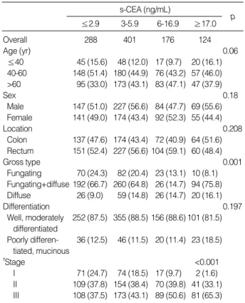
Of the 989 patients, 288 (29.1%) had s-CEA below 3 ng/mL, 401 (40.5%) had s-CEA of 3-6 ng/mL, 176 (17.8%) had s- CEA of 6-17 ng/mL, and 124 (12.5%) had s-CEA over 17 ng/mL. The survival rates in these four subgroups were 85.3%, 70.0%, 64.2%, and 55.2%, respectively (Fig. 1). The four subgroups did not differ significantly with respect to age, sex, location of primary tumor or cellular differentiation. We found, however, that, as s-CEA level increased, the gross pat- tern of diffuse type (p=0.007) and stage III (p<0.001) also increased (Table 1).
Mean preoperative s-CEA levels with respect to AJCC stage were 4.01 ng/mL (range, 1.0-37.5 ng/mL) in stage I, 11.07 ng/mL (range, 0.7-556.0 ng/mL) in stage II and 23.10 ng/mL (rage, 0.7-2510.0 ng/mL) in stage III, but these dif-
ferences did not achieve statistical significance (p=0.07). When we determined s-CEA levels relative to T and N categories, we found that mean s-CEA level increased progressively by N category (p<0.001), but there was no correlation between mean s-CEA level and T category.
Using the above-described cutoff levels, a significant dif-
s-CEA (ng/mL)
≤2.9 3-5.9 6-16.9 ≥17.0 p
Overall 288 401 176 124
Age (yr) 0.06
≤40 45 (15.6) 48 (12.0) 17 (9.7) 20 (16.1) 40-60 148 (51.4) 180 (44.9) 76 (43.2) 57 (46.0)
>60 95 (33.0) 173 (43.1) 83 (47.1) 47 (37.9)
Sex 0.18
Male 147 (51.0) 227 (56.6) 84 (47.7) 69 (55.6) Female 141 (49.0) 174 (43.4) 92 (52.3) 55 (44.4)
Location 0.208
Colon 137 (47.6) 174 (43.4) 72 (40.9) 64 (51.6) Rectum 151 (52.4) 227 (56.6) 104 (59.1) 60 (48.4)
Gross type 0.001
Fungating 70 (24.3) 82 (20.4) 23 (13.1) 10 (8.1) Fungating+diffuse 192 (66.7) 260 (64.8) 26 (14.7) 94 (75.8) Diffuse 26 (9.0) 59 (14.8) 26 (14.7) 20 (16.1)
Differentiation 0.197
Well, moderately 252 (87.5) 355 (88.5) 156 (88.6) 101 (81.5) differentiated
Poorly differen- 36 (12.5) 46 (11.5) 20 (11.4) 23 (18.5) tiated, mucinous
�Stage <0.001
I 71 (24.7) 74 (18.5) 17 (9.7) 2 (1.6) II 109 (37.8) 154 (38.4) 70 (39.8) 41 (33.1) III 108 (37.5) 173 (43.1) 89 (50.6) 81 (65.3)
*No in parentheses are %, �AJCC 6th edition.
Table 1.Clinicopathological characteristics of patients with respect to s-CEA concentration
Survivals
1.0 0.9 0.8 0.7 0.6 0.5 0.4 0.3 0.2 0.1 0.0
0 20 40 60 80 100 120 140
Months p=0.001
CEA; <3 ng/mL CEA; 3-6 ng/mL CEA; 6-17 ng/mL CEA≥17 ng/mL
Fig. 1.Disease-free survival curves of colorectal cancer patients relative to preoperative s-CEA levels. Survival is significantly relat- ed to preoperative s-CEA levels.
Survivals
1.0 0.9 0.8 0.7 0.6 0.5 0.4 0.3 0.2 0.1 0.0
0 20 40 60 80 100 120 140
Months
p=0.007 CEA; <3 ng/mL CEA; 3-6 ng/mL CEA; 6-17 ng/mL CEA≥17 ng/mL
Fig. 2.Preoperative s-CEA levels and disease-free survival rate of stage III colorectal cancer patients.
626 I.J. Park, H.C. Kim, C.S. Yu, et al.
ference in survival was observed only in patients with stage III tumors (p=0.007), not in those with stages I and II (Fig.
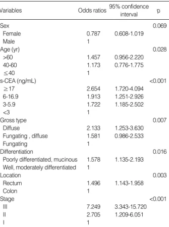
2). By multivariate analysis, preoperative s-CEA level using these new cutoff values was an independent prognostic factor of survival (p<0.001), as were age (p=0.028), tumor stage (p<
0.001), differentiation (p=0.016), and gross type (p=0.007), location (p=0.003) and preoperative s-CEA (p<0.001) (Table 2).
DISCUSSION
Since its first description, the clinical importance of s-CEA in the management of colorectal cancer patients has been investigated extensively, and it has been reported to predict recurrence or metastasis of this disease (3-6, 9-11). There were several drawbacks to these previous studies, however, includ- ing the use of arbitrary cutoff levels. In most studies, survival and recurrence rates were compared between patients with normal s-CEA level and those with elevated s-CEA (9-11), but these groups were not further stratified. In addition, the normal level of s-CEA was set arbitrarily, leading to insuffi- cient determination of the prognostic function of s-CEA level.
To correct these possible errors, we stratified s-CEA levels by comparing survival curves of patients below and above each s-CEA level, starting at 1 ng/mL and increasing by increments of 1 ng/mL.
We selected 3, 6, and 17 ng/mL s-CEA as practical cutoff points, which highly discriminate between higher and lower risk groups with respect to survival. In contrast, our evalua- tion did not attribute any discriminatory function to any other s-CEA level. It is interesting result considering we did not consider the importance of serum CEA, which confined with- in the normal range usually. Thus, regardless of pathologic stage, these patients could be stratified into subgroups with different prognoses based on their preoperative s-CEA levels, with patients having the highest preoperative s-CEA levels (i.e., >17 ng/mL) having the highest risk of metastasis or recur- rence.
CEA is expressed throughout life in normal adult tissues, including the colon, stomach, tongue, cervix, and prostate (1, 12). In the normal colon, CEA may be associated with cellular differentiation, and the degree of expression may be related to differentiation status (13). In colon cancer, CEA appears to provide a variety of cellular functions, including adhesion, in both intracellular and CEA-matrix interactions (14-17), signal transduction, and cellular migration (18-20), suggesting that CEA has a role as a facilitator of tumor inva- sion and metastasis. On the molecular level, there appears to be no difference between normal colonic CEA and tumor CEA, although the possibility that subtle posttranslational modifications may create differences cannot be excluded (13).
Our clinical data suggest that CEA may act in a dose-depen- dent manner. Thus, enhanced expression of CEA may confer on cancer cells the ability to invade adjacent tissues and to metastasize to lymph nodes or distant organs. Even normal serum levels of CEA may enhance cancer cell invasiveness, giving rise to more aggressive tumor cells.
Tumor stage is the most important predictor of survival in colorectal cancer (5, 6), and s-CEA levels tended to increase progressively with the stage of disease (3). In the present study, preoperative s-CEA level could be correlated with survival only in patients with stage III tumors, not those with stages I and II. This result conflicts with an earlier report (4), in which disease-free survival of patients with Dukes B and C lesions was related to preoperative s-CEA level. Our finding, however, is in agreement with a study showing no such cor- relation in patients with Dukes B lesions (21). It is not clear whether these differences are due to selection factors, differ- ent pathologic staging, small sample sizes, or a combination of these factors. Moreover, when we attempted to coordinate T and N categories of AJCC stage with preoperative s-CEA levels, we found that N, but not T, category was correlated with s-CEA, suggesting a connection of the latter with nodal metastasis.
Our findings suggest that preoperative s-CEA levels could be used as a stratification parameter for identifying subsets of colorectal cancer patients with different prognoses. Especially, patients with preoperative s-CEA above 3.0 ng/mL should be considered at higher risk compared with those with below 3.0 ng/mL, although this value is within the normal range,
95% confidence interval
Variables Odds ratios p
Sex 0.069
Female 0.787 0.608-1.019
Male 1
Age (yr) 0.028
>60 1.457 0.956-2.220
40-60 1.173 0.776-1.775
≤40 1
s-CEA (ng/mL) <0.001
≥17 2.654 1.720-4.094
6-16.9 1.913 1.251-2.926
3-5.9 1.722 1.185-2.502
<3 1
Gross type 0.007
Diffuse 2.133 1.253-3.630
Fungating , diffuse 1.581 0.986-2.533
Fungating 1
Differentiation 0.016
Poorly differentiated, mucinous 1.578 1.135-2.193 Well, moderately differentiated 1
Location 0.003
Rectum 1.496 1.143-1.958
Colon 1
Stage <0.001
III 7.249 3.343-15.720
II 2.705 1.209-6.051
I 1
Table 2.Factors related to survival rate: Multivariate analysis using Cox’s hazard model
s-CEA as a Prognostic Factor 627
especially in patients with stage III tumors. In addition, a prospective study is needed to determine the clinical useful- ness of these cut-off values.
REFERENCES
1. Gold P, Freedman SO. Specific carcinoembryonic antigens of the human digestive system. J Exp Med 1965; 122: 467-81.
2. Bendardaf R, Lamlum H, Pyrhonen S. Prognostic and predictive molecular markers in colorectal carcinoma. Anticancer Res 2004;
24: 2519-30.
3. Palmqvist R, Engaras B, Lindmark G, Hallmans G, Tavelin B, Nils- son O, Hammarstrom S, Hafstrom L. Prediagnostic levels of carci- noembryonic antigen and CA 242 in colorectal cacner: a matched case-control study. Dis Colon Rectum 2003; 46: 1538-44.
4. Wanebo HJ, Rao B, Pinsky CM, Hoffman RG, Stearns M, Schwartz MK, Oettgen HF. Preoperative carcinoembryonic antigen level as a prognostic indicator in colorectal cancer. N Engl J Med 1978; 299:
448-51.
5. Moertel CG, O’Fallon JR, Go VL, O’Connell MJ, Thynne GS. The preoperative carcinoembryonic antigen test in the diagnosis, stag- ing, and prognosis of colorectal cancer. Cancer 1986; 58: 603-10.
6. Carriquiry LA, Pineyro A. Should carcinoembryonic antigen be used in the management of patients with colorectal cancer? Dis Colon Rectum 1999; 42: 921-9.
7. Wiratkapun S, Karemer M, Sheow-Choen F, Ho YH, Eu KW. High preoperative serum carcinoembryonic antigen predicts metastatic recurrence in potentially curative colonic cancer: results of a five- year study. Dis Colon Rectum 2001; 44: 231-5.
8. Kim JC, Hwang DY, Kim BS, Min YL, Lee MH, Park KC. Changes of serum Careinoembryonic antigen in patients with colorectal can- cer. J Korean Cancer Assoc 1992; 24: 880-4.
9. Wolmark N, Fisher B, Wieand HS, Henry RS, Lerner H, Legault- Poisson S, Deckers PJ, Dimitrov N, Gordon PH, Jochimsen P. The prognostic significance of preoperative carcinoembryonic antigen levels in colorectal cancer. Results from NSABP (National Surgical Adjuvant Breast and Bowel Project) clinical trials. Ann Surg 1984;
199: 375-82.
10. Kim DW, Ryu MH, Kim TY, Heo DS, Bang YJ, Park JK, Kim NK.
A multivariate analysis of prognostic factors in colorectal cancer. J Korean Med Assoc 2003; 46: 268-74.
11. Harrison LE, Guillem JG, Paty P, Cohen AM. Preoperative carci- noembryonic antigen predicts outcomes in node-negative colon can- cer patients: a multivariate analysis of 572 patients. J Am Coll Surg 1997; 185: 55-9.
12. Prall F, Nollau P, Neumaier M, Haubeck HD, Drzeniek Z, Helmchen U, Loning T, Wagener C. CD66a (BGP) an adhesion molecule of the carcinoembryonic antigen family, is expressed in epithelium, endothelium, and myeloid cells in a wide range of normal human tissues. J Histochem Cytochem 1996; 44: 35-41.
13. Bjerner J, Lebedin Y, Bellanger L, Kuroki M, Shively JE, Varaas T, Nustad K, Hammarstrom S, Bormer OP. Protein epitopes in carci- noembryonic antigen. Report of the ISOBM TD8 Workshop. Tumor Biol 2002; 23: 249-62.
14. Benchimol S, Fuks A, Jothy S, Beauchemin N, Shirota K, Stanners CP. Carcinoembryonic antigen, a human tumor marker, functions as an intercellular adhesion molecule. Cell 1989; 57: 327-34.
15. Pignatelli M, Durbin H, Bodmer WF. Carcinoembryonic antigen functions as an accessory adhesion molecule mediating colon epithe- lial cell-collagen interactions. Proc Natl Acad Sci USA 1990; 87:
1541-5.
16. Obrink B. CEA adhesion molecules: Multifunctional proteins with signal regulatory properties. Curr Opin Cell Biol 1997; 9: 616-26.
17. Prado IB, Laudanna AA, Carneiro CR. Susceptibility of colorectal- carcinoma cells to natural-killer-mediated lysis: relationship to CEA expression and degree of differentiation. Int J Cancer 1995; 61: 854- 60.
18. Kim JC, Rho SA, Park KC. Adhesive function of carcinoembryonic antigen in the liver metastasis of KM-12c colon carcinoma cell line.
Dis Colon Rectum 1997; 40: 946-53.
19. Stanners CP, DeMarte L, Rojas M, Gold P, Fuks A. Opposite func- tions for two classes of genes of the human carcinoembryonic anti- gen family. Tumour Biol 1995; 16: 23-31.
20. von Kleist S, Miguel I, Halla B. Possible function of CEA as cell- contact inhibitory molecule. Anticancer Res 1995; 15: 1889-94.
21. Goslin R, Steele G Jr, MacIntyre J, Mayer R, Sugarbaker P, Cleghorn K, Wilson R, Zamcheck N. The use of preoperative plasma CEA levels for the stratification of patients after curative resection of col- orectal cancers. Ann Surg 1980; 192: 747-51.