© 2012 The Korean Academy of Medical Sciences.
This is an Open Access article distributed under the terms of the Creative Commons Attribution Non-Commercial License (http://creativecommons.org/licenses/by-nc/3.0) which permits unrestricted non-commercial use, distribution, and reproduction in any medium, provided the original work is properly cited.
pISSN 1011-8934 eISSN 1598-6357
Unintended Cannulation of the Subclavian Artery in a 65-Year- Old-Female for Temporary Hemodialysis Vascular Access:
Management and Prevention
Ultrasound-guided cannulation of a large-bore catheter into the internal jugular vein was performed to provide temporary hemodialysis vascular access for uremia in a 65-yr-old woman with acute renal failure and sepsis superimposed on chronic renal failure. Despite the absence of any clinical evidence such as bleeding or hematoma during the procedure, a chest x-ray and computed tomographic angiogram of the neck showed that the catheter had inadvertently been inserted into the subclavian artery. Without immediately removing the catheter and applying manual external compression, the arterial misplacement of the hemodialysis catheter was successfully managed by open surgical repair. The present case suggests that attention needs to be paid to preventing iatrogenic arterial cannulation during central vein catheterization with a large-bore catheter and to the management of its potentially devastating complications, since central vein catheterization is frequently performed by nephrologists as a common clinical procedure to provide temporary hemodialysis vascular access.
Key Words: Hemodialysis; Complication; Central Venous Catheterization Jeong-Im Choi, Sung-Gun Cho,
Joo-Hark Yi, Sang-Woong Han, and Ho-Jung Kim
Renal Division, Department of Internal Medicine, Hanyang University Guri Hospital, Guri, Korea Received: 5 March 2012
Accepted: 7 June 2012 Address for Correspondence:
Joo-Hark Yi, MD
Renal Division, Department of Internal Medicine, Hanyang University Guri Hospital, 153 Gyeongchun-ro, Guri 471-701, Korea
Tel: +82.31-560-2186, Fax: +82.31-567-5666 E-mail: joohark@hanyang.ac.kr
http://dx.doi.org/10.3346/jkms.2012.27.10.1265 • J Korean Med Sci 2012; 27: 1265-1268
CASE REPORT
Nephrology
INTRODUCTION
Central vein cannulation (CVC) using a large-bore catheter (≥ 7.0 Fr) is widely used in renal patients for hemodynamic monitor- ing, rapid large-volume replacement, parenteral nutrition or antibiotic administration, and hemodialysis (HD). In particular, the creation of temporary vascular access by CVC for HD under ultrasound (US) guidance, preferably using the internal jugular vein (IJV), is increasingly accepted as a straightforward proce- dure by nephrologists and the so-called “the new nephrologist”
trained in nephrology-related procedures. The aim is to mini- mize procedure-related delays in initiating immediate HD ther- apy, improve the convenience of those patients, and enhance the doctor-patient relationship (1). However, although this pro- cedure has helped numerous renal patients with uremia, it oc- casionally has serious complications, and can even lead to fatal outcomes. Therefore, through described our case, nephrologists and nephrology-trainees should not only be thoroughly trained in the temporal HD catheter (THC) placement, but should also be well-informed about how to prevent and manage the proce- dure-related complications such as arterial injury and arterial misplacement.
CASE DESCRIPTION
A 65-yr-old woman with an underlying chronic kidney disease was presented to the emergency room (ER) with uremia because of sepsis accompanied by oliguria and severe metabolic acido- sis on August 29, 2008. To begin emergency HD, we attempted to insert a 7.5 French (Fr) catheter into the right IJV under ultra- sound guidance. The initial puncture with an 18-gauge needle into the right IJV using real-time US guidance by the short-axis method was successful with no pulsatile arterial back flow.
Thereafter, the rest of the procedure with removal of the ultra- sound transducer was performed without any resistance to ad- vancing the guide wire and HD catheter by the Seldinger tech- nique. No immediate complications such as rapidly expanding hematoma, pain, dyspnea, shock, or signs of neurologic deficits were noticed during insertion of the THC. However, a routine post-cannulation chest X-ray suggested that the catheter tip had been misplaced into the right subclavian artery (SCA) (Fig. 1), and subsequent computed tomographic (CT) angiography with contrast enhancement revealed that the catheter had indeed passed through the right IJV and been misplaced into the right SCA (Fig. 2). We assumed that the puncture needle had initially been inserted into the right IJV, but had been unintentionally
Choi J-I, et al. • Unintended Arterial Cannulation of Hemodialysis Catheter
1266 http://jkms.org http://dx.doi.org/10.3346/jkms.2012.27.10.1265 advanced into the right SCA after the absence of arterial back
flow had been established and before inserting the guide wire.
The HD catheter into the right SCA was left in place without an immediate catheter removal and direct external compres- sion, because of the uncontrollable arterial bleeding that might
result from application of the pull/pressure technique. Follow- ing consultation with a vascular surgeon, the surgical explora- tion in the operating room revealed that the HD catheter had transected the right IJV and the right SCA. The catheter was re- moved and the SCA injury was surgically repaired without com- plications. An angiogram under real-time fluoroscopy revealed no further bleeding or fistula between right IJV and right SCA (Fig. 3). Also, subsequent evaluation of neurological status fol- lowing open surgical repair and angiography for any intra-arte- rial catheter-related thrombi or emboli, revealed no neurologic deficit. The patient underwent an uneventful hospital course with antibiotic therapy and intermittent hemodialysis through the THC into the right femoral vein, as required. HD was stopped on the 12th hospital day.
DISCUSSION
Among the various mechanical complications of CVC, unin- tended arterial cannulation has been reported to occur in up to 8% of the cases (2), and has been particularly noted in patients at high risks due to obesity, short neck, emergency puncture and hemodynamic instability with severe hypotension or low hemoglobin saturation or coagulation defects, as well as in the absence of ultrasound guidance (3). Iatrogenic arterial injury to patients following percutaneous CVC with a large-bore catheter (≥ 7.0 Fr) has resulted in various devastating complications such as arteriovenous fistula, arterial dissection, pseudoaneurysm, airway obstruction with massive cervical bleeding and hema- toma, shock from hemothorax, stroke from arterial thrombosis Fig. 1. Chest X-ray following central vein cannulation for a temporary hemodialysis
vascular access via the right internal jugular vein showing that the catheter might have been misplaced in the right subclavian artery (arrows).
Fig. 3. Angiogram following open surgical repair showing absence of visible bleeding and of any arterio-venous fistula of the right subclavian artery (arrows).
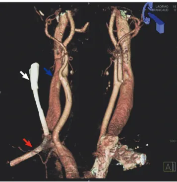
Fig. 2. Three-dimensional reconstruction of computed tomographic angiography of the neck through the inadvertently inserted catheter confirming that it (white arrow) had penetrated the vessel wall of the right internal jugular vein (blue arrow) and had been inserted into the right subclavian artery (red arrow).
Choi J-I, et al. • Unintended Arterial Cannulation of Hemodialysis Catheter
http://jkms.org 1267
http://dx.doi.org/10.3346/jkms.2012.27.10.1265 or cerebral emboli, and even fatality (4).
Compared to traditional blind central venous catheter place- ment using superficial anatomical landmarks, catheter place- ment under US guidance achieved an initial high success rates including fewer needle attempts, rapid vein localization and fewer complications (3, 5, 6). In fact, the use of US-guided CVC lowered the risk of complications by up to 73% (7). However, inadvertent arterial trauma or cannulation under US-guided CVC, as in our case, still occurs (7). There is evidence that nov- ice operators of ultrasonograms may lose track of the punctur- ing needle tip when using the short-axis method only, rather than both short- and long-axis trials, and this can lead to the accidental arterial cannulation (8). As in our case, accidental advancing of the puncture needle into an artery prior to inser- tion of the guide wire, with no further tracing under US guid- ance after confirmation of absence of pulsatile blood return or puncture of the right IJV, has been reported (9). Thus, the CVC of a large-bore catheter under US guidance is not always guar- anteed as a safe procedure free from the risk of inadvertent ar- terial cannulation.
Once inadvertent arterial cannulation of a large-bore cathe- ter is suspected, immediate management of arterial injury fol- lowing its confirmation by imaging studies is required to avoid complications from prolonged arterial cannulation such as thrombus at the site of arterial injury. Compared to surgical specialties (3, 4), workers in the nephrology field have seldom proposed definite guidelines on how to manage this accidental arterial cannulation (10, 11). Scattered case reports have docu- mented accidental arterial catheterization of CVC, but they barely mentioned methods of managing these complications and how to predict the likely success of the chosen option, i.e., pull/pressure with immediate catheter removal and manual external compression, open surgical exploration with catheter removal, or arterial repair under direct vision, and endovascu- lar interventions.
The pull/pressure technique has been well recognized for re- sulting in serious complications such as massive hemorrhage leading to respiratory distress, pseudoaneurysm, arteriovenous fistula, and strokes (12-14). Also, cases of bleeding after pull/
pressure technique require urgent open surgical intervention.
The probability of serious complications is usually dependent on catheter diameter, time since catheter insertion and punc- ture site (3). A retrospective analysis of 30 cases of arterial mis- placement with large-bore cannula (≥ 7.0 Fr) showed that, un- like the pull/pressure technique, managements by immediate surgical exploration and artery repair under direct vision caused no complication at all and was considered the most effective and safe treatment (46% vs 0%, P = 0.004) (4). Furthermore, a retrospective analysis of 57 cases based on published series and case reports of carotid artery or SCA catheterizations with large- bore cannula revealed that the pull/pressure technique was as-
sociated with significantly more complications than surgical or endovascular repair (relative risk, 17.86; P < 0.001) (4). In the same study, a single patient in the surgical repair group (1/14) developed a post-surgery embolic stroke due to delayed surgi- cal intervention after cannulation of the carotid artery for more than 72 hr (15). Thus, prompt open surgical repair has been no- ticed to be safe, but its usual requirement for general anesthesia and even sternotomy cause some morbidity in patients with se- rious underlying diseases including uremia (3). Recently, less invasive endovascular interventions, such as use of a covered stent or percutaneous arterial closure device or balloon tam- ponade, have been reported (16-18). These options are ideal for managing arterial injuries that are difficult to expose or inac- cessible surgically, such as in the proximal carotid or subclavian artery below or behind the clavicle (4). Although one recent ret- rospective analysis for the endovascular interventions of 13 pa- tients of inadvertent SCA catheterization was promising with clinical success rate of 100% and without need for surgical re- construction, complications requiring additional stent-graft placement were still noticed in 4 patients (30.8%) (19). There- fore, any suspected arterial catheterization during CVC should be confirmed by radiologic methods, and the catheter should be left in place without use of the pull/pressure technique while arranging for open surgical repair or the endovascular interven- tion with skilled experience, particularly if the location appears surgically inaccessible (3, 4).
Since the avoidance of accidental arterial cannulation is clear- ly preferable to its managements in CVC with a large bore cath- eter, the following elements are needed: 1) a properly organized instruction program for CVC under US guidance; 2) supervision of novice physicians by an experienced or interventional expert in CVC; and 3) US-guided cannulation with real-time fluoros- copy in patients at high risk. An observational cohort study com- paring traditionally-trained residents for CVC versus simula- tion-trained residents showed that the simulation-based mas- tery program reduced complications related to CVC in actual patient care (20).
In summary, to prevent and manage unintended arterial cannulation during large-bore HD catheter placement, 1) can- nulation should be performed by well trained and competent physicians under US guidance as a standard of care, and with real-time fluoroscopy, for high-risk patients; 2) an operator should check for excessive or pulsatile backflow through the initial puncturing needle and the cannulated catheter, as well as any expanding local hematoma, dyspnea or pain; 3) chest X- ray should be performed to check the position of the catheter tip after cannulation; 4) in the event of accidental arterial mis- placement, CT angiography or conventional angiography should be immediately arranged to establish which artery is injured and to analyze the regional vascular anatomy; 5) open surgical repair or endovascular intervention, if surgery is unsuitable or
Choi J-I, et al. • Unintended Arterial Cannulation of Hemodialysis Catheter
1268 http://jkms.org http://dx.doi.org/10.3346/jkms.2012.27.10.1265 experienced ones for it are available, should be promptly ar-
ranged; and 6) after surgical repair or endovascular interven- tion, prompt clinical evaluation should be performed to detect procedures-related neurologic deficits such as strokes.
REFERENCES
1. O’neill WC. The new nephrologist. Am J Kidney Dis 2000; 35: 978-9.
2. Wisborg T, Flaatten H, Koller ME. Percutaneous placement of perma- nent central venous catheters: experience with 200 catheters. Acta Anaes- thesiol Scand 1991; 35: 49-51.
3. Pikwer A, Acosta S, Kölbel T, Malina M, Sonesson B, Akeson J. Manage- ment of inadvertent arterial catheterisation associated with central ve- nous access procedures. Eur J Vasc Endovasc Surg 2009; 38: 707-14.
4. Guilbert MC, Elkouri S, Bracco D, Corriveau MM, Beaudoin N, Dubois MJ, Bruneau L, Blair JF. Arterial trauma during central venous catheter insertion: Case series, review and proposed algorithm. J Vasc Surg 2008;
48: 918-25.
5. Leung J, Duffy M, Finckh A. Real-time ultrasonographically-guided inter- nal jugular vein catheterization in the emergency department increases success rates and reduces complications: a randomized, prospective study.
Ann Emerg Med 2006; 48: 540-7.
6. Randolph AG, Cook DJ, Gonzales CA, Pribble CG. Ultrasound guidance for placement of central venous catheters: a meta-analysis of the litera- ture. Crit Care Med 1996; 24: 2053-8.
7. Yonei A, Nonoue T, Sari A. Real-time ultrasonic guidance for percutane- ous puncture of the internal jugular vein. Anesthesiology 1986; 64: 830-1.
8. Blaivas M. Video analysis of arterial cannulation with dynamic ultra- sound guidance for central venous access. J Ultrasound Med 2009; 28:
1239-44.
9. Stone MB, Hern HG. Inadvertent carotid artery cannulation during ul- trasound guided central venous catheterization. Ann Emerg Med 2007;
49: 720.
10. el-Shahawy MA, Khilnani H. Carotid-jugular arteriovenous fistula: a
complication of temporary hemodialysis catheter. Am J Nephrol 1995;
15: 332-6.
11. Patel HV, Sainaresh VV, Jaqin SH, Kute VB, Godara S, Gumber MR, Munjappa B, Gera DN, Shah PR, Trivedi HL. Cartotid-jugular venous fistula: a case report of an iatrogenic complication following internal jugular vein catherization for hemodialysis access. Hemodial Int 2011;
15: 404-6.
12. McEnany MT, Austen WG. Life-threatening hemorrhage from inadver- tent cervical arteriotomy. Ann Thorac Surg 1977; 24: 233-6.
13. Shah PM, Babu SC, Goyal A, Mateo RB, Madden RE. Arterial misplace- ment of large-caliber cannulas during jugular vein catheterization: case for surgical management. J Am Coll Surg 2004; 198: 939-44.
14. Nicholson T, Ettles D, Robinson G. Managing inadvertent arterial cath- eterization during central venous access procedures. Cardiovasc Inter- vent Radiol 2004; 27: 21-5.
15. Brown CQ. Inadvertent prolonged cannulation of the carotid artery.
Anesth Analg 1982; 61: 150-2.
16. Basile A, Calcara G, Rapisarda F, Fatuzzo P, Desiderio C, Granata A, Patti MT. Aberrant right subclavian artery laceration due to internal jugular vein catheterization treated by stent-graft implantation. J Vasc Access 2010; 11: 80-2.
17. Yu H, Stavas JM, Dixon RG, Burke CT, Mauro MA. Temporary balloon tamponade for managing subclavian arterial injury by inadvertent cen- tral venous catheter placement. J Vasc Interv Radiol 2011; 22: 654-9.
18. Sharma M, Sakhuja R, Teitel D, Boyle A. Percutaneous arterial closure for inadvertent cannulation of the subclavian artery--a call for caution.
J Invasive Cardiol 2008; 20: E229-32.
19. Chemelli AP, Wiedermann F, Klocker J, Falkensammer J, Strasak A, Czermak BV, Waldenberger P, Chemelli-Steinguber IE. Endovascular management of inadvertent subclavian artery catheterization during subclavian vein cannulation. J Vasc Interv Radiol 2010; 21: 470-6.
20. Barsuk JH, McGaghie WC, Cohen ER, O’Leary KJ, Wayne DB. Simula- tion-based mastery learning reduces complications during central venous catheter insertion in a medical intensive care unit. Crit Care Med 2009;
37: 2697-701.