• 검색 결과가 없습니다.

Indication and surgical techniques of bypass choledochojejunostomy for intractable choledocholithiasis

N/A
N/A
Protected

Academic year: 2021

Share "Indication and surgical techniques of bypass choledochojejunostomy for intractable choledocholithiasis"

Copied!
6
0
0

로드 중.... (전체 텍스트 보기)

전체 글

(1)

Indication and surgical techniques of bypass

choledochojejunostomy for intractable choledocholithiasis

Shin Hwang

1

, Dong-Hwan Jung

1

, Sung Koo Lee

2

, and Myung-Hwan Kim

2

Departments of

1

Surgery and

2

Internal Medicine, Asan Medical Center, University of Ulsan College of Medicine, Seoul, Korea

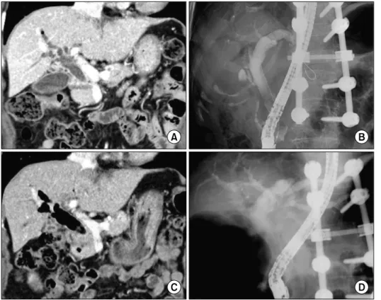
Despite development in endoscopic treatment and minimally invasive surgery for choledocholithiasis, there remains a small number of patients who require bypass Roux-en-Y choledochojejunostomy (RYCJ) because of the intractable occurrence of common bile duct (CBD) stones. We herein present the detailed procedures of open RYCJ customized for intractable choledocholithiasis. The first method is a side-to-end choledochojejunostomy with intraluminal closure of the distal CBD. This method was applied to a 79-year-old female patient who underwent endoscopic retrograde cholangiopancreatography (ERCP) more than 10 times in the past 14 years (Case No. 1). The distal CBD was explored through choledochotomy and then the distal CBD lumen was occluded with internal running sutures. A large-sized choledochojejunostomy was performed. The patient recovered uneventfully and has been doing well for the past 2 years. The second method is an end-to-end choledochojejunostomy with segmental CBD resection. It was applied to a 75-year-old male patient who underwent ERCP 9 times in the past 10 years (Case No. 2). The CBD was resected segmentally and a large-sized choledochojejunostomy was performed. The patient also recovered uneventfully and has been doing well for the past 2 years. In conclusion, the primary indication of bypass RYCJ is intractable chol- edocholithiasis which requires numerous sessions of endoscopic stone removal over a long period. Open RYCJ is the preferred procedure to date. If the papilla is patulous, the distal CBD should be occluded or resected to prevent reflux ascending cholangitis. We recommend to resect the intrapancreatic distal CBD if it is markedly dilated like chol- edochal cyst. (Ann Hepatobiliary Pancreat Surg 2021;25:259-264)

Key Words: Choledochojejunostomy; Endoscopic retrograde cholangiopancreatography; Benign stricture; Reflux;

Ascending cholangitis

Received: November 1, 2020; Revised: November 15, 2020; Accepted: November 15, 2020 Corresponding author: Shin Hwang

Department of Surgery, Asan Medical Center, University of Ulsan College of Medicine, 88 Olympic-ro 43-gil, Songpa-gu, Seoul 05505, Korea Tel: +82-2-3010-3930, Fax: +82-2-3010-6701, E-mail: shwang@amc.seoul.kr

Copyright Ⓒ 2021 by The Korean Association of Hepato-Biliary-Pancreatic Surgery

This is an Open Access article distributed under the terms of the Creative Commons Attribution Non-Commercial License (http://creativecommons.org/

licenses/by-nc/4.0) which permits unrestricted non-commercial use, distribution, and reproduction in any medium, provided the original work is properly cited.

Annals of Hepato-Biliary-Pancreatic Surgery ∙ pISSN: 2508-5778ㆍeISSN: 2508-5859

INTRODUCTION

Because of the recent development in endoscopic retro- grade cholangiopancreatography (ERCP) with endoscopic therapeutic intervention, the common bile duct (CBD) stone or choledocholithiasis has not been regarded as an eligible indication of surgical treatment any more.

1-9

How- ever, a small number of patients has experienced the re- peated occurrence of CBD stones, thus numerous sessions of endoscopic stone removal were required in such cases.

9

Such an intractable occurrence of CBD stones may be as- sociated with bottle-neck stasis of bile within the dilated CBD and reflux ascending cholangitis due to the patulous widening of the papilla. Bypass Roux-en-Y choledocho-

jejunostomy (RYCJ) can be a definitive treatment for in- tractable choledocholithiasis as it can prevent stone-pro- ducing bile stasis. RYCJ had been performed in a non- negligible number of patients with unresectable periam- pullary cancer and benign distal CBD stricture in the past;

however, currently, it is regarded as a surgical procedure

that is performed only in rare incidences. The surgical

procedure of RYCJ for intractable choledocholithiasis is

rather different from that for distal CBD stricture.

10,11

There exists an increasing number of case series regarding

laparoscopic or robotic RYCJ in the literature, but such

minimally invasive surgeries are not regarded as the pre-

ferred procedures primarily due to high medical cost and

high risk of surgical complications.

12-16

Thus, we herein

(2)

260 Ann Hepatobiliary Pancreat Surg Vol. 25, No. 2, May 2021 www.ahbps.org

Fig. 1. The preoperative finding of Case No. 1. Computed tomo- graphy (CT) and endoscopic retrograde cholangiopancreato- graphy (ERCP) taken 7 years before surgery (A, B) show marked dilatation of the com- mon bile duct with the patulous sphincter. CT scan and ERCP taken 1 month before surgery (C, D) show further dilatation of the common bile duct filled with air and sludges, indicating reflux ascending cholangitis.

The spine was externally fixed 11 years before due to spine fracture.

present the detailed procedures of open RYCJ customized for intractable choledocholithiasis.

SIDE-TO-END CHOLEDOCHO- JEJUNOSTOMY WITH INTRALUMINAL

CLOSURE OF THE DISTAL CBD

If the CBD is not very largely dilated, segmental re- section of the CBD is not essential to perform RYCJ.

Considering that RYCJ is usually performed for very old-aged patients who cannot tolerate repeated endoscopic stone removal any more, the aim of the surgery is focused on effective biliary bypass drainage with minimal risk of operative complications.

Just after cholecystectomy, the CBD is incised longi- tudinally to open the lumen. The distal CBD is explored with stone forceps, by which the opening status of the papilla can be examined. If the papilla is patulous permit- ting free passage of the curved stone forceps, the distal CBD should be closed to prevent reflux ascending cholan- gitis. The anterior wall of the CBD should be excised to make a large opening that would be suitable for anasto- mosis.

The detailed surgical procedures applied to a 79-year-

old female patient who underwent ERCP more than 10 times in the past 14 years (Case No. 1) (Fig. 1) are as follows: after cholecystectomy, the mid-portion of the CBD was incised longitudinally and the distal CBD was explored with a curved stone forceps. As the sphincter of Oddi was patulous, the lumen of the distal CBD at the upper board of the pancreas was securely closed with 5-0 Prolene running sutures. Subsequently, a part of the ante- rior wall of the CBD was elliptically excised to make a large opening that enabled a large choledocho- jejunostomy. A Roux-en-Y jejunal limb was anastomosed to the CBD opening in a side-to-side fashion (Fig. 2). The patient recovered uneventfully (Fig. 3) and has been doing well without any biliary problems for the past 2 years.

On follow-up imaging studies, a 1.5 cm-sized remnant portion of the distal CBD was visible within the pancreas head.

END-TO-END CHOLEDOCHO- JEJUNOSTOMY WITH SEGMENTAL

RESECTION OF THE CBD

If the CBD is markedly dilated, the abovementioned lu-

minal closure can leave a large blind-end space within the

(3)

Fig. 2. Intraoperative photograph showing the procedure of side-to-end choledochojejunostomy with intraluminal closure of the distal common bile duct (CBD) in Case No. 1. (A) The dilated CBD is visible after cholecystectomy. (B) The mid-portion of the CBD is incised longitudinally, showing markedly thickened CBD wall indicating repeated episodes of cholangitis. (C) The distal CBD is explored with curved stone forceps to examine the opening status of the sphincter. (D, E) The lumen of the distal CBD at the upper board of the pancreas is securely closed with 5-0 Prolene running sutures twice. The suture knots are made at the outer surface of the CBD (arrow). A part of the anterior wall of the CBD is excised to widen the opening.

(F-H) A Roux-en-Y jejunal limb is anastomosed to the CBD opening in a side-to-side fashion.

Fig. 3. Postoperative findings at two weeks after surgery in Case No. 1. (A) Computed tomography scan shows uneventful drainage of the bile from to proximal common bile duct (CBD) to the jejunal limb. The distal CBD is occluded (arrow). (B) Hepato- biliary scintigraphy shows a normal biliary excretion rate of 92.5% at 90 minutes. The distal CBD is occluded (arrow).

intrapancreatic bile duct as like a choledochal cyst, which can be a source of stone formation or pancreatitis. Thus, it is reasonable to remove the dilated CBD portion which can remain as a small blind-end lumen at the intra- pancreatic bile duct.

The detailed surgical procedures applied to a 75-year-old male patient who underwent ERCP 9 times in the past 10 years (Case No. 2) (Fig. 4) are as follows: after chol- ecystectomy, the CBD was incised longitudinally and the distal CBD was explored with a curved stone forceps.

Multiple CBD stones and sludges mixed with food materi-

als were removed from the distal CBD. As the distal CBD was markedly dilated and the papilla was patulous, the distal CBD was deeply dissected into the pancreas paren- chyma until the narrow portion of the intrapancreatic bile duct, and then transected and securely repaired with 5-0 Prolene sutures. The proximal CBD was also transected at the level of the right hepatic artery to make a large proximal bile duct opening. A Roux-en-Y jejunal limb was anastomosed to the CBD opening in an end-to-side fashion (Fig. 5). The patient recovered uneventfully (Fig.

6) and has been doing well with any biliary problems for

(4)

262 Ann Hepatobiliary Pancreat Surg Vol. 25, No. 2, May 2021 www.ahbps.org

Fig. 4. The preoperative finding of Case No. 2. Computed tomo- graphy (CT) and direct cholan- giography taken 10 years before surgery (A, B) show marked dilatation of the common bile duct with an obstructing mass at the sphincter (arrows), which was later proven to be a benign stricture. CT scan and magnetic resonance cholangiography taken 1 month before surgery (C, D) show that the common bile duct is filled with stones and sludges (arrows).

Fig. 5. Intraoperative photograph showing the procedure of end-to-end choledochojejunostomy with segmental resection of the common bile duct (CBD) in Case No. 2. (A) The dilated CBD is visible after cholecystectomy. (B) The mid-portion of the CBD is incised longitudinally, showing multiple CBD stones and sludges. (C) The distal CBD is dissected and transected at the narrow portion of the intrapancreatic bile duct, and then securely closed with 5-0 Prolene sutures. (D) The proximal CBD was also transected at the level of the right hepatic artery to make a large proximal bile duct opening. (E, F) A Roux-en-Y jejunal limb is anastomosed to the CBD opening in an end-to-side fashion.

the past 2 years. On follow-up imaging studies, a small portion of the remnant distal CBD was identified within the pancreas head.

DISCUSSION

In the era of prevailing minimally invasive surgeries

with laparoscopic or robotic approaches, open RYCJ is

(5)

Fig. 6. Postoperative findings at 10 days after surgery in Case No. 2. (A) Computed tomogra- phy shows uneventful drainage of the bile from to proximal common bile duct to the jejunal limb. (B) Hepatobiliary scinti- graphy shows a biliary excre- tion rate of 89.2% at 90 min- utes.

not a matter of concern.

12-16

Moreover, recent develop- ment in ERCP with endoscopic therapeutic intervention has led choledocholithiasis to be placed beyond the terri- tory of surgical indication.

1-9

On the contrary, we should not forget that there remains a small number of patients who still require open RYCJ because of the intractable oc- currence of CBD stones. Since such a surgical procedure has been rarely performed, many young surgeons major- ing in hepatobiliary surgery do not have enough experi- ence on the indications and surgical techniques for open RYCJ in the cases of patients with intractable choledo- cholithiasis.

The principles for surgical techniques toward achieving complication-free RYCJ include avoidance of unnecessary dissection of the CBD, making a large-sized biliary-en- teric anastomosis according to the fluid dynamics, and prevention of reflux ascending cholangitis. To avoid un- necessary dissection of the CBD, the luminal occlusion technique was used instead of segmental bile duct re- section in Case No. 1. As the wall of the dilated CBD is usually thickened over 2 mm, the elliptical side-wall excision should be performed to make a large opening since a simple longitudinal incision can be eventually nar- rowed due to the slit closing effect.

Unlike the abovementioned two points, some back- ground knowledge is necessary to understand why reflux ascending cholangitis should be prevented. If RYCJ is performed without interruption of distal CBD lumen, food materials would pass in a retrograde manner through the patulous papilla, which can be a source of frequent as- cending cholangitis and CBD stone formation. Based on our experience with 8 cases of conventional side-to-end

RYCJ without distal CBD interruption, episodes of reflux ascending cholangitis occurred repeatedly in at least 2 patients. A prospective study to determine the incidence of duodenobiliary reflux and acute cholangitis after place- ment of self-expanding metal stent across the main duode- nal papilla revealed that reflux of duodenal contents is a universal phenomenon.

17

A patulous papilla following multiple sessions of ERCP is vulnerable to reflux similar to the papilla state with a self-expanding metal stent. On the contrary, distal CBD interruption is not necessary for patients with distal CBD stenosis from benign or malig- nant diseases.

In the cases of usual patients with intractable chol- edocholithiasis, segmental bile duct resection is theoret- ically not necessary. However, if the CBD is markedly en- larged, leaving a large lumen within the distal CBD can make a blind-ended pool inducing infection or stone for- mation by food material regurgitation through the patulous ampulla. Thus, it is reasonable to resect the excessively dilated portion of the distal CBD, as demonstrated in Case No. 2.

We had used a refined technique of clustered bili-

ary-enteric anastomosis when the bile duct opening was

greater than 2 cm in diameter. Routine Roux-en-Y jejunal

limb was prepared and pulled up through the retrocolic

tunnel. A longitudinal incision was made at the ante-

mesenteric border of the jejunal limb and the posterior

wall anastomosis was performed with 2 or 3 segmented

continuous running sutures with 5-0 Prolene. Anterior

wall anastomosis was performed through multiple inter-

rupted sutures.

18

With the application of such customized

techniques for intractable choledocholithiasis, until date,

(6)

264 Ann Hepatobiliary Pancreat Surg Vol. 25, No. 2, May 2021 www.ahbps.org

we did not experience any noticeable surgical complica- tions yet 15 patients with age of 63-82 years.

In conclusion, we contemplate that the primary in- dication of bypass RYCJ is intractable choledocholithiasis which requires numerous sessions of ERCP over a long period. So far, open RYCJ has been the preferred proce- dure as it is much more reliable than the minimally in- vasive procedures regarding patient safety. If the papilla is patulous, distal CBD should be occluded or resected to prevent reflux ascending cholangitis. We recommend to resect the intrapancreatic distal CBD if it is markedly di- lated like a choledochal cyst because food material can regurgitate through the patulous ampulla.

CONFLICT OF INTEREST

None of the authors has any conflict of interest.

ORCID

Shin Hwang: https://orcid.org/0000-0002-9045-2531 Dong-Hwan Jung: https://orcid.org/0000-0001-5984-023X Sung Koo Lee: https://orcid.org/0000-0003-4155-4416 Myung-Hwan Kim: https://orcid.org/0000-0003-0992-6734

AUTHOR CONTRIBUTIONS

Conceptualization: SH. Data curation: SH, DHJ. Formal analysis: SH. Methodology: SH, SKL, MHK. Project ad- ministration: SH. Visualization: SH. Writing - original draft: SH. Writing - review & editing: All.

REFERENCES

1. Costi R, Gnocchi A, Di Mario F, Sarli L. Diagnosis and manage- ment of choledocholithiasis in the golden age of imaging, endos- copy and laparoscopy. World J Gastroenterol 2014;20:13382- 13401.

2. Nathanson LK, O'Rourke NA, Martin IJ, Fielding GA, Cowen AE, Roberts RK, et al. Postoperative ERCP versus laparoscopic choledochotomy for clearance of selected bile duct calculi: a

randomized trial. Ann Surg 2005;242:188-192.

3. Urbach DR, Khajanchee YS, Jobe BA, Standage BA, Hansen PD, Swanstrom LL. Cost-effective management of common bile duct stones: a decision analysis of the use of endoscopic retro- grade cholangiopancreatography (ERCP), intraoperative cholangio- graphy, and laparoscopic bile duct exploration. Surg Endosc 2001;

15:4-13.

4. Pring CM, Skelding-Millar L, Goodall RJ. Expectant treatment or cholecystectomy after endoscopic retrograde cholangiopancreato- graphy for choledocholithiasis in patients over 80 years old?

Surg Endosc 2005;19:357-360.

5. Maple JT, Ikenberry SO, Anderson MA, Appalaneni V, Decker GA, Early D, et al. The role of endoscopy in the management of choledocholithiasis. Gastrointest Endosc 2011;74:731-744.

6. Shim CS. How should biliary stones be managed? Gut Liver 2010;

4:161-172.

7. Sugiyama M, Atomi Y. Endoscopic sphincterotomy for bile duct stones in patients 90 years of age and older. Gastrointest Endosc 2000;52:187-191.

8. Moon JH, Cha SW, Ryu CB, Kim YS, Hong SJ, Cheon YK, et al. Endoscopic treatment of retained bile-duct stones by using a balloon catheter for electrohydraulic lithotripsy without chol- angioscopy. Gastrointest Endosc 2004;60:562-566.

9. Sugiyama M, Suzuki Y, Abe N, Masaki T, Mori T, Atomi Y.

Endoscopic retreatment of recurrent choledocholithiasis after sphincterotomy. Gut 2004;53:1856-1859.

10. Kadaba RS, Bowers KA, Khorsandi S, Hutchins RR, Abraham AT, Sarker SJ, et al. Complications of biliary-enteric anastomoses.

Ann R Coll Surg Engl 2017;99:210-215.

11. Vogt DP, Hermann RE. Choledochoduodenostomy, chol- edochojejunostomy or sphincteroplasty for biliary and pancreatic disease. Ann Surg 1981;193:161-168.

12. Hori T, Aisu Y, Yamamoto M, Yasukawa D, Iida T, Yagi S, et al. Laparoscopic approach for choledochojejunostomy. Hepato- biliary Pancreat Dis Int 2019;18:285-288.

13. Hori T. Comprehensive and innovative techniques for laparo- scopic choledocholithotomy: a surgical guide to successfully ac- complish this advanced manipulation. World J Gastroenterol 2019;25:1531-1549.

14. Han HS, Yi NJ. Laparoscopic Roux-en-Y choledochojejunostomy for benign biliary disease. Surg Laparosc Endosc Percutan Tech 2004;14:80-84.

15. Ruurda JP, van Dongen KW, Dries J, Borel Rinkes IH, Broeders IA. Robot-assisted laparoscopic choledochojejunostomy. Surg Endosc 2003;17:1937-1942.

16. Benzie AL, Sucandy I, Spence J, Ross S, Rosemurgy A. Robotic choledochoduodenostomy for benign distal common bile duct stricture: how we do it. J Robot Surg 2019;13:713-716.

17. Misra SP, Dwivedi M. Reflux of duodenal contents and chol- angitis in patients undergoing self-expanding metal stent placement. Gastrointest Endosc 2009;70:317-321.

18. Hwang S, Ha TY, Song GW, Jung DH. Cluster hepaticojejuno-

stomy with radial spreading anchoring traction technique for se-

cure reconstruction of widely opened hilar bile ducts. Korean J

Hepatobiliary Pancreat Surg 2016;20:66-70.

수치

Fig. 1. The preoperative finding  of Case No. 1. Computed  tomo-graphy (CT) and endoscopic  retrograde  cholangiopancreato-graphy (ERCP) taken 7 years  before surgery (A, B) show  marked dilatation of the  com-mon bile duct with the patulous  sphincter
Fig. 3. Postoperative findings at  two weeks after surgery in Case  No. 1. (A) Computed tomography  scan shows uneventful drainage  of the bile from to proximal  common bile duct (CBD) to the  jejunal limb
Fig. 4. The preoperative finding  of Case No. 2. Computed  tomo-graphy (CT) and direct  cholan-giography taken 10 years before  surgery (A, B) show marked  dilatation of the common bile  duct with an obstructing mass  at the sphincter (arrows), which  was
Fig. 6. Postoperative findings at  10 days after surgery in Case  No. 2. (A) Computed  tomogra-phy shows uneventful drainage  of the bile from to proximal  common bile duct to the jejunal  limb

참조

관련 문서

Post-surgical use of radioiodine (131I) in patients with papillary and follicular thyroid cancer and the issue of remnant ablation: a consensus

Method We obtained blood samples from 49 patients who underwent surgical treatment for esophageal squamous cell cancer (ESCC) We performed ctDNA analysis with

Endoscopic spine surgery has become a practical, minimally invasive technique for decompression in patients with spinal disc herniation or stenosis.. However,

The aims of this study are to evaluate the endoscopic modality for assessment of severity of ulcerative colitis during follow up and to know whether there

Percutaneous endoscopic cervical discectomy (PECD) is an effective minimally invasivesurgery for soft cervical disc herniation in properly selected cases..

This showed that the surgical outcome was successfulin 269 patients (98.89%),butthe revision surgery due to surgicalrelapse was performed in three patients.Of 152 patients

Recently, for lumbar disc herniation, Trans-Sacral Epiduroscopic Laser Decompression (SELD) has been a minimally invasive procedure before surgery.. Especially,

Polypoid lesions of the gallbladder; Report of 111 cases with surgical reference to operative indication.. Polypoid lesion of GB: Indication of carcinoma