Effects of Myelophycus Simplex Papenfuss Methanol Extract on Adipocyte Differentiation and Adipogenesis in 3T3-L1 Preadipocytes
Hyang Suk Kim1, Da Hye Kwon2, Ji Min Cheon2, Eun Ok Choi3, Ji Hyun Kim3, Min Ho Han4, Yung Hyun Choi1,4,6, Byung Woo Kim5,6 and Hye Jin Hwang2,6*
1Anti-aging Research Center, Dong-eui University, Busan 614-714, Korea
2Department of Food and Nutrition, Dong-eui University, Busan 614-714, Korea
3Department of Food and Nutrition, Pusan National University, Busan 609-735, Korea
4Department of Oriental Medicine, Dong-eui University, Busan 614-714, Korea
5Department of Life Science and Biotechnology, Dong-eui University, Busan 614-714, Korea
6Blue Bio Industry RIC., Dong-eui University, Busan 614-714, Korea
Received December 3, 2014 /Revised December 12, 2014 /Accepted January 6, 2015
Myelophycus simplex Papenfuss is distributed over the northern Pacific and southern coast of Korea, and is a member of the brown algae family. The objective of this study was to investigate the effect of M. simplex methanol extract on adipocyte differentiation and adipogenesis in 3T3-L1 preadipocytes.
Treatment with M. simplex methanol extract significantly suppressed terminal differentiation of 3T3-L1 preadipocytes in a dose-dependent manner, as confirmed by a decrease in lipid droplet content ob- served by Oil Red O staining. Also, the M. simplex methanol extract significantly suppressed the tri- glyceride content of 3T3-L1 preadipocytes in a dose-dependent manner. Treatment with 300 and 500 μg/ml of M. simplex methanol extract caused a 42% and 76% reduction in lipid droplet content, respectively. In order to understand the anti-adipogenic effects of M. simplex methanol extract, the changes in the expression of several adipogenic transcription factors, including peroxisome pro- liferator-activated receptor (PPAR) γ-cytidine-cytidine-adenosine-adenosine-thymidine (CCAAT) /enhancer binding protein (C/EBP) α and β, were investigated using immunoblotting. M. simplex suppressed the expression of PPARγ, C/EBPα, and C/EBPβ proteins compared with control. Therefore, the results of this study suggest that M. simplex methanol extract inhibits adipocyte differentiation and thus may have applications as a potential source for an anti-obesity functional food agent.
Key words : Adipogenesis, Myelophycus simplex Papenfuss, triglyceride, 3T3-L1
*Corresponding author
*Tel : +82-51-890-1594, Fax : +82-51-890-2646
*E-mail : [email protected]
This is an Open-Access article distributed under the terms of the Creative Commons Attribution Non-Commercial License (http://creativecommons.org/licenses/by-nc/3.0) which permits unrestricted non-commercial use, distribution, and reproduction in any medium, provided the original work is properly cited.
Journal of Life Science 2015 Vol. 25. No. 1. 62~67 DOI : http://dx.doi.org/10.5352/JLS.2015.25.1.62
서 론
비만은 체내에 지방이 과잉 축적되어 있는 상태를 말하며 adipogenesis (지방세포 분화) 과정에 의하여 생성되는 지방세 포의 비대 및 과형성에 의한 중성지방의 축적에 의하여 유발 되는 것으로 알려져 있다[1, 18]. 이러한 지방세포 내에 과도한 지방의 축적은 당뇨병, 고혈압, 심혈관 질환 및 암을 포함한 만성질병 유발의 원인이 되기도 한다[7].
지방세포 분화과정은 세포형태, 호르몬 감수성 및 유전자 발현의 다양한 변화가 동반되는 복합적인 과정이며, 이 과정 에 관여하는 중요한 인자로는 peroxisome proliferator acti-
vated receptor γ (PPARγ)와 CCAAT/enhancer-binding pro- tein α (C/EBPα)가 있다[3, 15]. 지방세포 분화과정의 핵심 조 절인자인 PPARγ와 C/EBPα는 서로 상호작용하여 상승효과 를 가지며 분화 초기에 발현되는 C/EBPs에 의해 유전자 발현 이 증가하고, 지방세포가 분화되는 동안 adiponectin과 glu- cose transporter 4 (GLUT4), fatty acid synthethase와 같은 지방세포 분화과정의 terminal marker를 조절한다[8, 14].
Adipogenesis 경로에서 PPARγ와 C/EBPα의 상위에 위치하 는 C/EBPβ와 C/EBPδ는 세포분열 유도물질과 호르몬의 자극 에 의해 분화 초기에 빠르게 증가되어 PPARγ와 C/EBPα의 발현을 증가시키고 분화의 후기에는 감소한다[11, 13]. C/EBP β와 C/EBPδ 이외에도 PPARγ와 C/EBPα의 상위에 위치한 다양한 지방세포 형성 유도 및 억제 조절인자들이 분화의 초 기와 후기에 발현되거나 감소되면서 adipogenesis를 조절한 다[13, 19].
바위수염(Myelophycus simplex Papenfuss)은 갈조류로서 넓 은 미역쇠과에 속하며, 갈색 또는 흑갈색의 바늘 모양으로 외 가닥이며 여러 개가 모여 다발을 이룬다. 크기를 보면 높이
5~15 cm, 굵기 1~2 mm 정도이며, 사상형 또는 비틀어진 형태 로 구성되어 있다. 바위수염의 주된 분포지는 북태평양 서안 과 우리나라의 남부해안 전역 및 일본에 분포하는 것으로 알 려져 있으며 봄철 조간대 상부에 불등풀가사리와 함께 생육하 는 것으로 조사되어 있다[10]. 갈조류는 항암과 항염증 생리활 성을 갖는 fucoidan과 laminarin이라는 각각의 성분을 함유하 는 것으로 알려져 있다[9, 16]. 바위수염도 fucoidan을 함유하 여 여러 생리활성을 가질 것으로 보이나, 현재까지는 바위수 염의 기능성에 대한 연구는 신경세포 보호효과에 대한 연구 [17] 외에는 거의 없는 실정이다.
따라서 본 연구에서는 현재 식용으로 이용되지 않고 있으며 기능성이 거의 연구되지 않은 소재인 갈조류 바위수염을 이용 하여, 이의 항비만 관련 활성을 탐색하고자 하여 비만예방 기 능성 식품소재로서의 가능성을 검증해보고자 하였다.
재료 및 방법
시료
본 연구에 사용된 바위수염 메탄올 추출물(Myelophycus simplex Papenfuss methanol extracts)은 제주 유용생물자원추 출물은행(Jeju Bio-Resource Extract Bank, Jeju, Korea)에서 구 입하여 이용하였다.
실험재료
본 실험에서 단백질 분석을 위하여 사용된 PPARγ, C/EBP α, C/EBPβ 및 actin 항체는 Santa Cruz Biotechnology Inc.
(Santa Cruz, CA, USA) 및 Cell Signaling Technology (Beverly, Ma, USA)에서 구입하였다. Immunoblotting을 위해 2차 항체로 사용된 peroxidase-labeled donkey anti-rabbit 및 peroxidase-labeled sheep anti-mouse immunoglobulin은 Amersham Life Science Corp. (Arlington Heights, IL, USA) 에서 구입하였다. 또한 3T3-L1 지방전구세포의 분화를 위하여 사용된 insulin, dexamethasone 및 IBMX와 지방세포 내 tri- glyceride 생성을 확인하기 위하여 사용된 Oil Red O 시약은 Sigma-Aldrich (St. Luis, MO, USA)에서 구입하였다.
세포배양
실험에 사용된 3T3-L1 지방전구세포는 90%의 Dulbecco’s Modified EaDCRT Media (DMEM, Gibco-BRL, Grand Island, NY, USA), 10%의 bovine calf serum (BCS, Gibco BRL) 및 1%의 penicillin 및 streptomycin (Gibco BRL)이 포함된 배지 를 사용하여 37℃, 5% CO2조건 하에서 배양하였다. FBS는 잔 존하는 보체성분을 불활성화 시키기 위해 실온에서 녹인 후 heat inactivation (50℃ water bath에서 30분간 가열)하여 사 용하였다. 세포수의 증식에 따른 과밀도 현상을 해소하기 위 하여 성장배지의 교환을 매 48시간마다 실시하여 적정수의
세포를 유지하였다.
MTT assay
각 물질들의 세포독성 측정과 더불어 실험에 사용될 농도 범위 결정하기 위하여 MTT assay를 실시하였다. 세포 배양용 6 well plate에 3T3-L1 지방전구세포를 분주하여 confluent 상 태까지 배양한 후 바위수염 메탄올 추출물을 100, 300, 500 μg/ml의 농도로 처리하였다. 72시간 후 희석된 0.5 mg/ml 농도의 tetrazolium bromide salt (MTT, Amresco, Solon, OH, USA)를 200 μl씩 분주하고 2시간 동안 CO2 incubator에서 배 양시킨 다음 배지와 MTT 시약을 깨끗하게 제거하였다.
DMSO를 1 ml씩 분주하여 각 well에 생성된 formazan을 모두 녹인 후 96 well plate에 200 μl씩 옮겨서 ELISA reader (Molecular Devices, Sunnyvale, CA, USA)로 540 nm에서 흡 광도를 측정하였다. 각 세포에 대한 독성은 각각의 대조군의 평균 흡광도 값을 구하여 평균 흡광도 값에 대한 백분율로 나타내었다.
지방세포 분화유도
3T3-L1 지방전구세포를 세포 배양용 6 well plate에서 con- fluent 상태까지 배양한 후 10%의 fetal bovine serum (FBS, Gibco BRL)이 포함된 분화배지로 교환하여 2일간 더 배양하 였다. 배양한 후 10 μg/ml insulin, 1 μM dexamethasone 및 0.5 mM 3-isobutyl-1-methylxanthine (IBMX)을 첨가하여 2일 간 배양하였다. 그 후 매 2일마다 10 μg/ml insulin이 포함된 배지로 교환하였으며, 8일간 지방전구세포의 분화를 유도하 였다.
Oil Red O staining
지방전구세포 분화시 형성된 지방의 함량을 확인하기 위하 여 Oil Red O 염색을 실시하였다. 준비된 3T3-L1 지방전구세 포를 PBS로 세척한 후 3.7% formalin으로 10분간 고정하고 60% isopropanol을 이용하여 세척한 다음 Oil Red O solution 을 처리하여 실온에서 20분 간 염색하였다. 염색 후 Oil Red O solution을 제거하고 PBS로 4회 세척한 다음 염색된 세포를 위상차 현미경을 이용하여 관찰하였다. 또한, Oil Red O 염색 후 정량적 분석을 위하여 100% isopropanol을 이용하여 지방 을 추출한 후 96 well plate에 200 μl씩 옮겨서 ELISA reader로 500 nm에서 흡광도를 측정하였고, 대조군의 흡광도 값에 대한 백분율로 나타내었다.
Western blot analysis
동일한 방법으로 준비된 3T3-L1 지방전구세포에 적당량의 lysis buffer [25 mM Tris-Cl (pH 7.5), 250 mM NaCl, 5 mM EDTA, 1% NP-40, 1 mM phenylmethanesulfonylfluoride (PMSF), 5 mM dithiothreitol (DTT)] 를 첨가하여 4℃에서 1시
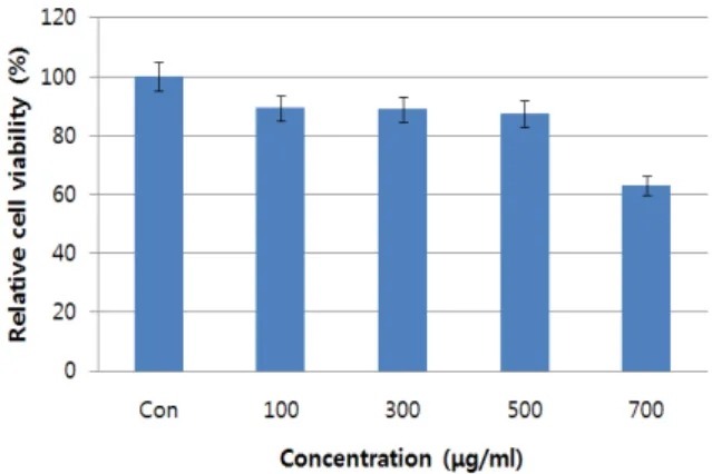
Fig. 1. Effect of Myelophycus simplex Papenfuss methanol extract on the cell viability in 3T3-L1 mouse preadipocytes by the MTT assay. 3T3-L1 cells were cultured with various concentrations Myelophycus simplex Papenfuss methanol extract for 72 hr. Data were expressed as percentage of control. Con; untreated cells.
간 동안 반응시킨 후, 14,000 rpm으로 30분간 원심 분리하여 상층액에 있는 총 단백질을 분리하였다. 상층액의 단백질 농 도는 Bio-Rad 단백질 정량 시약(Bio-Rad, Hercules, CA, USA) 과 그 사용방법에 따라 정량 한 다음 동량의 Laemmli sample buffer (Bio-Rad)를 섞어서 sample을 만들었다. 동량의 sam- ple을 sodium dodecyl sulphate (SDS)-polyacrylamide gel을 이용하여 전기영동으로 분리한 후, PVDF membrane (Bio- Rad, USA)으로 electroblotting에 의해 전이시켰다. 분리된 단 백질이 전이된 PVDF membrane을 5% skim milk를 처리하여 비특이적인 단백질들에 대한 blocking을 실시하고 1차 항체를 처리하여 상온에서 2시간 이상 또는 4℃에서 over night 시킨 다음 PBS-T로 세척(10분간 4번)하고 처리된 1차 항체에 맞는 2차 항체(PBS-T로 1:1,000으로 희석하여 사용)를 사용하여 상 온에서 1시간 정도 반응시켰다. 반응이 끝난 후 암실에서 Enhanced Chemiluminoesence (ECL) solution (Amersham Life Science Corp.)을 적용시킨 다음 X-ray film에 감광시켜 특정 단백질의 발현 양을 분석하였다.
Statistical analysis
모든 실험 결과는 Statistical Package for the Social Sciences (SPSS) 통계 프로그램을 이용하여 평균(mean)±표준편차 (S.D.)로 나타냈다. 각 실험군의 분석 항목별 통계의 유의성은 Student t-test와 Duncan's multiple range test를 이용하여 p<0.05 수준에서 검증하였다.
결과 및 고찰
바위수염 메탄올 추출물이 3T3-L1 세포 생존율에 미치는 영향
3T3-L1 지방전구세포에서 바위수염 메탄올 추출물의 세포
독성을 조사하기 위해서 100~700 μg/ml의 농도에서 MTT as- say 실험을 수행하였다. 바위수염 메탄올 추출물을 처리하였 을 때 대조군에 비해 세포 증식의 변화 정도를 퍼센트로 계산 하여 바위수염 메탄올 추출물이 세포독성을 나타내는지 조사 하였다. 바위수염 메탄올 추출물의 농도에 따른 3T3-L1 세포 의 성장에 미치는 영향을 MTT assay로 분석한 결과는 Fig. 1과 같다. 100~500 μg/ml의 농도에서는 80% 이상의 비슷한 수준 의 세포생존율을 보였다. 따라서 생존율 및 증식에 큰 영향을 미치지 않는 500 μg/ml 농도까지를 실험 조건으로 설정하였 다(Fig. 1).
바위수염 메탄올 추출물이 3T3-L1 세포의 lipid droplet 및 TG 생성에 미치는 영향
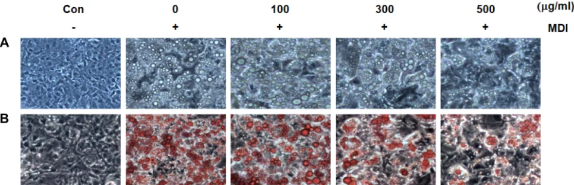
바위수염 메탄올 추출물의 처리가 지방전구세포의 지방세 포 분화 및 triglyceride 축적에 미치는 영향을 확인하기 위하 여 바위수염 메탄올 추출물을 적정농도로 처리하여 지방전구 세포에서 성숙지방세포로 분화를 유도하였다. 분화를 유도한 후 Oil Red O 염색 전후로 구분하여 지방구 생성 정도를 위상 차 현미경으로 관찰하였다. Fig. 2에서 볼 수 있듯이 바위수염 메탄올 추출물을 처리하지 않고 분화 유도하였을 때 세포질 내 지방구 형성이 활발하게 유발되는 것을 관찰하였다. 하지 만 바위수염 메탄올 추출물을 농도별로 처리하였을 때 지방구 의 형성이 처리 농도 의존적으로 억제되었으며, 특히 500 μg/
ml의 처리군에서 현저하게 감소되었다. 또한 바위수염 메탄올 추출물을 100, 300, 500 μg/ml 처리한 후의 중성지방의 양은, 바위수염 메탄올 추출물을 처리하지 않았을 때보다 35%, 42%, 76%로 농도 의존적으로 억제되는 것을 확인하였다(Fig. 3). 따 라서 바위수염 메탄올 추출물이 3T3-L1 지방전구세포에서 지 방세포로의 분화 억제 효과가 높았음을 알 수 있었다.
갈조류의 항비만 효과에 대한 실험으로는 톳을 대상으로 한 연구에서[2] 유기용매(EtOAc, n-BuOH, H2O, EtOH, CH2Cl2) 를 달리하여 추출한 각각의 분획물이 3T3-L1 지방전구세포에 서 지방구(lipid droplet) 및 중성지방의 생성을 억제하는 효과 가 나타났고, 특히 H2O 처리군에서 이러한 현상이 가장 강하 게 나타났다. 바위수염에 포함된 fucoidan의 항비만 효과에 대한 연구[12]에서 보면 3T3-L1 지방 세포에 용량 의존적으로 fucoidan을 처리한 후 Oil Red O 염색을 이용하여 지방구 형 성에 미치는 영향을 본 결과, 200 μg/ml의 농도에서 높은 지질 억제 활성을 보였다고 보고되었다(p<0.001). 또한 제주도에 서 식하는 식물자원들로부터 유용한 항비만 소재를 개발하기 위 한 기초연구에서는[5] 300 여종의 식물 추출물들을 대상으로 3T3-L1 전구지방세포의 분화억제 활성을 탐색하여, 지방분화 억제활성이 우수한 3종(미역쇠, 진귤, 제주조릿대)의 추출물들 과 미역쇠와 진귤에서 분리한 fucoxanthin, sinensetin의 항비 만 효과를 보고하였다. 또한 감태의 폴리페놀 추출물(SPN)의 지방억제 효과에 관한 연구에서[20] 고지방식이를 급여한 마
A
B
Fig. 2. A) Effect of Myelophycus Simplex Papenfuss methanol extract on the microscopic morphological changes of differentiated 3T3-L1 mouse preadipocytes. Differentiation of confluent 3T3-L1 mouse preadipocytes was initiated with MDI (0.5 mM 3-iso- butyl-1-methyl-xanthine, 1 μM dexamethasone, and 10 μg/ml insulin) and maintained DMEM-10% FBS medium in the absence or presence of Myelophycus simplex Papenfuss methanol extract. After day 8, differentiating 3T3-L1 cells were visualized by light microscopy. Magnification, ×200. B) Inhibitory effect of Myelophycus Simplex Papenfuss methanol extract on lipid droplet accumulation of differentiated 3T3-L1 mouse preadipocytes. Differentiation of confluent 3T3-L1 mouse preadipocytes was initiated with MDI and maintained DMEM-10% FBS medium in the absence or presence of Myelophycus Simplex Papenfuss methanol extract. After day 8, cells were fixed and stained with Oil Red O to visualize lipid droplets by light microscopy.
Magnification, ×200.
Fig. 4. Effects of Myelophycus simplex Papenfuss methanol ex- tracts on the levels of adipogenic transcription factors protein expression in differentiated 3T3-L1 mouse pre- adipocytes. Differentiation of confluent 3T3-L1 mouse preadipocytes was incubated with the absence or pres- ence of Myelophycus simplex Papenfuss methanol extracts for 8 day after initiated with MDI. Cells were lysed and cellular proteins were separated by SDS-polyacrylamide gels and transferred onto nitrocellulose membranes. The membranes were probed with the indicated antibodies.
Proteins were visualized using an ECL detection system.
Actin was used as an internal control.
Fig. 3. Inhibitory effect of Myelophycus simplex Papenfuss meth- anol extract on triglyceride accumulation of differ- entiated 3T3-L1 mouse preadipocytes. Triglyceride con- tents were determined by Oil Red O staining after treat- ment of the absence or presence of Myelophycus simplex Papenfuss methanol extract. The rates of triglyceride contents were measured at λ=500 nm wavelength by the ELISA reader. Data are expressed as the means ± SD of three separate experiments.
우스에 SPN과 dieckol 투여가 총콜레스테롤, 중성지방, LDL 콜레스테롤 수준을 상당히 감소시켰고, 또한 3T3-L1의 pre- adipocytes를 이용한 Oil Red O 염색에서 SPN, dieckol 모두 현저히 3T3-L1지방 세포의 축적을 억제한 것으로 보고되었다.
바위수염 메탄올 추출물이 adipogenic transcription factor 발현에 미치는 영향
바위수염 추출물이 adipogenic transcription factor들의 발 현에 어떠한 영향을 미치는지를 단백질 수준에서 확인하여
Fig. 4에 나타내었다. 분화유도인자인 insulin, dexamethasone 및 IBMX를 처리하여 분화를 유발하였을 경우 PPARγ, C/EBP α 및 C/EBPβ의 발현이 현저하게 증가하는 것으로 나타났으 며, 이러한 분화유도 과정에서 바위수염 추출물을 처리한 결 과 100 μg/ml의 농도보다 500 μg/ml의 처리 군에서 PPARγ, C/EBPα 및 C/EBPβ의 발현이 단백질 수준에서 감소하였다.
갈조류가 3T3-L1 지방세포에서 비만 관련 유전자 발현에 미치는 영향을 본 연구에서[4] 곰피를 분획(MeOH, CH2Cl2, EtOAc, BuOH)하였을 때 CH2Cl2 분획 추출물이 지방축적을 저해하는 효과가 가장 높았고, 농도 의존적으로 PPARγ, C/
EBPα도 감소하는 것으로 보고되었다. 또한 미역의 지방세포 분화억제에 대한 연구에서[6] 미역 에탄올 추출물은 지방세포 에서 triglyceride의 축적을 40% 감소시켰으며, 미역 에탄올추 출물의 농도가 높아질수록 PPARγ와 leptin의 발현이 억제되 었음이 보고된 바 있다.
본 연구 결과 갈조류인 바위수염은 지방전구세포에서 adi- pogenic transcription factor인 PPARγ, C/EBPα 및 C/EBPβ 의 발현을 효과적으로 억제하여 지방세포로의 분화를 억제시 키는 것으로 나타나, 항비만 건강 기능 식품 소재로서의 가치 가 충분하다고 본다.
감사의 글
본 연구는 2014년 동의대학교 교내연구비(2014AA214)와 산업통상자원부▪부산광역시 지원 동의대학교 블루바이오 소재 개발 및 실용화 지원 센터(RIC 08-06-07)에 의해 연구되 었으며 이에 감사드립니다.
References
1. Chen, Z., Torrens, J. I., Anand, A., Spiegelman, B. M. and Friedman J. M. 2005. Krox20 stimulates adipogenesis via C/EBPbeta-dependent and -independent mechanisms. Cell Metab. 1, 93-106.
2. Choi, E. O., Kim, H. S., Han, M. H., Choi, Y. H., Kim, B.
W., Hwang, J. A. and Hwang, H. J. 2012. Effects of Hizikia fusiforme extracts on adipocyte differentiation and adipo- genesis in 3T3-L1 preadipocytes. J. Life Sci. 22, 1399-1406.
3. Darlington, G. J., Ross, S. E. and MacDougald, O. A. 1998.
The role of C/EBP genes in adipocyte differentiation. J. Biol.
Chem. 273, 30057-30060.
4. Jung, H. A., Jung, H. J., Jeong, H. Y., Kwon, H. J., Kim, M. S. and Choi, J. S. 2014. Anti-adipogenic activity of the edible brown alga Ecklonia stolonifera and its constituent fu- costerol in 3T3-L1 adipocytes. Arch. Pharm. Res. 37, 713-720.
5. Kang, S. I. 2012. Anti-obesity effects of Petalonia binghamiae, Citrus sunki, and Sasa quelpaertensis. Doctoral dissertation.
Jeju National University, Jeju.
6. Kim, H. J., Kang, C. H. and Kim, S. K. 2012. Anti-adipogenic effect of Undaria pinnatifida extracts by ethanol in 3T3-L1 adipocytes. J. Life Sci. 22, 1052-1056.
7. Kopelman, P. G. 2000. Obesity as a medical problem. Nature 404, 635-643.
8. Morrison, R. F. and Farmer, S. R. 2000. Hormonal signaling and transcriptional control of adipocyte differentiation. J.
Nutr. 130, 3116S-3121S.
9. Noda, H., Amano, H., Arshima, K., Hashimoto, S. and Nisizawa, W. 1989. Studies on the antitumor activity of ma- rine algae. Bull. Japan Soc. Sci. Fish. 55, 1259-1264.
10. Oh, B. G., Lee, J. W. and Lee, H. B. 2002. A summer marine benthic algal flora and community of Uninhabited islands in Haenamgun, southern coast of Korea. J. Kor. Fish. Soc.
35. 57-63.
11. Owen, T. A., Aronow, M., Shalhoub, V., Barone, L. M., Wilming, L., Tassinari, M. S., Kennedy, M. B., Pockwinse, S., Lian, J. B. and Stein, G. S. 1990. Progressive development of the rat osteoblast phenotype in vitro: reciprocal relation- ships in expression of genes associated with osteoblast pro- liferation and differentiation during formation of the bone extracellular matrix. J. Cell Physiol. 143, 420-430.
12. Park, M. K., Jung, U. and Roh, C. 2011. Fucoidan from ma- rine brown algae inhibits lipid accumulation. Mar. Drugs.
9. 1359-1367.
13. Roncari, D. A., Lau, D. C. and Kindler, S. 1981. Exaggerated replication in culture of adipocyte precursors from mas- sively obese persons. Metabolism 30, 425-427.
14. Rosen, E. D. and Macdougald, O. A. 2006. Adipocyte differ- entiation from the in side out. Nat. Rev. Mol. Cell Biol. 7, 885-896.
15. Rosen, E. D., Hsu, C. H., Wang, X., Sakai, S., Freeman, M.
W., Gonzalez, F. J. and Spiegelman, B. M. 2001. C/EBPα induces adipogenesis through PPARγ: a unified pathway.
Genes Dev. 16, 22-26.
16. Schwartsmann, G., Brondani da Rocha, A., Berlinck, R. G.
and Jimeno, J. 2001. Marine organisms as a source of new anticancer agents. Lancet Oncol. 2, 221-225.
17. Shi, D. B., Han, E. H. and Park, S. S. 2014. Cytoprotective effects of phaeophyta extracts from the coast of Jeju island in HT-22 mouse neuronal cells. J. Kor. Soc. Food Sci. Nutr.
43, 224-230.
18. Visscher, T. L. and Seidell, J. C. 2001. The public health im- pact of obesity. Annu. Rev. Publ. Health 22, 355-375.
19. Wu, Z., Xie, Y., Bucher, N. L. and Farmer, S. R. 1995.
Conditional ectopic expression of C/EBP beta in NIH-3T3 cells induces PPAR gamma and stimulates adipogenesis.
Genes Der. 9, 2350-2363.
20. Yeo, A. R., Lee, J., Tae, I. H., Park, S. R., Cho, Y. H., Lee, B. H., Shin, H. C., Kim, S. H. and Yoo, Y. C. 2012. Anti-hy- perlipidemic effect of polyphenol extract (Seapolynol(™)) and Dieckol isolated from Ecklonia cava in in vivo and in vitro models. Prev. Nutr. Food Sci. 17, 1-7.
초록:바위수염 메탄올 추출물이 3T3-L1 지방전구세포의 분화에 미치는 영향 김향숙1․권다혜2․천지민2․최은옥3․김지현3․한민호4․최영현4,6․김병우5,6․황혜진2,6*
(1동의대학교 항노화연구소, 2동의대학교 식품영양학과, 3부산대학교 식품영양학과, 4동의대학교 한의학과,
5동의대학교 생명응용학과, 6블루바이오 소재개발 및 실용화지원센터)
본 연구는 바위수염 메탄올 추출물이 3T3-L1 지방세포의 분화 및 지방생성의 억제에 미치는 영향을 탐색하고 자 하였다. 바위수염 메탄올 추출물의 농도에 따른 3T3-L1 세포의 성장에 미치는 영향을 MTT assay로 분석한 결과 100~500 μg/ml의 농도에서는 80% 이상의 비슷한 수준의 세포생존율을 보였다. 바위수염 메탄올 추출물을 처리하지 않고 분화를 유도하였을 경우에 세포질 내 지방구의 형성이 활발하게 유발되는 것으로 관찰되었으며, 바위수염 메탄올 추출물에 의한 지방구의 형성이 처리 농도 의존적으로 억제 되는 것을 확인하였다. 또한 바위수 염 메탄올 추출물을 100, 300, 500 μg/ml 처리한 후의 중성지방의 양은 바위수염 메탄올 추출물을 처리하지 않았 을 때보다 35%, 42%, 76%로 농도 의존적으로 억제되는 것을 확인하였다. Insulin, dexamethasone 및 IBMX를 처 리하여 분화를 유발하였을 경우 PPARγ, C/EBPα 및 C/EBPβ의 발현이 현저하게 증가되었으며, 이러한 분화유도 과정에서 바위수염 추출물을 처리한 결과 100 μg/ml의 농도보다 500 μg/ml의 처리군에서 PPARγ, C/EBPα 및 C/EBPβ의 발현이 단백질 수준에서 감소하였다. 본 연구결과 바위수염 메탄올 추출물은 lipid droplet 및 TG 생성 을 감소시킴으로써 지방세포로의 분화를 억제시키는 것으로 나타나, 항비만 식품 소재로서의 개발 가능성이 있는 것으로 사료된다.