online©ML Comm REVIEW
Vascular Neurology 2009;1:38-45 ISSN 2092-6855
Neurovascular Coupling and Its Involvement in Alzheimer’s Disease
Jinho Kim, Yong Jeong
Department of Bio and Brain Engineering, KAIST, Daejeon, Korea
신경-혈관 연결과 알츠하이머병에서의 역할
KAIST 바이오및뇌공학과
김 진 호·정 용
Received May 3, 2009 Accepted May 22, 2009 Correspondence Yong Jeong, MD, PhD Department of Bio and Brain Engineering, KAIST, 335 Gwahank-ro, Yuseong-gu, Daejeon 305-701, Korea Tel +82-42-350-4324 Fax +82-42-350-4380 E-mail [email protected]
Neurovascular coupling (NVC) is the regulation of cerebrovasculature in regard to neuronal activity. It is the core basis of functional magnetic resonance imaging (fMRI) and recently is thought to be involved in various neurodegenerative diseases. Endothelial cells (EC), vascular smooth muscle cells (VSMC), astrocytes, neurons (interneurons), and pericytes are known to be major players in NVC, and together are termed “neurovascular unit.” The functions of ECs are to release various vasoactive factors, and to detect shear stress of blood flow to maintain basal tone. They are also thought to be involved in relaying vascular regulatory signals to upper level vessels. VSMCs and pericytes exert the actual contractile activity. Astrocytes react to glu- tamate released from synaptic activity and convey the information to their end-feet to induce vasodilatation or vasoconstriction, possibly depending on the metabolic state. Interneurons can release various vasoactive factors to nearby astrocytes, VSMCs or ECs. The dynamics of NVC are important in normal brain functions. Also increasing evidence suggests that impaired NVC would be involved in Alzheimer’s disease. Vascular Neurology 2009;1:38-45 Key Words Neurovascular coupling, Alzheimer’s disease.
신경혈관연결이란 무엇이며 왜 중요한가?(What is Neurovascular Coupling, and Why is it Important?)
신경세포 활성화에 따라 뇌혈류량이 변화한다는 사실은 약 120년 전부터 알려져 왔다.1 조직의 활성에 따른 혈류 량의 변화-정확히는 증가-를 functional hyperemia 또는 metabolic hyperemia라 부르며, 이는 근육, 장, 심장 등 다른 조직에서도 관찰할 수 있는 작용이다. 그 중 특히 뇌 에서는 별도로 저장되는 에너지원이 없기 때문에 functio- nal hyperemia의 중요성이 더 크며, 이런 중요성은 전체 몸무게의 2%밖에 차지하지 않는 뇌가 심박출량의 20%가 량을 받는다는 점으로부터도 알 수 있다. 뇌에서의 functio- nal hyperemia는 신경세포의 활성에 따른 혈관의 변화이 기 때문에 신경혈관연결(neurovascular coupling, NVC)
이라 부른다. NVC는 fMRI 연구에서 특정 task에 의한 뇌 활성부위를 유추하는 방법의 기본 원리이기도 하다. fMRI 에서 사용하는 신호를 BOLD(blood oxygen level depen- dent) 신호라 하며, 특정 부위 신경세포(군)의 활성화가 그 부위 뇌혈관의 혈류량을 증가시키고 혈액의 산화헤모글로 빈의 양을 증가시킨다는 관찰로부터 신경세포 활성화 부위 를 유추하는 방법으로 이용되고 있다.
신경혈관연결에는 신경세포(neuron)와 혈관평활근세포 (vascular smooth muscle cell, VSM)외에도, 성상교세포 (astrocyte), 혈관내피세포(vascular endothelial cell, EC), 혈관주위세포(pericyte) 등이 관여하며, 이들을 하나로 묶 어 신경혈관단위(neurovascular unit)라고 부른다(Fig. 1).
혈관평활근세포와 혈관내피세포는 주위의 성상교세포의 end- feet과 뇌혈관장벽(blood-brain barrier)을 형성하며 혈관 평활근세포가 없는 모세혈관에는 수축기능이 있는 pericyte
가 관여한다. 그리고 직접적인 신호처리 및 다른 신경혈관 단위체로의 신호전달은 신경세포(neuron, 특히 중간뉴런 interneuron)들이 맡고 있다. 뇌혈관의 구조는 대뇌피질을 겉면을 따라 주행하는 pial artery에서 penetrating arterio- le이 뇌실질내로 들어가고 이 arteriole로부터 capillary가 분주되는 형태이다(Fig. 1). 본고에서는, 이들 각각이 어떤 신호전달 과정을 통해 신경혈관연결에 영향을 주는지, 그 리고 신경혈관연결이 퇴행성 뇌질환에 대해 갖는 중요성에 대해 알아보고자 한다.
신경혈관연결에서의 신호전달 (Signaling in Neurovascular Coupling)
혈관내피세포와 혈관평활근세포 사이의 신호전달
혈관평활근세포는 거의 모든 신체부위의 arteriole에서
혈관의 직경 및 혈류량을 변화시킨다. 혈관의 직경(r)과 혈 류량(˙Q) 사이에는 4승 비례의 관계가 있기 때문에(Poise- uille’s Law, ˙Q∝πr4/8ηL) 혈관 직경이 10%만 늘어나도 그 혈관을 흐르는 혈류량은 약 46%가량 증가한다. 혈관 내피세포는 혈관내강을 덮고 있으며 바깥쪽의 혈관평활근 세포의 수축을 조절함으로써 functional hyperemia에 중 요한 역할을 한다.
혈관내피세포는 혈관의 가장 안쪽을 한 층으로 둘러싸고 있다. 각 세포들은 gap junction으로 연결되어 있으며 각 세 포 사이 20 nm 정도의 intercellular cleft에는 junctional strand가 존재한다. Junctional strand가 얼마나 발달되어 있는지가 내피세포의 투과 정도를 결정하는데, 뇌혈관의 내 피세포는 junctional strand가 많이 발달하여 물질이동이 제한적인 뇌혈관장벽을 이룬다. 혈관내피세포 자체는 혈관 내경에 직접적인 영향을 주지않지만, 혈관평활근에 일산
Scale bar 50 μm
SR101 OGB-1 Merged
Scale bar 50 μm Scale bar 50 μm Scale bar 50 μm
A B
C
Figure 1. Structure of Cerebrovasculature and Neurovascular Unit. (A) Penetrating arterioles are branched out from pial arteries running along cortical surface, and capillaries are further branched out from arterioles. Endothelial cells (orange) form the innermost layer of the vessels, and vascular smooth muscle cells (green) envelope endothelial cells and show contractile activity in arterioles and venules. As- trocyte end-feet (blue) can fully or partially enclose vascular smooth muscle cells in arterioles and venules, and regulate vascular smooth muscle cell activity. Pericytes (white) wrap up brain capillaries, while interneurons (yellowish green) signal microvessels, and both of them can regulate vessel diameter. (B) Using two-photon laser scanning microscopy, cerebrovasculature was imaged after injecting 2MDa FITC-dex- tran into tail vein of a B6 mouse. Left panel is a depth-coding image (0--400 μm) and right panel is z-axis maximum projection image, and both of them confirm cerebrovasculature drawn in (A). Scale bar=50 μm. (C) After injecting astrocyte-specific dye sulforhodamine 101 (SR101) and calcium-specific dye Oregon green BAPTA-1 AM (OGB-1) into cerebral cortex, an image was taken by two-photon laser scanning microscope. Red shows astrocyte end-foot, green shows calcium signal, and both envelope arteriole (black). Scale bar=50 μm.
화질소(NO), endothelin 등의 혈관조절인자(vasoactive fac- tor)를 분비함으로써 간접적으로 수축 정도를 조절한다. 혈 관내피세포는 혈류량의 증가에 따른 shear stress의 증가 를 감지하여 혈관수축인자를 분비하기도 하는데, 생체에서 는 기본적인 혈류에 의한 shear stress가 있으므로 내피 세포는 혈관의 basal tone을 유지하는 데 큰 역할을 하며 이때 endogenouse NO synthase(eNOS)를 통한 혈관확 장인자인 NO가 관여한다.
혈관평활근세포는 실제로 수축 운동을 하는 filament 구 조를 가지고 있다. 이 세포는 막전위차의 변화 또는 수용기 의 작용에 따른 세포 내 칼슘이온 농도 변화를 신호로 받아 들여 수축 또는 이완을 한다. 단순히 칼슘이온 농도의 변 화뿐만이 아니라 칼슘이온에 대한 민감도 변화 또한 수축/
이완 운동에 영향을 준다. 혈관평활근세포는 세포막에는 막 전위의 변화를 유도하는 이온채널들이 존재하는데 이 중 K+ 채널이 과분극화 유도에, Ca2+ 채널이 칼슘이온 농도 증 가에 중요하게 작용한다. 증가된 칼슘은 calmodulin 복합 체 형성을 유도하여 myosin light chain(MLC) kinase를 활성화시키고, 이는 myosin light chain을 인산화시켜 연 결다리를 형성하여 수축을 시킨다. 칼슘은 또한 protein kin- ase C와 rhoA kinase를 활성화시켜 MLC phosphatase 를 억제함으로써 수축을 유지한다.
혈관평활근세포와 내피세포 사이는 myoendothelial gap junction(또는 heterocellular gap junction)으로 연결되어 있다. 이 gap junction과 내피세포 사이의 gap junction은 국소부위의 혈류량 증가에 따른 근위부 혈관의 확장에 중 요한 역할을 하는 것으로 생각되고 있다. 즉 국소부위의 혈 류량 증가를 위해서는 그 부위의 혈관뿐 아니라 그 근위부 에서의 혈관의 확장이 동반되어야 하기 때문이다. 그 기전
을 보면 첫째 앞서 언급한 gap junction을 통한 신호전달 이 있다(Fig. 2A) 특정 부위의 혈관평활근세포가 탈분극 (또는 과분극)되면, 이 전기적 신호는 myoendothelial gap junction을 통해 그 부위의 내피세포로 전달되고, 이 신호 는 다시 내피세포 사이의 gap junction을 통해 근위 내피 세포까지 전달될 수 있다. 그 상위 혈관에서 다시 myoen- dothelial gap junction을 통해 역으로 내피세포의 신호가 혈관평활근세포로 전달되어 그 위치의 혈관에서 탈분극(또 는 과분극)이 나타나게 된다. 둘째로는 혈류량 자체에 의 할 수 있다(Fig. 2B). 국소부위의 혈류량 증가는 그 혈관 의 근위에 해당하는 혈관에서도 마찬가지로 혈류량 증가를 유도하며, 이렇게 증가된 혈류량은 그 근위 혈관의 내피세 포에 shear stress를 증가시키게 된다. 이는 다시 eNOS 에 대한 자극으로 이어서 혈관 확장을 유도할 수 있다.
신경세포와 성상교세포 사이의 신호전달
성상교세포는 신경세포의 시냅스와 근접해있고, end- feet으로 혈관과 직접 닿아있다는 구조적 특성으로 인해 오 래 전부터 신경혈관연결의 주요 인자로 인식되어 왔다.2 성 상교세포는 활동전위를 이용한 신호전달을 하진 않지만, 막 전위의 변화가 일어남에 따라 Ca2+ 이온의 농도 변화가 나 타나며 이 신호는 end-feet으로 이동하기도 하고, 주위 성 상교세포로 이동하기도 한다.3 Ca2+ 신호의 성상교세포 간 이 동은 Ca2+ 증가로 인해 세포 밖으로 유리된 ATP 및 ATP 의 분해로 인해 생성되는 adenosine이 그 세포 자신뿐만 아니라 주위 다른 세포의 P2 수용체 또는 A2B 수용체에 작 용하여 세포 내 Ca2+ 증가를 유도함으로써 이루어진다.4
신경세포 활성 이후의 성상교세포 Ca2+ signal이 뇌혈관 을 조절한다는 직접적인 근거는 Zonta 등의 논문에서 밝혀
Figure 2. Possible Mechanisms of Relaying Local Vessel Dilatation to Upper Level Vessels. A: Local microvessel dilatation stimulates en- dothelial cell (orange) possibly in a form of an electric signal, and this is conveyed to endothelial cells of upper level vessels via gap junctions (blue) between each endothelial cells. At the upper level, this signal can be transferred to vascular smooth muscle cells (green) via myoen- dothelial gap junctions (purple) to generate dilatation. B: Local arteriole (or microvessel) dilatation leads to an increase in flow (F↑) locally which causes an increase in flow in the upper level vessels, which then in turn increases shear stress. Shear stress stimulates endothelial cells to release vasodilatatory factors such as NO, and this induces relaxation of vascular smooth muscle cells to dilate the vessel.
(A)
(B) Shear Stress↑
F↑
F↑
F↑
Capillary
Arteriole Pial artery
A B
졌다.5 랫드의 뇌피질 절편에서 공초점 현미경으로 성상교 세포의 Ca2+ 농도 변화와 DIC 영상으로 혈관의 직경변화 를 관찰한 결과 성상교세포의 Ca2+ signal이 end-feet과 닿아있는 혈관을 확장시킴을 보였다. 이러한 자극은 신경 세포 활성시 시냅스에 유리된 glutamate가 주위의 성상교 세포의 metabotrophic glutamate receptor(mGluR)에 작 용하여 진행되는 것으로 보인다. mGluR에 의해 증가된 성 상교세포의 Ca2+은 end-feet까지 전달되어 cyclooxyge- nase(COX)를 활성화시켜 혈관확장물질인 prostaglandin E2(PGE2)를 유리하여 작용하는 것으로 생각된다.6 즉, 신 경세포의 활성화, mGluR 활성화 또는 직접적인 성상교세 포의 자극으로 인한 Ca2+ 농도 증가가 COX 활성을 통해 PGE2를 생성하여 혈관 확장을 유도한다는 결론을 내릴 수 있었다. 또한 in vivo 실험에서도 mGluR의 antagonist들 이 감각신경자극에 의한 뇌혈류량 증가를 억제하는 것으로 밝혀졌다. 한편 Mulligan과 MacVicar(2004)는 랫드와 형 질전환 마우스의 뇌절편에서 성상교세포 end-feet의 Ca2+
신호가 혈관을 수축시킨다는 상반되는 결과를 보고하였다.7 이들은 caged-Ca2+의 photolysis를 통해 성상교세포를 자 극하면 혈관이 수축함을 보였으며, 이는 phospholipase A2 (PLA2) 저해제와 cytochrome P450 억제제에 의해 차단 되어 수축과정에 이들이 관여한다고 보고하였다. 즉, PLA2 는 증가된 Ca2+에 반응해 arachidonic acid를 생성하며, 이 는 혈관평활근세포 내에서 cytochrome P450 4A(CYP4A) 에 의해 vasoconstrictor인 20-hydroxyeicosatetraenoic acid(20-HETE)를 생성하여 혈관 수축을 유도한다는 것 이다. 이상의 결과를 통해 성상교세포의 mGluR을 통한 Ca2+
신호가 혈관 이완 또는 수축에 관여하는 신호전달체계를 Fig. 3에 표시하였다.
두 논문의 상반된 결과는 실험 방법의 차이로 설명할 수 있다.3 뇌조직 절편에서는 in vivo 상황과는 달리 혈액의 흐 름이 없다. 혈액의 흐름은 내피세포에 shear stress로 작 용하고, 이로 인해 vasoconstrictor가 분비되어 basal tone 을 가지게 된다. 뇌조직 절편은 혈액의 흐름이 없으므로 혈 관은 실제보다 더 확장된다. 따라서 혈관의 확장을 관찰하 기 위한 실험에서는 basal tone을 유지하는 것이 필요하고, 이를 위해 NO synthase 억제제인 L-NAME을 투여한다.
실제로 Zonta 등의 실험에서는 L-NAME을 투여한 후 실 험을 진행하였고, Mulligan과 MacVicar는 L-NAME이 없 는 상태에서 실험을 하였다.5 Mulligan과 MacVicar도 L- NAME이 있는 상황에서는 mGlu 길항제인 t-ACPD가 혈 관의 확장을 유도하는 현상을 관찰하였다. Ex vivo 실험 의 이러한 모순점 및 한계는 in vivo 실험의 필요성을 불
러왔다.
Takano 등이 시행한 in vivo 실험에서, caged Ca2+의 photolysis 또는 신경세포 자극은 성상교세포의 Ca2+ sig- nal을 통해 혈관 확장을 유도하였고, 이는 mGluR과 COX에 의존적이었다.8 결론적으로 ex vivo에서는 혈관 확장과 수 축이 다 나타날 수 있으나, in vivo에서는 주로 혈관 확장 만 관찰되었다.2
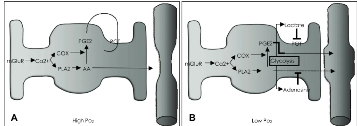
이런 결과들은 성상교세포의 혈관 조절 기능이 조건에 따 라 다를 수 있음을 보여준다. MacVicar의 연구진은 최근 성상교세포의 혈관 조절방향이 뇌의 대사에 의해 영향을 받을 수 있다고 보고하였다.9 뇌조직 절편에서 성상교세포 활성이, 높은 산소 분압에서는 혈관 수축을, 낮은 산소 분 압에서는 혈관 확장을 유도함을 보였다. 이들은, 높은 산소 분압에서는 생성된 PGE2가 prostaglandin transporter (PGT)를 통해 성상교세포로 재유입되어 혈관을 수축시키 고, 낮은 산소 분압에서는 TCA 회로보다는 당분해 과정을 통해 생성된 젖산이 PGT를 억제하고 같이 생성된 adeno- sine이 A2A receptor를 통해 혈관평활근내의 Ca2+ 농도 를 낮춰, arachidonic acid에 의한 혈관수축기능을 무마시 키는 것으로 보인다(Fig. 4).
신경세포와 성상교세포 사이의 glutamate를 통한 신호전 달이 신경혈관연결에 매우 중요한 역할을 하는 것은 분명 하다. 현재까지 in vivo에서는 주로 성상교세포의 혈관 확 장 기능만이 밝혀졌지만, 성상교세포는 분명 자극에 의해 혈관을 수축시킬 수 있는 기능도 가지고 있다. 이러한 기능 이 정상적 또는 병적인 상황에서 어떤 역할을 하는지는 추 후에 밝혀야 할 숙제로 남아 있다.
Ca2+
mGluR
Glu PLA2
COX AA
CYP4 20-HETE
PGE2 vasodilatation
vasoconstriction
Figure 3. Vessel Regulation by Astrocytes. Glutamate released and spilled over from a synapse binds to metabotrophic glutamate re- ceptor (mGluR) of nearby astrocytes, which increases intracellular Ca2+ concentration. Increased Ca2+ concentration stimulates phos- pholipase A2 (PLA2) to generate arachidonic acid (AA), and in- creased Ca2+ concentration also stimulates cyclooxygenase (COX) to produce prostaglandin E2 (PGE2) which acts as a vasodilator.
AA can be delivered to vascular smooth muscle cells, in which cy- tochrome P450 4A converts AA to 20-hydroxyeicosatetraenoic acid (20-HETE) which acts as a vasoconstrictor.
중간뉴런(Interneuron)의 역할
앞서 살펴보았듯이 신경세포간의 신호전달은 glutamate 를 통한 성상교세포의 자극으로 이어져 혈관을 조절할 수 있다. 억제성 신경전달물질인 GABA성 중간뉴런은 또 다 른 기전으로 혈관을 조절할 수 있음이 밝혀졌다.10 Hamel 그룹은 랫드 대뇌절편에서 혈관 주변의 중간뉴런에 전기자 극을 주었을 때 혈관이 수축 또는 확장하는 것을 관찰하였 다. 이들은 RT-PCR을 방법으로 확인한 결과 GAD65와 GAD67을 전사하는 GABA 중간뉴런임을 확인하였다. 혈 관 조절의 방향이 중간뉴런이 분비하는 혈관작용성 인자의 종류에 따라 결정될 것이라는 가설 아래, single cell RT- PCR로 각각의 GABA 중간뉴런이 어떤 물질의 mRNA를 전사하는지 알아보았다. 그 결과 VIP(vasoactive intesti- nal peptide)를 전사하는 중간뉴런은 지속적인 혈관 확장을, NOS(nitric oxide synthase)와 NPY(neuropeptide Y)를 같이 전사하는 중간뉴런은 가역적 확장을, SOM(somatos- tatin)만을 전사하는 중간뉴런은 가역적 수축을 시키는 것 을 보았다. 조절되는 혈관은 중간뉴런 주변으로 한정되어 있었고, 주위 성상교세포, 내피세포, 혈관평활근세포들의 수 용체들을 RT-PCR로 알아본 결과 NPY, VIP, SOM, CCK 에 대한 수용체들을 가지고 있음을 확인하였다. 이상의 관 찰들을 통해 저자들은 신경세포의 활성이 GABA 중간뉴런 을 통해 혈관을 직/간접적으로 조절할 수 있는 가능성을 제 시하였다. GABA 중간뉴런의 혈관조절 능력은 소뇌 절편 을 이용한 연구에서도 관찰되었다.11
혈관주위세포(Pericyte)의 역할
모세혈관은 내피세포 단층으로 구성되어 있으므로 주위 에 혈관평활근세포가 없어서 수축/이완이 없기 때문에 모
세혈관전 소동맥(precapillary arteriole)에 의해 혈류랑이 조절된다고 생각되었으나, 뇌의 모세혈관의 기저막에는 수 축/이완이 가능한 혈관주위세포가 존재하여 혈류량을 조절 하는 것으로 생각된다. 또한 noradrenaline성 신경말단의 65% 정도가 실제로 소동맥이 아닌 모세혈관에 분포함이 알 려져 있어 모세혈관 자체에서도 혈류 조절이 일어날 수 있 음을 유추해 볼 수 있다.12 이러한 증거는 2006년 Attwell 그룹이 망막 및 소뇌 절편에서 실험한 결과로부터 얻어졌 다.13 망막 모세혈관 주위의 혈관주위세포에 직접 전기자 극을 주었을 때 혈관주위세포의 수축으로 인한 모세혈관의 수축을 관찰하였고, 이 신호는 주위의 다른 혈관주위세포 로도 전달되어 주위의 모세혈관 또한 수축되는 것이 관찰 되었다. ATP 또는 P2Y 수용체 효현제인 UTP 등을 투여 하였을 때도 같은 현상을 관찰하였다. 소뇌 절편에서는 노 르아드레날린에 의한 혈관주위세포의 수축과 glutamate에 의해 이러한 수축이 회복됨이 관찰되었다. 즉, 모세혈관 자 체에서도 수축가능한 혈관주위세포를 통한 국소적인 혈류 조절과 함께 근위 소동맥으로의 신호 전달을 통한 혈류량 조절의 기전이 존재하는 것으로 생각된다.
이상의 실험적 결과들을 토대로 신경혈관연결을 담당하 는 단위체들의 역할을 Fig. 5에 제시하였다.
알츠하이머병과 신경혈관연결 이상
최근 들어 신경혈관연결이 퇴행성 뇌질환과 깊은 관련이 있을 것이라는 의견들이 제시되고 있다.14,15 알츠하이머병 은 병리적으로 아밀로이드 단백질의 침착에 의한 아밀로이 드반(amyloid plaque)과, 타우 단백질의 인산화 및 응집 으로 인한 신경섬유반(neurofibrillary tangle)을 특징적으
Figure 4. Astrocyte Regulates Vessel Diameter Differently Based on Metabolic Condition. A: In high Po2conditions, prostaglandin E2(PGE2) is produced and released but prostaglandin transporters (PGTs) reuptake it. On the other hand, released arachidonic acid (AA) constricts the vessel. B: In low Po2 conditions, glycolysis is stimulated resulting in lactate and adenosine being released as a byproduct. Lactate inhibits PGTs, therefore PGE2 can diffuse to the vessel to dilate it. Also, adenosine binds to adenosine A2A receptor to reduce Ca2+ concentration in vascular smooth muscle cells, diminishing the action of AA on the cell. The result is vessel dilatation.
mGluR Ca2+
PGE2
AA PGT
PLA2 COX
High Po2
mGluR Ca2+
PGE2 PGT
PLA2 COX
Adenosine Glycolysis
Lactate
Low Po2
A B
로 하는 퇴행성 질환이다. 기전으로는 가족성 알츠하이머병 환자들을 통해 밝혀진 APP, PS1 또는 PS2 유전자의 변이 를 근거로 하여 아밀로이드 cascade 가설이 널리 받아들 여지고 있다.16 최근에 알츠하이머병에 여러 가지 혈관성 요인이 관여함이 밝혀지고 있다. 역학적으로도 혈관성 질환 의 주요 위험인자들인 고혈압, 당뇨, 고콜레스테롤혈증 등 이 알츠하이머병에도 주요한 위험요소임이 알려졌으며17 알 츠하이머병에서도 순수하게 알츠하이머병에 의한 요인 외에 도 혈관성 요소가 임상증상이나 병의 진행 등에 중요하게 관여함이 알려졌다. 또한 혈관성 치매는 뇌혈관 기능 부전, 허혈성 손상 등의 혈관성 요인 외에도 아밀로이드 축적 등 이 관여함이 알려졌다. 또한 알츠하이머병 환자들의 뇌동맥 에서 더 심한 동맥 경화증이 보이는 것으로 알려져 알츠하 이머 병인에 혈관성 요인들이 기존에 생각되던 것보다는 큰 것으로 생각되고 있다.18
여러 가능한 기전들 가운데에는 아밀로이드 베타(Aβ) 단백질이 관여하는 것으로 생각된다. 우선 Aβ가 신경세 포만이 아니라 혈관에도 영향을 준다는 연구결과가 있다.
APPSWE 형질전환 마우스에서 6개월 이후에 인지 저하가, 9~12개월째 아밀로이드반이 형성되는 반면에19-21 2~3개 월째부터 뇌혈관 조절에 변화가 관찰되었다. 주된 변화로 는 뇌혈관 자가조절(autoregulation)에 장애가 일어나 혈 관내피 의존적인 혈관확장인자(endothelium-dependent vasodilator)에 의한 뇌혈류량 증가가 감소된 점과22,23 코 털 자극에 의한 체성 감각 피질로의 혈류 증가가 감소된 점24 등이다. 합성 Aβ 단백질의 대뇌 피질 국소 처리가 뇌혈관 조절이상을 초래하며 전신투여의 경우에도 안정시 뇌혈류 량이 줄어드는 점 등도 관찰되었다.25,26 이들의 관찰에서는 Aβ 올리고머 중 짧은 형태인 Aβ1-40이 긴 형태의 Aβ1-42 보다 뇌혈관에 더 큰 영향을 준다는 점도 알려졌다. 이 점 은 특히 알츠하이머병에서의 뇌혈관 조절 이상이 아밀로이
드 플라크 형성 이후에 신경세포 사멸에 의한 2차적인 결 과가 아닐 수 있음을 시사한다.
Aβ의 뇌혈관에 대한 영향은 주로 활성산소종(reactive oxygen species, ROS) 생성에 의존한다고 생각된다. ROS 는 산소분자의 전자 겉껍질에 전자가 추가되면서 생성되는 superoxide anion(O2·-
), hydrogen peroxide(H2O2) and hydroxyl radical(H2O-) 등의 반응성이 높은 물질들을 말 한다. ROS는 단백질, 인지질, DNA등 여러 세포 고분자에 손상을 주어 세포 사멸에까지 이르게 할 수 있다.27 APP 형질전환 마우스에서 3~6개월에 혈관 산화 스트레스가 관 찰되었고,28 이들에서의 혈관조절이상이 산화 방지제 또는 자유 라디칼 제거 효소인 supreoxide dismutase의 과발현 에 의해 되돌려질 수 있음이 확인되었다.22,25,28 ROS를 생 성하지 않는 Aβ의 돌연변이 형태는 뇌혈관에 영향을 주지 않는 것도 관찰되었다.25 Aβ는 미세아교세포 및 성상교세 포에서 NADPH oxidase를 통해 ROS를 생성하거나,29-31 Aβ의 침전된 형태는 자체적으로 ROS를 생성하기도 한 다.32 또한 알츠하이머 환자의 뇌에서 NADPH oxidase 활 성이 증가됨이 관찰되었다.33 이렇게 생성된 ROS는 여러 가지 방법으로 뇌혈관에 영향을 주는데, 가장 큰 효과는 주요 혈관확장인자인 일산화질소(NO)와의 급격한 반응으 로 인한 NO 농도의 감소와 이차적으로 질산 과산화아세틸 (ONOO-)에 인한 작용으로 보인다. 이러한 작용은 성상교 세포 뿐 아니라 신경혈관연결을 구성하는 여러 구성 요소 들에 작용할 것으로 생각된다.
알츠하이머병에서의 혈관조절이상은 Aβ의 제거에도 영 향을 준다. 뇌혈관장벽(BBB)을 통한 물질의 이동은 중추 신경계(CNS)의 용해성 Aβ의 농도에 결정적인 역할을 한 다.34 Aβ 이동을 담당하는 BBB 물질로 receptor for ad- vanced glycation end products (RAGE)26와 low-den- sity lipoprotein receptor-related protein(LRP)35,36가 있으며, Aβ의 항상성을 유지하는 단백질로는 apolipopro- tein(apo)E와 apoJ가 있다.37,38 LRP는 Aβ를 뇌실질에서 혈장으로, RAGE는 Aβ를 혈장에서 뇌실질로 이동시키며, apoE와 apoJ는 Aβ 단백질의 분해, 올리고머화(oligome- rization), aggregation, 생성 등에 관여한다. 혈관신생이상 또는 혈관내피세포의 노화로 인한 LRP의 감소나 RAGE의 증가, apoE, apoJ의 이상 등은 BBB를 통한 Aβ 제거를 방해한다. 이로 인한 뇌실질의 Aβ 증가는 ROS 생성 등 을 통해 혈관 및 신경세포 등에 다양한 영향을 끼칠 수 있 다. 최근 Zlokovic 연구진의 한 연구에 의하면 알츠하이머 환자의 뇌혈관내피세포에서 호메오박스 유전자 MEOX2의 발현이 저하되어있음이 알려졌다.39 MEOX2 유전자는 혈
ASTROCYTE INTERNEURON
PERICYTE
VSMC EC
• Release vasoactive factors such as NO &
endothelin
• Basal tone
• Relaying signal to upper level vessels
• Releasing vasoactive factors such as VIP, NO, NPY & SOM
• Contraction at capillary walls
• Arteriole diameter regulation regarding vasoactive factors
mGluR Ca2+ COX PLA2
PGE2 CYP4A
20-HETE
Figure 5. Integrated Action of Neurovascular Unit. Endothelial cell (EC), vascular smooth muscle cell (VSMC), astrocyte, interneuron and pericyte can work as a whole or partially together to regulate blood vessel diameter. See text for detail explanation.
관 분화 조절인자로, 이 유전자가 발현하는 단백질인 GAX 를 알츠하이머 환자의 뇌혈관내피세포에 주입한 결과 혈관 신생을 자극하고 LRP를 증가시켰다. MEOX2를 삭제한 유 전자조작 마우스는 뇌미세혈관 밀도와 안정시 뇌혈류량이 줄었고, LRP의 감소로 BBB를 통한 Aβ 유출이 줄어들었 다. 이는 앞선 연구들과 마찬가지로 알츠하이머병에 대한 혈관 중심적 치료 방법의 적합성에 대한 근거가 된다.
결 론
혈관내피세포, 혈관평활근세포, 신경세포, 성상교세포, 혈 관주위세포 간의 복잡하고 정교한 신호전달은 뇌활동에 필 요한 에너지원 보충과 부산물 제거에 필수적이다. 뇌에는 다른 조직처럼 에너지 저장고로서 작용하는 요소가 없다는 점은 신경혈관연결의 중요성을 더욱 부각시킨다. 이처럼 정 상적인 인지 활동에 필수불가결한 신경혈관연결에 이상이 생길 경우 여러 질환이 나타날 수 있음은 당연하다. 특히 신경혈관연결의 이상이 알츠하이머병에서 단순한 결과가 아 니라 병의 원인의 일부로 작용할 수 있다는 점을 뒷받침할 수 있는 증거들을 살펴보았다. 그 중심에는 Aβ 제거의 저 하와 뇌실질 내 Aβ의 증가, 이로 인한 ROS 생성의 증가 와 그에 따른 혈관조절인자의 활성 억제 및 신경혈관단위 들의 손상 등이 있다. ROS가 기본적으로 노화에서 중심적 인 역할을 하며, 알츠하이머병뿐만이 아니라 파킨슨병, 운 동신경병(루게릭병), 헌팅턴병 등 여러 퇴행성 뇌질환을 일 으키는 주요 인자로 알려짐에 따라,40 다른 퇴행성 뇌질환 에서도 신경혈관연결이 치료 표적이 될 가능성이 있다.
Acknowledgments
This study was supported by a grant of the Korea Healthcare tech- nology R&D Project, Ministry for Health, Welfare & Family Affairs, Republic of Korea (A080945).
REFERENCES
1. Roy CS, Sherrington CS. On the Regulation of the Blood-supply of the Brain. J Physiology 1890;11:85-108,117.
2. Iadecola C, Nedergaard M. Glial regulation of the cerebral microvas- culature. Nat Neurosci 2007;10:1369-1376.
3. Haydon PG, Carmignoto G. Astrocyte control of synaptic transmission and neurovascular coupling. Physiol Rev 2006;86:1009-1031.
4. Koehler RC, Roman RJ, Harder DR. Astrocytes and the regulation of cerebral blood flow. Trends in neurosci 2009;32:160-169.
5. Zonta M, Angulo MC, Gobbo S, Rosengarten B, Hossmann KA, Po- zzan T, et al. Neuron-to-astrocyte signaling is central to the dynamic control of brain microcirculation. Nature neuroscience 2003;6:43-50.
6. Bezzi P, Carmignoto G, Pasti L, Vesce S, Rossi D, Rizzini BL, et al.
Prostaglandins stimulate calcium-dependent glutamate release in ast- rocytes. Nature 1998;391:281-285.
7. Mulligan SJ, MacVicar BA. Calcium transients in astrocyte endfeet cause cerebrovascular constrictions. Nature 2004;431:195-199.
8. Takano T, Tian GF, Peng W, Lou N, Libionka W, Han X, et al. As- trocyte-mediated control of cerebral blood flow. Nat Neurosci 2006;
9:260-267.
9. Gordon GR, Choi HB, Rungta RL, Ellis-Davies GC, MacVicar BA.
Brain metabolism dictates the polarity of astrocyte control over ar- terioles. Nature 2008;456:745-749.
10. Cauli B, Tong XK, Rancillac A, Serluca N, Lambolez B, Rossier J, et al. Cortical GABA interneurons in neurovascular coupling: relays for subcortical vasoactive pathways. J Neurosci 2004;24:8940-8949.
11. Rancillac A, Rossier J, Guille M, Tong XK, Geoffroy H, Amatore C, et al. Glutamatergic control of microvascular tone by distinct GABA neurons in the cerebellum. J Neurosci 2006;26:6997-7006.
12. Cohen Z, Molinatti G, Hamel E. Astroglial and vascular interactions of noradrenaline terminals in the rat cerebral cortex. J Cereb Blood Flow Metab 1997;17:894-904.
13. Peppiatt CM, Howarth C, Mobbs P, Attwell D. Bidirectional control of CNS capillary diameter by pericytes. Nature 2006;443:700-704.
14. Zlokovic BV. Neurovascular mechanisms of Alzheimer’s neurodege- neration. Trends in neurosciences 2005;28:202-208.
15. Iadecola C. Neurovascular regulation in the normal brain and in Al- zheimer’s disease. Nature reviews 2004;5:347-360.
16. Hardy J, Selkoe DJ. The amyloid hypothesis of Alzheimer’s disease:
progress and problems on the road to therapeutics. Science 2002;297:
353-356.
17. de la Torre JC. Alzheimer disease as a vascular disorder: nosological evidence. Stroke 2002;33:1152-1162.
18. Roher AE, Esh C, Kokjohn TA, Kalback W, Luehrs DC, Seward JD, et al. Circle of willis atherosclerosis is a risk factor for sporadic Al- zheimer’s disease. Arterioscler Thromb Vasc Bio 2003;23:2055-2062.
19. Hsiao K, Chapman P, Nilsen S, Eckman C, Harigaya Y, Younkin S, et al. Correlative memory deficits, Abeta elevation, and amyloid pla- ques in transgenic mice. Science 1996;274:99-102.
20. Westerman MA, Cooper-Blacketer D, Mariash A, Kotilinek L, Ka- warabayashi T, Younkin LH, et al. The relationship between Abeta and memory in the Tg2576 mouse model of Alzheimer’s disease. J Neu- rosci 2002;22:1858-1867.
21. Kawarabayashi T, Younkin LH, Saido TC, Shoji M, Ashe KH, You- nkin SG. Age-dependent changes in brain, CSF, and plasma amyloid (beta) protein in the Tg2576 transgenic mouse model of Alzheimer’s disease. J Neurosci 2001;21:372-381.
22. Iadecola C, Zhang F, Niwa K, Eckman C, Turner SK, Fischer E, et al.
SOD1 rescues cerebral endothelial dysfunction in mice overexpressing amyloid precursor protein. Nature Neurosci 1999;2:157-161.
23. Niwa K, Kazama K, Younkin L, Younkin SG, Carlson GA, Iadecola C. Cerebrovascular autoregulation is profoundly impaired in mice overexpressing amyloid precursor protein. Am J Physiol 2002;283:
H315-H323.
24. Niwa K, Younkin L, Ebeling C, Turner SK, Westaway D, Younkin S, et al. Abeta 1-40-related reduction in functional hyperemia in mouse neocortex during somatosensory activation. PNAS 2000;97: 9735- 9740.
25. Niwa K, Carlson GA, Iadecola C. Exogenous A beta1-40 reproduces cerebrovascular alterations resulting from amyloid precursor protein overexpression in mice. J Cereb Blood Flow Metab 2000;20:1659- 1668.
26. Deane R, Du Yan S, Submamaryan RK, LaRue B, Jovanovic S, Hogg E, et al. RAGE mediates amyloid-beta peptide transport across the blood-brain barrier and accumulation in brain. Nat Med 2003;9:907-913.
27. Orrenius S, Gogvadze V, Zhivotovsky B. Mitochondrial oxidative st- ress: implications for cell death. Annu Rev Pharmacol Toxicol 2007;
47:143-183.
28. Park L, Anrather J, Forster C, Kazama K, Carlson GA, Iadecola C.
Abeta-induced vascular oxidative stress and attenuation of functional hyperemia in mouse somatosensory cortex. J Cereb Blood Flow Me- tab 2004;24:334-342.
29. Abramov AY, Canevari L, Duchen MR. Beta-amyloid peptides induce mitochondrial dysfunction and oxidative stress in astrocytes and death of neurons through activation of NADPH oxidase. J Neurosci 2004;
24:565-575.
30. Bianca VD, Dusi S, Bianchini E, Dal Pra I, Rossi F. Beta-amyloid ac- tivates the O-2 forming NADPH oxidase in microglia, monocytes, and neutrophils. A possible inflammatory mechanism of neuronal damage in Alzheimer’s disease. J Biol Chem 1999; 274:15493-15499.
31. Parvathenani LK, Tertyshnikova S, Greco CR, Roberts SB, Robertson B, Posmantur R. P2X7 mediates superoxide production in primary microglia and is up-regulated in a transgenic mouse model of Alzhei- mer’s disease. J Biol Chem 2003;278:13309-13317.
32. Hensley K, Carney JM, Mattson MP, Aksenova M, Harris M, Wu JF, et al. A model for beta-amyloid aggregation and neurotoxicity based on free radical generation by the peptide: relevance to Alzheimer di- sease. Proc Natl Acad Sci USA 1994;91:3270-3274.
33. Shimohama S, Tanino H, Kawakami N, Okamura N, Kodama H, Yamaguchi T, et al. Activation of NADPH oxidase in Alzheimer’s disease brains. Biochem Biophys Res Commun 2000;273:5-9.
34. Zlokovic BV. Clearing amyloid through the blood-brain barrier. J Ne- urochem 2004;89:807-811.
35. Deane R, Wu Z, Sagare A, Davis J, Du Yan S, Hamm K, et al. LRP/
amyloid beta-peptide interaction mediates differential brain efflux of Abeta isoforms. Neuron 2004;43:333-344.
36. Shibata M, Yamada S, Kumar SR, Calero M, Bading J, Frangione B, et al. Clearance of Alzheimer’s amyloid-ss(1-40) peptide from brain by LDL receptor-related protein-1 at the blood-brain barrier. J clin Invest 2000;106:1489-1499.
37. Fagan AM, Watson M, Parsadanian M, Bales KR, Paul SM, Hol- tzman DM. Human and murine ApoE markedly alters A beta meta- bolism before and after plaque formation in a mouse model of Alzhe- imer’s disease. Neurobiol Dis 2002;9:305-318.
38. DeMattos RB, Cirrito JR, Parsadanian M, May PC, O’Dell MA, Taylor JW, et al. ApoE and clusterin cooperatively suppress Abeta levels and deposition: evidence that ApoE regulates extracellular Abe- ta metabolism in vivo. Neuron 2004;41:193-202.
39. Wu Z, Guo H, Chow N, Sallstrom J, Bell RD, Deane R, et al. Role of the MEOX2 homeobox gene in neurovascular dysfunction in Alzhei- mer disease. Nature Medicine 2005;11:959-965.
40. Lin MT, Beal MF. Mitochondrial dysfunction and oxidative stress in neurodegenerative diseases. Nature 2006;443:787-795.