Archives of Aesthetic Plastic Surgery
111
Arch Aesthet Plast Surg
창상처치에 반응하지 않는 국한성 베게너육아종증에 의해 발생한 다발성 안면부 궤양: 미용적 측면
송승한 · 김순제 · 김주학 · 강낙헌 충남대학교 의과대학 성형외과학교실
Chronic Ulcerating Lesions due to Limited Form of Wegener’s Granulomatosis on the Face: Cosmetic Consideration
Seung Han Song, M.D., Soon Je Kim, M.D., Joo Hak Kim, M.D., Nak Heon Kang, M.D.
Department of Plastic & Reconstructive Surgery, College of Medicine, Chungnam National University, Daejeon, Korea
Wegener’s granulomatosis (WG) is a systemic disease characterized by necrotizing granulomas and vasculitis involving the upper and lower respiratory tract as well as the kidneys. Limited form of WG usually involves the head and neck, lacks renal involvement, and may not progress to generalized disease. We report the case of limited form of WG who presented not systemic symptom but several times relapsed multiple ulcerating lesions on the face, uveitis and keratoconjunctivitis. A 23 year-old female initially presented with ulcerative skin lesions on the left cheek and nose. The skin lesion had commenced as an ulcerative and nodulopapular lesion on her right cheek initially, 8 months ago. Subsequently, there was progression of the disease to her left cheek and nose. The patient was treated with oral prednisolone and oral cyclophosphamide. Two weeks later, skin lesion had started to heal, oral prednisolone tapered to 15 mg. Eight weeks later, all of skin lesions were healed well. With silicone gel sheets and Laser therapies, we gained excellent cosmetic results. In the aesthetic aspect, early recognition of rare variants of limited form of WG, facial chronic ulcerative wounds that are nonresponsive to conservative treatment, is very important as appropriate therapy can prevent facial mutilation. (Archives of Aesthetic Plastic Surgery 18: 111, 2012) Key Words: Wegener granulomatosis, Uveitis, Keratoconjunctivitis
Ⅰ. INTRODUCTION
Wegener’s granulomatosis is a progressive disease causing necrotizing vasculitis involving the medium and small blood vessels, which can induce broad inflammation in multi-organ at the end without proper treatment. Even the cause of Wegen- er’s granulomatosis is unknown, it is known as anti-neutrophil cytoplasmic antibodies (ANCAs)-associated autoimmune
disease. The estimated annual prevalence is about 10 cases per a million, and it commonly affects upper and lower esopha- gus and kidney in 50’s. Typical manifestations of Wegener’s granulomatosis include necrotizing granulomatosis in upper and lower respiratory tracts, systemic vasculitis and nephritis.
Whereas, limited Wegener's granulomatosis has mild internal symptoms but mainly in head and neck with good prognosis.
The skin lesion in typical form is usually seen on the lower ex- tremities, while being seen on the face in limited form, which lasts for several months sometimes and can be spread systemi- cally into multi-organ if not treated properly.
The authors met a patient with limited Wegener’s granulo- matosis presented with multiple skin ulcerations on the face lasting for several months, and we had good cosmetic results with only small scars owing to early detection and appropriate treatment, so that we reported this case with other references.
Received May 26, 2012 Revised June 7, 2012 Accepted June 8, 2012
Address Correspondence: Nak Heon Kang, M.D., Department of Plastic & Reconstructive Surgery, Chungnam National University Hospital, 640 Daesa-dong, Jung-gu, Daejeon 301-721, Korea. Tel: +82- 42-280-7380, Fax: +82-42-280-7384, E-mail: [email protected]
Vol. 18, No. 2, 111-114, 2012
Ca se R ep or t
112
Arch Aesthet Plast Surg
Archives of Aesthetic Plastic Surgery Vol. 18, No. 2, 2012
ment, since the lesion was improved, we reduced PD to 15 mg.
The skin lesions were recovered to normal appearance after simultaneous wound management and medical treatment for 8 weeks, and ophthalmologic symptoms were gone with nor- malized ESR (Fig. 3). We stopped PD at this point and reduced CYP to 50 mg, which was maintained for 6 months with this dose. By using silicone gel sheet and Laser therapies, the skin lesions on the face were healed without noticeable scars. Until now, no specific complication has developed (Fig. 4).
Ⅲ. DISCUSSION
Wegener’s granulomatosis is an autoimmune disease devel- oping vasculitis commonly involving upper and lower airway, and kidney. In 1931, Klinger firstly reported a patient with severe sinusitis, nephritis, and sporadic vasculitis, and at the first time Wegener defined the patients with granulomatous necrotizing vasculitis in upper and lower respiratory system or glomerular nephritis as Wegener’s granulomatosis.1Although the cause of Wegener’s granulomatosis is unknown, anti-neu- trophil cytoplasmic antibodies (ANCAs) are associated and it is known that it involves commonly small and medium vessels.2 It is a rare disease and the exact prevalence is not known, but the estimated prevalence was reported to be about 10 cases per a million. About 15% of Wegener's granulomatosis develop in 19
Ⅱ. CASE
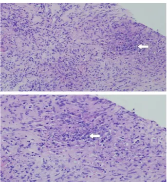
A 23-year-old female presented to out-patient department with chronic skin ulcerations on the bilateral cheek and na- sal regions. Eight months ago, the lesion was developed on the right cheek first, and she visited local private clinic and it seemed to be healed with scar left, but new additional lesions were developed on the left cheek and nose (Fig. 1). History tak- ing with the patient told us that uveitis and keratoconjunctivitis were also found at the similar time when skin ulcer developed and she visited to several ophthalmology clinics, but it was not recovered. And also there was a suppurative rhinorrhea but not upper and lower respiratory symptoms, and physical examina- tion showed only erosion on the nasal mucosa, but not other findings. On the blood and urine test, there was no specific finding except of elevated ESR. Thick exudate, which was like pus gangrene, was seen in the skin lesion, but no bacteria was cultured. Cytoplasmic(c)-ANCA was elevated and histologic finding revealed inflammation in the capillary vessel, multiple interstitial necrosis, and chronic granulomatous inflammation (Fig. 2). Considering all of the findings, we questioned about chronic facial skin ulcer lasting for several months and accom- panied uveitis and keratoconjunctivitis not responded to usual treatment and we suspected Wegener's granulomatosis. During the early period of the treatment, vaseline gauze, wet dress- ing, and EGF application were done, but no sign of recovery.
Later on, the lesion was diagnosed as WG, immunosuppressive treatment and local dressing was carried out and it started to improve. Afterwards, the wound became healed, and silicone gel sheet and Fraxel Laser was applied subsequently.
As a treatment, 75 mg of cyclophosphamide (CYP) and 30 mg of prednisolone (PD) were administered for one week, and then CYP was increased by 100 mg. At 2 weeks after the treat-
Fig. 1. Right side wounds of the face were almost healed. Newly developed lesions were made on her nose and left side of the cheek.
Fig. 2. Biopsy of the left cheek ulcerative lesion. The skin showed deep penetrating ulcer with vasculitis. (Hematoxylin and eosin stain,
×200) It showed necrotizing vasculitis and chronic granulomatous inflammation (Arrow). (Hematoxylin and eosin stain, ×400)
Seung Han Song, et al.: Limited Form of Wegener’s Granulomatosis
113
Arch Aesthet Plast Surg
test is a one of evidence for Wegener’s granulomatosis but not an essential factor for diagnosis. It is 50% or so to show all of interstitial necrosis on the histologic examination, vasculitis, and granulomatous inflammation. Wegener’s granulomatosis cannot be diagnosed only by blood test or histologic examina- tion, but it is diagnosed by overall interpretation of symptoms, response to treatment, blood test and histologic examination.3,8 The authors suspected Wegener’s granulomatosis from recur- rent skin necrosis lasting for a long time, interstitial necrosis, and granulomatous inflammation, and started treatment with PD and CYP, which induced good response.
The mean life span of the patients with Wegener’s granu- lomatosis in 1950's was around 5 months, yet it has been increased owing to use of PD and CYP since 1970's. Usually improvement of the symptoms is seen from initial treatment, and the treatment has to be continued for at least 1 year until complete remission of the symptoms.8In this case, skin lesion was improved from 2 weeks after the treatment, and we kept the maintaining dose although the symptoms were improved at 8 weeks. Systemic antibiotics therapy might be used in some cases, yet it has no effect on skin lesion in this case, so that we used it restrictively to prevent second infection in immunosup- pressive state.
It is crucial to start treatment early in limited Wegener’s granulomatosis accompanying chronic skin ulcer without bac- teria cultured and not induced by malignant tumor or trauma.
Contrary to typical Wegener’s granulomatosis, it, as an atypical type, presents only upper and lower respiratory disorders with- out renal disorder, or it shows ocular symptoms, skin and mu- cosal lesion, or no symptom sometimes. Particularly, in the case with negative for ANCA, it is difficult to diagnose Wegener’s granulomatosis, thus the treatment with immunosuppressive agents may be delayed and Wegener’s granulomatosis can be spread into multiple organ.7 There are many disease needed to years old or less with a mean age of 50 years, but rarely develops
it before adolescent. There is no difference according to gender, and it occurs more in white people than black people. It is divid- ed into systemic and localized form and localized form does not always progress to systemic form and it has better prognosis.3
Skin lesion is accompanied in about 50% of patients diag- nosed as Wegener's granulomatosis, which is not specific and develops commonly on the elbow, knee and cheek. It shows various forms from large ulcer similar to gangrenous pus to subcutaneous nodule, hemorrhagic bullae, pus formation, vesicle, spot, and hemorrhagic spot.4
Periocular inflammation in Wegener’s granulomatosis reaches to about 50~60% of all, it is reported, and it is found as an initial symptom, which varies as conjunctivitis, episcleritis, scleritis, granulomatous sclerotic uveitis, ciliary vasculitis, reti- nal vasculitis, obstruction of nasolacrimal duct, dacryocystitis, and pseudotumor. Even though many other disease with mi- croscopic multiple vasculitis involve to periocular region, the incidence rate is very low and more rarely is it seen as an initial symptom. For instance, polyarteritis nodosa and Churg-Strauss syndrome are rare to involve into ocular region, and even ocu- lar involvement of rheumatic arthritis is relatively common than previously mentioned disease, ocular symptoms such as scleritis is seen in progressive or old lesion but rarely as an ini- tial symptoms like Wegener's granulomatosis.5
Wegener’s granulomatosis can be suspected when the fol- lowing results are seen; positive ANCA on blood test, struc- tural deformity on CT or MRI, inflammation and ulcer in the mucosa, or interstitial necrosis, vasculitis and granulomatous inflammation on histologic examination.6,7 The sensitivity and specificity of ANCA are 91% and 99% in active Wegener's gran- ulomatosis respectively, but it is negative in about 20% of usual Wegener’s granulomatosis. In this case, c-ANCA was elevated and ESR increased as well. However, elevated ANCA on blood Fig. 3. Eight weeks after treatment. The wounds on her left cheek
and nose improved without any spread on other sites. Fig. 4. Two and half years after treatment. The wounds on her face were almost healed without noticeable scars.
114
Arch Aesthet Plast Surg
Archives of Aesthetic Plastic Surgery Vol. 18, No. 2, 2012
treated based on its fundamental cause. With that process, it can bring out an excellent aesthetic outcome.
REFERENCES
1. Fauci AS, Wolff SM: Wegener's granulomatosis: studies in eigh- teen patients and a review of the literature. Medicine (Baltimore) 52: 535, 1973
2. Leavitt RY, Fauci AS: Wegener’s granulomatosis. Curr Opin Rheu- matol 3: 8, 1991
3. Vischio JA, McCrary CT: Orbital Wegener's granulomatosis: a case report and review of the literature Clin Rheumatol 27: 1333, 2008
4. Handfield-Jones SE, Parker SC, Fenton DA, Newton JA, Greaves MW: Wegener’s granulomatosis presenting as pydermagangreno- sum. Clin Exp Dermatol 17: 197, 1992
5. Pakrou N, Selva D, Leibovitch: Wegener’s granulomatosis: oph- thalmic manifestations and management. Semin Arthritis Rheu- matol 35: 284, 2006
6. Muhle C, Reinhold-Keller E, Richter C, Duncker G, Beigel A, Brinkmann G, Gross WL, Heller M: MRI of the nasal cavity, the paranasal sinuses and orbits in Wegener's granulomatosis. Eur Radiol 7: 566, 1997
7. Barksdale SK, Hallahan CW, Kerr GS, Fauci AS, Stern JB, Travis WD: Cutaneous pathology in Wegener's granulomatosis. Am J Surg Pathol 19: 161, 1995
8. Kuchel J, Lee S: Cutaneous Wegener's granulomatosis: a variant or atypical localized form? Australas J Dermatol 44: 129, 2003 be differentiated from typical Wegener's granulomatosis (Table
1). Among granulomatous disease with skin lesion similar to this case, sarcoidosis has no skin necrosis, berylliosis has a his- tory of exposure to beryllium, and midline granuloma is trans- mitted aggressively into facial soft tissue, wheareas Wegener’s granulomatosis is not aggressive and it is transmitted sporadi- cally. Inflammatory granulomatosis diseases, such as tubercu- losis, histoplasmosis, blastomycosis, coccidioidomycosis, and syphilis, can be differentiated by culturing the bacteria.1
In conclusion, it is important to remind Wegener’s granu- lomatosis in the case with recurrent skin ulcer lasting for a long time and to take thorough physical examination leading to quick diagnosis. And then immunosuppressive treatment and conventional therapy according to symptoms should be performed to reduce unnecessary excision operation for ulcer and to prevent transmission into other organ and renal failure resulting in death. We performed treatment with combination therapy of PD and CYP in a patient with Wegener’s granulo- matosis presented with multiple skin ulcers in facial region last- ing for several months, and it was healed with slight scar tissue left without other complications. In the aesthetic aspect, WG causes repetitive ulceration and nodulopapular lesions, which result in depressed scar and pigmentation. Wounds without any reasons, such as a tumor or trauma, and not responsive to conservative dressing, should be systemically evaluated and
Table 1. Diseases Frequently Included in the Differential Diagnosis of Wegener’s Granulomatosis Predominantly vasculitic diseases
Periarteritisnodosa Hypersensitivity angiitis Systemic lupus erythematosus Scleroderma
Dermatomyositis SjÖgren’s syndrome Henoch-SchÖnleinpurpura Giant cell arteritis
Arteritis in association with rheumatoid arthritis Predominantly granulomatous diseases
Sarcoidosis Berylliosis Midline granuloma
Mixed granulomatosis and vasculitic diseases Allergic granulomatosis
Loeffler’s syndrome Eosinophilic pneumonia
Infectious granulomatous diseases Tuberculosis
Histoplasmosis Blastomycosis Coccidioidomycosis Syphilis
Pulmonary-renal syndromes Goodpasture’s Syndrome
Streptococcal pneumonia with glomerulonephritis Neoplastic diseases
Nasopharyngeal lymphomas and sarcomas Midline malignant reticulosis
Primary and metastatic lung disease
Hodgkin’s disease and lymphoma involving the lung
Fauci AS, Wolff SM.: Wegener's granulomatosis: studies in eighteen patients and a review of the literature. Medicine (Baltimore) 52: 535-61, 1973