• 검색 결과가 없습니다.

Development of a Coronary Aneurysm at a Sirolimus-Eluting Stent-Implanted Lesion in a Patient With Churg-Strauss Syndrome

N/A
N/A
Protected

Academic year: 2022

Share "Development of a Coronary Aneurysm at a Sirolimus-Eluting Stent-Implanted Lesion in a Patient With Churg-Strauss Syndrome"

Copied!
4
0
0

로드 중.... (전체 텍스트 보기)

전체 글

(1)

559

Print ISSN 1738-5520 / On-line ISSN 1738-5555 Copyright © 2011 The Korean Society of Cardiology CASE REPORT

http://dx.doi.org/10.4070/kcj.2011.41.9.559

Open Access

Development of a Coronary Aneurysm at a Sirolimus-Eluting

Stent-Implanted Lesion in a Patient With Churg-Strauss Syndrome

Yujung Cho, MD

1

, Hyunmin Choe, MD

1

, Bo Ram Kang, MD

1

, Min Yong Park, MD

1

, Joon Hyung Doh, MD

1

, Jae-Jin Kwak, MD

1

, Bo Young Yoon, MD

2

, June Namgung, MD

1

, Sung Yun Lee, MD

1

, and Gam Hur, MD

3

1

Departments of Internal Medicine, Vision 21 Cardiac and Vascular Center,

2

Rheumatology and

3

Radiology, Ilsan Paik Hospital, Goyang, Korea

ABSTRACT

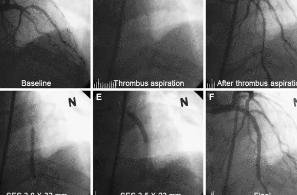
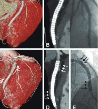
A coronary aneurysm (CA) can occur in sirolimus-eluting stent (SES)-implanted coronary lesions. Although several possi- ble mechanisms have been suggested, the precise pathogenesis of a CA in SES-implanted lesions is still unknown. We report a patient with Churg-Strauss syndrome who underwent successful percutaneous coronary intervention with SES and then experienced a CA in an SES-implanted coronary lesion. We describe the CA characteristics through the use of coronary angi- ography, coronary 64-multidetector computed tomography, and intravascular ultrasound and discuss the etiological factors for the CA in this patient. (Korean Circ J 2011;41:559-562)

KEY WORDS: Coronary aneurysm; Churg-Strauss syndrome.

Received: January 27, 2011 Revision Received: March 22, 2011 Accepted: March 28, 2011

Correspondence: Hyunmin Choe, MD, Department of Internal Medi- cine, Vision 21 Cardiac and Vascular Center, Ilsan Paik Hospital, 2240 Daehwa-dong, Ilsanseo-gu, Goyang 411-706, Korea

Tel: 82-31-910-7833, Fax: 82-31-910-7829 E-mail: hmchoi49@naver.com

• The authors have no financial conflicts of interest.

cc

This is an Open Access article distributed under the terms of the Cre- ative Commons Attribution Non-Commercial License (http://creativecom- mons.org/licenses/by-nc/3.0) which permits unrestricted non-commer- cial use, distribution, and reproduction in any medium, provided the origi- nal work is properly cited.

Introduction

The sirolimus-eluting stent (SES) has provided excellent clinical outcomes by significantly reducing the number of major adverse cardiac events.

1)2)

However, SESs can cause un- expected vascular complications, such as stent thrombosis

3)

and a coronary aneurysm (CA)

4)

at SES-implanted coronary lesions. Furthermore, systemic necrotizing vasculitis, such as Churg-Strauss syndrome (CSS), can involve the native coro- nary artery in the form of coronary ectasia or an aneurysm. Al- though the precise mechanisms of CA development at SES- implanted coronary lesions are still unclear, both SESs and CSS can precipitate a CA by weakening the coronary vessel walls.

5)

We report a case of CA at an SES-implanted coronary lesion in a patient with CSS and describe the lesion features

using several imaging modalities.

Case

A 43 year old man presented with ongoing resting chest pain for the past 2 hours. He had been diagnosed with CSS at another general hospital 4 months earlier. He met the diag- nostic criteria for CSS

6)

with asthma, allergic sinusitis, hype- reosinophilia, and systemic vasculitis of the skin, and was re- ceiving oral prednisone and cyclophosphamide. He had no other coronary risk factors. An initial electrocardiogram sh- owed an ST-segment elevation from the V2 to V6 leads. Di- agnostic coronary angiography (CAG) revealed a total oc- clusion of the mid-left anterior descending (LAD) coronary artery (Fig. 1A). After a manual intracoronary aspiration th- rombectomy (Export; Medtronic, Santa Rosa, CA, USA) at the mid LAD lesion (Fig. 1B), a subsequent CAG revealed dif- fuse irregular and significant stenosis from the mid to distal LAD with restoration of thrombolysis and myocardial in- farction grade 3 (Fig. 1C). A successful primary percutaneous coronary intervention (PCI) was performed with 2 SESs (Cy- pher; Cordis Johnson and Johnson, Warren, NJ, USA) placed from the mid to proximal LAD (diameter×length: 3.0×33 mm, and 3.5×23 mm) at a pressure of 18 atm (Fig. 1D and E).

The final CAG showed good angiographic results at the LAD

lesion (Fig. 1F). The patient was discharged on the third day

(2)

560 Coronary Aneurysm at Sirolimus-Eluting Stent-Implanted Lesion

with no cardiac symptoms and was prescribed 75 mg clopi- dogrel and 100 mg aspirin daily.

A follow-up CAG was performed at 14 months after the primary PCI. We substituted coronary 64-multidetector com- puted tomography (MDCT) for conventional CAG. Three-di- mensional (3-D) surface reconstruction images from the co- ronary 64-MDCT revealed good positioning of the two over- lapping SESs with no neointimal hyperplasia (Fig. 2A). Addi- tionally, a volume-rendering image on coronary 64-MDCT showed no neointimal hyperplasia or stent malapposition (Fig. 2B) compared to the final result.

He was readmitted to our hospital 39 months later with atypical chest discomfort for the past 10 days. We performed a follow-up coronary 64-MDCT and CAG. The follow-up 3-D surface reconstruction view on coronary 64-MDCT disclosed a fusiform like diffuse enlargement of the LAD at the SES-implanted lesion. (Fig. 2C), and the volume-render- ing image on coronary 64-MDCT showed a crescent-like shape to contrast dye filling in around the stent border (Fig.

2D). A follow-up CAG showed a diffuse multiple conglom- erate contrast filling in along the stent border without in-stent restenosis (Fig. 2E). An intravascular ultrasound revealed significant gaps between the SES struts and the vessel wall and an enlargement of the external elastic membrane with a maximum diameter of 6.59 mm in the proximal portion, 6.42 mm in the mid portion, and 6.82 mm in the distal portion (Fig. 3). We then had the patient undergo an exercise tread- mill test to confirm myocardial ischemia; however, the result

was negative. Finally, we decided to treat the patient with me- dical therapy. The patient is under careful observation and is receiving dual antiplatelet therapy.

Discussion

A CA is defined as a segment with a diameter greater than 1.5 times the normal adjacent artery segment and can be cla- ssified as fusiform or saccular.

7)

A CA at an SES-implanted site accompanies stent malapposition. Although the precise mechanism of a CA at an SES-implanted site is unclear, sever- al possible mechanisms, including acute vessel damage during coronary intervention, localized hypersensitivity to the poly- mer, and vascular remodeling as a result of high drug con- centrations have been postulated.

8)

The CAG and coronary 64-MDCT findings in the present case suggested that the CA development may have been related to hypersensitivity to the SES at the contact sites between the vessel wall and the st- ent surface. Because we did not find a CA on the baseline CAG, the SES may have played an important role in CA develop- ment. In contrast, vascular inflammatory responses occur- ring in patients with CSS may not affect the development of a CA, because no elevated eosinophils were found on the complete blood count, and serum C-reactive protein was not elevated in this patient. Only one case of acute myocardial infarction has been reported due to plaque rupture in a patient with CSS.

9)

Furthermore, previous studies indicate that coro- nary artery involvement in patients with CSS is uncommon.

10)

A

D

B

E

C

F

Fig. 1. A: baseline cranial-view coronary angiography (CAG) demonstrated total occlusion of the mid left anterior descending (LAD) artery.

B: the manual thrombus aspiration catheter is shown. C: restoration of LAD blood flow after thrombosuction. D and E: two overlapping siro-

limus-eluting stents (SES) were implanted from the distal to the proximal LAD artery lesion after balloon dilation. F: final CAG shows the

successfully implanted SESs, good thrombolysis, and myocardial infarction flow.

(3)

Yujung Cho, et al. 561

Sirolimus has an anti-inflammatory property that inhibits the local inflammatory response at the stent-implanted lesion site. Sirolimus may not only inhibit neointimal growth but also endothelial depletion at the stent-implanted site. Endothelial depletion caused by sirolimus may trigger the development of a CA secondary to smooth muscle cell degeneration.

The natural course and the appropriate treatment for a CA remain uncertain. However, it is certain that patients with a CA need prolonged aspirin and dual antiplatelet therapy. Surgi- cal treatment is indicated for patients with a significant CA ob- structive lesion or evidence of embolization leading to myo- cardial ischemia and progressive enlargement of the saccular type of CA leading to rupture. Because a CA at an SES-im- planted coronary lesion may have a benign clinical course, it is very important to choose the optimal treatment plan carefully.

However, it has not been demonstrated that localized exag- gerated hypersensitivity due to sirolimus, polymers, or stent metal triggers the development of a CA. The development of the CA in this patient seemed to be due to more complex pa- thogenesis than expected, so an in-depth investigation would confirm the association between localized hypersensitivity vasculitis and incomplete endothelization.

Fig. 2. A and B: a three-dimensional surface reconstruction and volume rendering image view of 64-multidetector computed tomog- raphy (MDCT) 14 months after primary percutaneous coronary intervention revealed good configuration of two overlapping siroli- mus-eluting stents (SES) and no in-stent restenosis compared with the final coronary angiography (CAG). C: follow-up three-dimensio- nal surface reconstruction view of coronary 64-MDCT at 39 months revealed diffuse enlargement of the left anterior descending (LAD) artery from the distal to the proximal stent border (triple white ar- rows). D: volume-rendering image shows crescent-like shaped con- trast dye filling out of the stent border with a good configuration of the two overlapping SESs (triple white arrows). E: follow-up CAG shows diffuse multiple conglomeration contrast filling along the st- ent border without in-stent restenosis (triple black arrows).

A

C

B

D E

Fig. 3. Intravascular ultrasound assessment revealed a markedly enlarged left anterior descending (LAD) external elastic membrane (EEM) around the sirolimus-eluting stents (SES) struts. Maximal EEM size of the LAD was 6.59 mm at the proximal (p) portion, 6.42 mm at the mid (m) portion, and 6.82 mm at the distal (d) portion.

REFERENCES

1) Windecker S, Remondino A, Eberli FR, et al. Sirolimus-eluting and paclitaxel-eluting stents for coronary revascularization. N Engl J Med 2005;353:653-62.

2) Nienaber CA, Akin I, Schneider S, et al. Clinical outcomes after siro- limus-eluting, paclitaxel-eluting, and bare metal stents (from the first phase of the prospective multicenter German DES.DE registry). Am J Cardiol 2009;104:1362-9.

3) Daemen J, Wenaweser P, Tsuchida K, et al. Early and late coronary stent thrombosis of sirolimus-eluting and paclitaxel-eluting stents in routine clinical practice: data from a large two-institutional cohort study. Lancet 2007;369:667-78.

4) Bavry AA, Chiu JH, Jefferson BK, et al. Development of coronary an- eurysm after drug-eluting stent implantation. Ann Intern Med 2007;

146:230-2.

5) Mavrogeni S, Manoussakis MN, Karagiorga TC, et al. Detection of coronary artery lesions and myocardial necrosis by magnetic reson- ance in systemic necrotizing vasculitides. Arthritis Rheum 2009;61:

1121-9.

6) Masi AT, Hunder GG, Lie JT, et al. The American College of Rheuma-

tology 1990 criteria for the classification of Churg-Strauss syndrome

(4)

562 Coronary Aneurysm at Sirolimus-Eluting Stent-Implanted Lesion (allergic granulomatosis and angiitis). Arthritis Rheum 1990;33:

1094-100.

7) Murthy PA, Mohammed TL, Read K, Gilkeson RC, White CS. MD- CT of coronary artery aneurysms. Am J Roentgenol 2005;184(3 Suppl):S19-20.

8) Alfonso F, Pérez-Vizcayno MJ, Ruiz M, et al. Coronary aneurysms af- ter drug-eluting stent implantation: clinical, angiographic, and intra-

vascular ultrasound findings. J Am Coll Cardiol 2009;53:2053-60.

9) Wagner AD, Meyer GP, Rihl M, et al. Acute coronary syndrome as- sociated with Churg-Strauss syndrome. Vasc Health Risk Manag 2007;

3:775-9.

10) Noth I, Strek ME, Leff AR. Churg-Strauss syndrome. Lancet 2003;

361:587-94.

수치

Fig. 1. A: baseline cranial-view coronary angiography (CAG) demonstrated total occlusion of the mid left anterior descending (LAD) artery
Fig. 3. Intravascular ultrasound assessment revealed a markedly  enlarged left anterior descending (LAD) external elastic membrane  (EEM) around the sirolimus-eluting stents (SES) struts

참조

관련 문서

• Laws of conservation of mass, momentum, and energy can be stated in the differential form, applicable at a point. •

Serum uric acid levels and risk for vascular disease in patients with metabolic syndrome... Prevalence if the metabolic syndrome in a Turkish

The stain field associated with a dislocation can in certain cases provide a favorable interaction with the strain field of the martensite nucleus, such that one of the

1 John Owen, Justification by Faith Alone, in The Works of John Owen, ed. John Bolt, trans. Scott Clark, "Do This and Live: Christ's Active Obedience as the

Prevention of coronary and stroke events with atorvastatin in hypertensive patients who have average or lower-than-average cholesterol concentrations, in

(c) the same mask pattern can result in a substantial degree of undercutting using substantial degree of undercutting using an etchant with a fast convex undercut rate such

The stain field associated with a dislocation can in certain cases provide a favorable interaction with the strain field of the martensite nucleus, such that one of the

ƒ Monosaccharide may be present in the form of Monosaccharide may be present in the form of a linear or ring structure. ƒ In solution it is in the form of