181
<Received:February 9, 2012, Revised (1st: April 12, 2012, 2nd: May 20, 2012, 3rd: June 11, 2012), Accepted:June 11, 2012>
Corresponding to:Sung Jae Choi, Department of Internal Medicine, Korea University Ansan Hospital, 123, Jeokgeum-ro, Danwon-gu, Ansan 425-707, Korea. E-mail:csjmd@ hotmail.com
pISSN: 2093-940X, eISSN: 2233-4718
Copyright ⓒ 2013 by The Korean College of Rheumatology
This is a Free Access article, which permits unrestricted non-commerical use, distribution, and reproduction in any medium, provided the original work is properly cited.
Division of Rheumatology, Department of Internal Medicine, Korea University Anam Hospital, Division of Rheumatology, Department of Internal Medicine, Korea University Guro Hospital3,
Department of Internal Medicine, Korea University Anam Hospital4, Seoul, Korea
Wegener's granulomatosis (WG) classically consists of necrot- izing granulomatous inflammation of the upper and/or lower respiratory tract, necrotizing glomerulonephritis, and an au- toimmune necrotizing systemic vasculitis affecting predom- inantly small vessels. We report a case of WG with central nervous system (CNS) involvement. WG is being diagnosed through pulmonary nodule biopsy. A small nodular lesion in the left posterior basal ganglia of brain being highly suspi- cious for granulomatosis was detected by MRI. After IV pulse
cyclophosphamide and oral corticosteroid treatment for over 4 months, clinical manifestations and CNS lesions in brain MRI is improved. WG might have multiple granulomatous le- sions which could be misdiagnosed due to malignancy. CNS involvement in WG is rare but careful evaluation is necessary when there are suspicious symptoms or lesions in CNS.
Key Words. Wegener's granulomatosis, Central nervous system involvement, Antineutrophil cytoplasmic antibody- associated vasculitis
Introduction
Wegener's granulomatosis (WG) is an autoimmune disease which involves various organ systems (1). It classically con- sists of necrotizing granulomatous inflammation of the upper and/or lower respiratory tract, necrotizing glomerulonephritis, and an autoimmune necrotizing systemic vasculitis affecting predominantly small vessels (2). This disease has a variety of presentations but central nervous system (CNS) involvement in WG is rare at initial presentation.
We describe here a patient with WG who presented initially with dyspnea, hemoptysis, and headache which could be mis- diagnosed as lung cancer with brain metastasis. To our knowl- edge, this is the first WG case with multiple pulmonary nod- ules and cerebral parenchymal nodule reported in Korea.
Case Report
A 63-year-old Mongolian man with dyspnea, hemoptysis and
headache was hospitalized in a tertiary medical center in January 2011. He was a current smoker, and had smoked one pack per day for 30 years. Three months prior to his admission, he had developed a cough resistant to the usual antitussive medication. Two months after the onset of cough, a small amount of hemoptysis, dyspnea, and headache developed.
On physical examination, the patient was acutely ill-looking, with a blood pressure of 110/60 mmHg, a heart rate of 80 beats/min, and body temperature of 36.8oC. The general ex- amination revealed normal except for crackling in the whole lung field.
Laboratory results were as following: hemoglobin 12.7 g/dL (normal range: 12.6∼17.4 g/dL), WBC count 9,920/μL (normal range: 4,500∼11,000/μL), platelet 449×103/μL (normal range: 150∼400×103/μL), AST 73 IU/L (normal range: 0∼45 IU/L), ALT 86 IU/L (normal range: 0∼50 IU/L), erythrocyte sedimentation rate >120 mm/hr, C-reactive protein
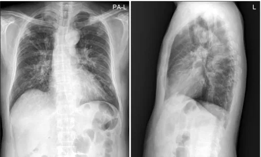
Figure 1. Chest X ray (PA and lateral view) shows nodular opa- cities and cavitary lesions in both upper lung fields and patchy opa- cities in both lower lung fields.
Figure 2. Chest CT shows multiple cavities and nodules in both lung fields and focal consolidation is also noted in upper right lobe.
25.496 mg/dL (normal range: 0.02∼0.3 mg/dL), in urine analy- sis WBC 1∼4/HPF (normal range: 0∼4/HPF) and protein neg- ative, serum urea nitrogen 12.8 mg/dL (normal range: 3∼24 mg/dL), creatinine 0.86 mg/dL (normal range: 0.3∼1.6 mg/dL), prothrombin time 72% (normal range: 90∼130%), positive antineutrophil cytoplasmic antibodies (ANCA: screen- ing test by multiplex flow immunoassay), positive anti- proteinase-3 antibody, negative antimyeloperoxidase antibody, negative fluorescent antinuclear antibody (FANA), rheumatoid factor 118 IU/mL (normal range: <20 IU/mL) and a 24-hour urine protein value of 261.6 mg/day. Based on his clinical symptom and high prevalence of pulmonary tuberculosis in Korea, sputum study was done. Sputum Acid-fast bacilli (AFB) stain was repeated 3 times, all were negative and no organisms
were cultured. An initial chest X-ray showed nodular patchy opacities and cavitary lesions in both upper lung fields (Figure 1). Additionally, chest computed tomography (CT) showed multiple cavities and nodules in both lung fields (Figure 2).
Subsequently, we performed a biopsy of nodular pulmonary le- sion to rule out lung cancer, as well as F-18 fluorodeox- yglucose torso positron emission tomography (18FDG PET)-CT and brain magnetic resonance imaging (MRI) for metastasis work up. The tissue obtained through Video-Assisted Thoraco- scopic Surgery (VATS) showed necrotizing granuloma, scat- tered giant cells, and fibroblastic proliferation. Vasculitis was also present with neutrophils and lymphocytes infiltrating the wall of small arterioles (Figure 3). AFB stain of biopsied tissue was negative. Special immunostainings for CD34 and CD68
Figure 3. Hematoxylin and eosin stain of a lung nodule. Tissue obtained from VATS biopsy showed giant cells (black arrow) and necrotizing granulomatous vasculitis (white arrow).
Figure 4. 18FDG PET-CT (A) Multiple hyper-metabolic lesions in the nasal septum and in both lungs with similar metabolisms (B) Multiple hyper-metabolic lesions in the nasal septum with extension into the adjacent nasal mucosa with similar metabolisms to those of the lungs.
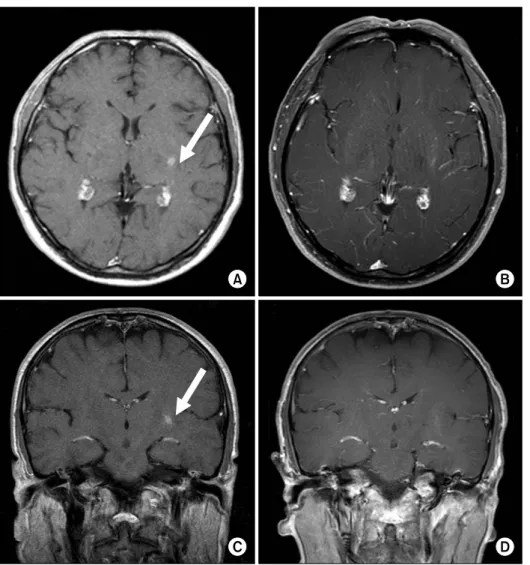
were positive, which implied the presence of giant cell asso- ciated vasculitis. 18FDG PET-CT showed multiple hyper-meta- bolic lesions in both lungs and the nasal septum (Figure 4A and B). There was no nasal bone destruction in con- trast-enhanced paranasal sinus CT (not shown here). An 8.2 mm-sized rim-enhancing small lesion in the left posterior basal ganglia was observed with brain MRI (Figure 5A and C). The result of brain MRI could not rule out malignant lesion, other infection, vasculitis or granulomatous lesion. But since the pathologic result of pulmonary nodule was consistent with WG, small cerebral enhancing nodule was thought to be a CNS in- volvement of WG. With all these results, the final diagnosis was WG involving paranasal sinus, lung, and probably brain parenchyma.
Treatment with prednisone (1 mg/kg) and intravenous (IV)
cyclophosphamide (15 mg/kg every 2∼3 weeks) has started.
After having received four cycles of cyclophosphamide pulse therapy for 12weeks, the patient complained of increased na- sal discharge and didn’t want to receive IV cyclophosphamide anymore. So the treatment regimen changed into oral cyclo- phosphamide (2 mg/kg) and low-dose steroid. Four months af- ter this immunosuppressive therapy, the parenchymal lesion in the left posterior basal ganglia disappeared in the follow-up brain MRI (Figure 5B and D). Brain MRI was followed-up again one year after initial diagnosis, and still there was no evidence of WG involvement (not shown here).
Discussion
WG is a rare autoimmune disease associated with gran- ulomatous inflammation and antineutrophil cytoplasmic anti-
Figure 5. T1-enhanced MR images before (A, C) and after (B, D) four times cyclophosphamide pulse the- rapy. Arrow indicates an 8.2 mm- sized rim-enhancing small nodule in the left posterior basal ganglia mi- micking metastatic cancer.
body-associated vasculitis, which mainly occurs in the upper and lower respiratory tract (1). Nervous system involvement was observed in 36.6% of microscopic polyangiitis, 50.8% of WG, and 76.0% of Churg-Strauss syndrome patients. Peripheral neuropathy is predominated in each type of ANCA-associated vasculitis (3). Peripheral nervous system involvement presents as polyneuropathy or mononeuritis multiplex which probably occur due to vasculitis of the vasa nervorum. 32.3% of WG patients with nervous system involvement had CNS involve- ment and most of them are cranial neuropathy and external ophthalmoplegia (70%) (4). Except for cranial neuropathy, CNS involvement of WG is usually presented by cerebral vasculitis such as hemorrhage (intracranial or subarachnoid), transient is- chemic attacks or ischemic infarction of cerebrum or spinal cord and arterial or venous thrombosis. Rarely granulomatous lesions can develop in intra-cerebral tissue (5). In WG, pons and basal ganglia were reported to be predominantly affected (6) as in this case. Brain imaging modalities such as CT or MRI in CNS involvement of WG could detect dural thickening
and enhancement, cerebral infarction, and MR signal abnormal- ities in the brain stem and white matter (7).
In this case, the mass lesion in the brain needed to be biop- sied for proper diagnosis and to rule out malignancies. But the brain lesion was too small to have a mass effect and also there was a risk of brain operation. In addition, the pathologic result of pulmonary nodule revealed vasculitis and necrotizing granulomas which were compatible with WG. So we planned to observe the response to the ongoing treatment instead of brain biopsy.
18FDG PET-CT is known to be a useful tool for distinguish- ing benign versus malignant lesions in oncology fields. In ad- dition, clinical utility of 18FDG PET-CT for the assessment of inflammatory and infectious diseases were increasingly reported. In patients of vasculitis involving large vessels such as giant cell arteritis or Takayasu arteritis, usefulness of
18FDG PET-CT has been reviewed (8) and there are also some case reports using PET-CT to facilitate the diagnosis of WG (9-11). Active inflammation of WG increases uptake of FDG.
initiate timely therapeutic intervention.
Summary
We have described a WG patient with parenchymal brain involvement. WG might have multiple granulomatous lesions which could be misdiagnosed as malignancy. CNS involvement in WG is rare but careful evaluation is needed when there are suspicious symptoms or lesions in CNS.
References
1. Schilder AM. Wegener's Granulomatosis vasculitis and granuloma. Autoimmun Rev 2010;9:483-7.
2. Lamprecht P, Gross WL. Wegener's granulomatosis. Herz 2004;29:47-56.
3. Zhang W, Zhou G, Shi Q, Zhang X, Zeng XF, Zhang FC. Clinical analysis of nervous system involvement in ANCA-associated systemic vasculitides. Clin Exp Rheu- matol 2009;27(1 Suppl 52):S65-9.
4. Nishino H, Rubino FA, DeRemee RA, Swanson JW, Parisi JE. Neurological involvement in Wegener's gran-
8. Fuchs M, Briel M, Daikeler T, Walker UA, Rasch H, Berg S, et al. The impact of 18F-FDG PET on the man- agement of patients with suspected large vessel vasculitis.
Eur J Nucl Med Mol Imaging 2012;39:344-53.
9. Ueda N, Inoue Y, Himeji D, Shimao Y, Oryoji K, Mitoma H, et al. Wegener's granulomatosis detected initially by in- tegrated 18F-fluorodeoxyglucose positron emission tomog- raphy/computed tomography. Mod Rheumatol 2010;20:
205-9.
10. Almuhaideb A, Syed R, Iordanidou L, Saad Z, Bomanji J. Fluorine-18-fluorodeoxyglucose PET/CT rare finding of a unique multiorgan involvement of Wegener's gran- ulomatosis. Br J Radiol 2011;84:e202-4.
11. Beggs AD, Hain SF. F-18 FDG-positron emission tomo- graphic scanning and Wegener's granulomatosis. Clin Nucl Med 2002;27:705-6.
12. Seo P, Min YI, Holbrook JT, Hoffman GS, Merkel PA, Spiera R, et al. WGET Research Group. Damage caused by Wegener's granulomatosis and its treatment: prospective data from the Wegener's Granulomatosis Etanercept Trial (WGET). Arthritis Rheum 2005;52:2168-78.