Letter to the Editor
Vol. 27, No. 2, 2015 235
Received March 13, 2014, Revised May 14, 2014, Accepted for publication June 23, 2014
Corresponding author: Jee-Bum Lee, Department of Dermatology, Chonnam National University Hospital, 42 Jebong-ro, Dong-gu, Gwangju 501-757, Korea. Tel: 82-62-220-6684, Fax: 82-62-222-4058, E-mail: [email protected]
This is an Open Access article distributed under the terms of the Creative Commons Attribution Non-Commercial License (http://
creativecommons.org/licenses/by-nc/3.0) which permits unrestricted non-commercial use, distribution, and reproduction in any medium, pro- vided the original work is properly cited.
Fig. 1. (A, B) Erythematous to brownish papules with porcelain- white central atrophy. (C) Recurrent oral aphthous ulcer.
http://dx.doi.org/10.5021/ad.2015.27.2.235
Degos Disease Associated with Behçet’s Disease
Young Jee Kim, Sook Jung Yun, Seung-Chul Lee, Jee-Bum Lee
Department of Dermatology, Chonnam National University Medical School, Gwangju, Korea
Dear Editor:
Degos disease (DD) is a rare, thrombo-occlusive vasculop- athy that primarily affect the skin, gastrointestinal tract, and central nervous system1. The cutaneous lesions pres- ent as characteristic papules with porcelain-white central atrophy and an erythematous raised border. Behçet’s dis- ease (BD) is a systemic vasculitis characterized clinically by recurrent oral aphthous and genital ulcers, cutaneous lesions, and iridocyclitis/posterior uveitis2. DD can be
found as an apparent idiopathic disease or as a surrogate clinical finding in some connective tissue diseases. Here, we report a patient with cutaneous manifestation of DD, associated with mucocutaneous and systemic BD lesions.
A 45-year-old woman had asymptomatic erythematous papules with central, porcelain-white colored umbilication on the trunk and both extremities for 20 years, and some of the lesions had healed to leave atrophic scars (Fig. 1A, B). She also had recurrent oral and genital ulcers for 20
Letter to the Editor
236 Ann Dermatol
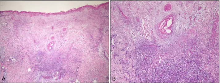
Fig. 2. Histopathologic finding showing a wedge-shaped dermal infarct with a broad base toward the epidermis. An occluded dermal vessel is visible at the base of the dermal infarct area, and a lymphohistiocytic inflammatory infiltrate is visible at the lateral and deeper margins of the dermal area (H&E; A: ×40, B: ×100).
years (Fig. 1C). She had been treated with prednisolone, dapsone, and colchicine for 2 years, but the cutaneous le- sions did not improve with BD treatments.
Histopathologic examination of a typical lesion showed a cell-poor, wedge-shaped area of dermal necrosis with ves- sel occlusion as well as dermal edema. Occluded vessels with thrombosis and vascular endothelial cell swelling were observed at the base of the wedge-shaped lesion, and the area was surrounded by lymphocytic infiltration (Fig. 2). Copious mucin deposition was visible in the der- mis, which also showed positive staining for Alcian blue (data not shown). The laboratory findings for antinuclear antibody, lupus anticoagulant, anticardiolipin antibodies, and antiphospholipid antibodies were all negative. The re- sults of the pathergy test were positive. The patient also had non-erosive asymmetric arthritis, and a 99m Tc-HDP 3-phase bone scan showed hot uptake. She had a scarring change on the retina due to recurrent chorioretinitis.
Colonoscopy showed no specific findings for DD or BD.
The patient was diagnosed with DD associated with BD.
The cutaneous lesions, which were not responsive to BD treatment, improved after 4 months of treatment with as- pirin and dipyridamole.
While the etiology of DD is unknown, evidence suggests a primary vaso-occlusive process. Vascular coagulopathy, endothelial cell damage, and an autoimmune process are considered the major pathogenic mechanisms3,4. In BD, an immune-mediated occlusive vasculitis is found in most sites of the active disease, and the endothelium appears to be the primary target5. The cutaneous lesions of the pres- ent patient were characteristic of DD and did not respond
to BD treatments. This indicates that, while DD can be as- sociated with BD, it is a distinct disease entity to BD.
To the best of our knowledge, although Sweet’s syndrome and pyoderma gangrenosum in BD have been reported in association with BD, our patient represents the first re- ported case of DD associated with BD. With the under- lying vaso-occlusive process of both DD and BD, it is rea- sonable for an association between DD and BD to exist.
Whether this patient had the same underlying mecha- nisms or the findings were coincidental is yet to be determined. We suggest that DD and BD are distinct dis- ease entities but that DD can be associated with BD.
REFERENCES
1. Thrash B, Patel M, Shah KR, Boland CR, Menter A. Cuta- neous manifestations of gastrointestinal disease: part II. J Am Acad Dermatol 2013;68:211.e1-33.
2. Oh SH, Lee JH, Shin JU, Bang D. Dermatological features in Behçet disease-associated vena cava obstruction. Br J Der- matol 2008;159:555-560.
3. Magro CM, Poe JC, Kim C, Shapiro L, Nuovo G, Crow MK, et al. Degos disease: a C5b-9/interferon-α-mediated endo- theliopathy syndrome. Am J Clin Pathol 2011;135:599-610.
4. Gilaberte Y, Coscojuela C, Lezaún A, Marigil MA. Degos disease associated with protein S deficiency. Br J Dermatol 2005;153:666-667.
5. Sağdiç K, Ozer ZG, Saba D, Türe M, Cengiz M. Venous lesions in Behçet's disease. Eur J Vasc Endovasc Surg 1996;
11:437-440.