• 검색 결과가 없습니다.

Intraductal Migration of a Breast Tissue Marker Placed under Ultrasound Guidance during COVID-Induced Delay of Surgery

N/A
N/A
Protected

Academic year: 2021

Share "Intraductal Migration of a Breast Tissue Marker Placed under Ultrasound Guidance during COVID-Induced Delay of Surgery"

Copied!
7
0
0

로드 중.... (전체 텍스트 보기)

전체 글

(1)

ABSTRACT

Breast tissue markers are common in current clinical practice and are susceptible to migration. Herein, we present the case of a 47-year-old woman with invasive breast carcinoma diagnosed through ultrasound-guided core biopsy, who underwent placement of a breast marker (HydroMARK
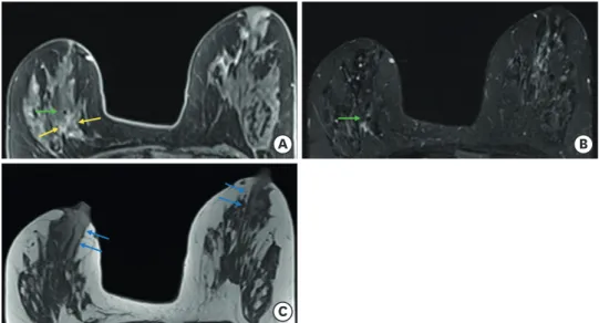
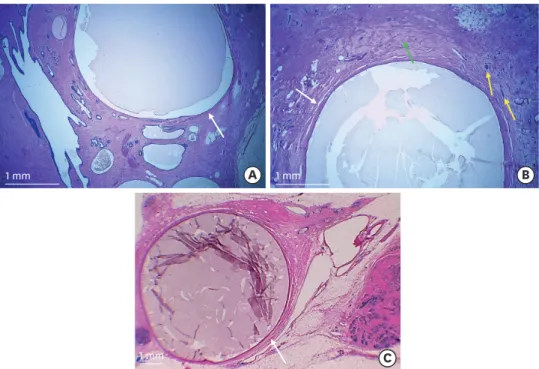
®

) under ultrasound guidance 30 days after core biopsy and with subsequent marker migration to the nipple. The correct position of the marker was documented by mammography after its placement and by magnetic resonance imaging (MRI) after neoadjuvant chemotherapy. Migration of the marker to the nipple was evident only by mammography on the day of surgery. We hypothesized that an intraductal path was the route of marker migration in this patient. Marked ductal ectasia evident on MRI and histopathologic examination supported this hypothesis. To the best of our knowledge, this is the first published case of intraductal migration of a breast tissue marker.

Keywords: Breast; Breast neoplasms

INTRODUCTION

Image-guided core biopsy followed by placement of a marker has become the standard of care of suspicious breast lesions [1].

Migration of a breast lesion marker is a known complication that is most commonly associated with vacuum-assisted biopsies [2]. Significant migration may hamper the achievement of clear margins after surgical removal. This is particularly relevant in breast cancer because of the high rates of clinical response after neoadjuvant systemic therapy achieved nowadays, often making markers the only landmark of the remaining lesion.

The mechanisms of migration described in the literature include the accordion effect, displacement by a hematoma, and migration through the biopsy track or through adipose breast tissue. The accordion effect is a common complication of vacuum-assisted biopsies and occurs along the axis of needle insertion immediately after breast decompression.

Marker displacement due to a hematoma resulting from biopsy may occur, owing to its mass

Case Report

Received: May 23, 2021 Revised: Jul 18, 2021 Accepted: Aug 10, 2021 Correspondence to Gisela Andrade

Department of Radiology, Hospital Prof.

Doutor Fernando Fonseca EPE, IC 19, 2720- 276, Amadora, Portugal.

E-mail: [email protected]

© 2021 Korean Breast Cancer Society This is an Open Access article distributed under the terms of the Creative Commons Attribution Non-Commercial License (https://

creativecommons.org/licenses/by-nc/4.0/) which permits unrestricted non-commercial use, distribution, and reproduction in any medium, provided the original work is properly cited.

ORCID iDs Gisela Andrade

https://orcid.org/0000-0001-8003-4232 André Pereira

https://orcid.org/0000-0002-2578-2075 Lucília Gonçalves

https://orcid.org/0000-0002-3902-0717 Cláudia Videira

https://orcid.org/0000-0002-1532-154X Conflicts of Interest

The authors declare that they have no competing interest.

Author Contributions

Conceptualization: Andrade G; Writing - original draft: Andrade G, Pereira A; Writing - review & editing: Andrade G, Gonçalves L, Videira C.

Gisela Andrade 1 , André Pereira 2 , Lucília Gonçalves 2 , Cláudia Videira 1

1

Department of Radiology, Hospital Prof. Doutor Fernando Fonseca EPE, Amadora, Portugal

2

Department of Pathology, Hospital Prof. Doutor Fernando Fonseca EPE, Amadora, Portugal

Intraductal Migration of a Breast Tissue Marker Placed under

Ultrasound Guidance during COVID-

Induced Delay of Surgery

(2)

effect. There is also a known association between marker migration and predominant fatty breast tissue, presumably due to less structural resistance to marker movement [3-5].

Many types of breast markers are commercially available with different sizes, shapes, and compositions. The most common materials are made of metal and embedded in hygroscopic material [6]. HydroMARK

®

(Mammotome, Cincinnati, OH, USA) is of this type. It is

composed of titanium or stainless steel embedded in a hydrophilic hydrogel. The metal component is permanently visible under conventional radiography and magnetic resonance imaging (MRI), and measures approximately 2 mm [6]. Hydrogels expand after placement by absorbing water and are then degraded via hydrolysis over time. When the hydrogel is expanded, sonographic detection is possible. Once hydrogel degradation occurred, only a smaller marker composed of metal remained [7].

CASE REPORT

A 47-year-old woman with no relevant medical history and no family antecedents of breast cancer experienced a lump in the right breast, without nipple discharge, and sought medical attention. Physical examination revealed a palpable mass. Ultrasonography (US) confirmed the presence of an irregular and hypoechoic solid mass in the right breast, in the inner lower quadrant at 4 o′clock (Figure 1). US also revealed a dilated breast duct around the lesion and marked ipsilateral retroareolar ductal ectasia with anechoic content. MRI demonstrated a suspicious mass enhancement at the corresponding location (Figure 1). No suspicious lymph nodes were found on US or MRI.

A B

C

Figure 1. Breast ultrasound and MRI. (A) Ultrasound of the right breast shows an irregular hypoechoic mass in the inner lower quadrant at 4 o′clock (yellow arrows). A dilated duct is in the vicinity of the lesion (blue arrow).

(B) Ultrasound of the right breast also reveals retroareolar ductal ectasia with anechoic content (green arrows).

(C) Axial T1-weighted fat-suppressed MRI image after gadolinium administration demonstrates a suspicious mass enhancement with 18 mm of maximum diameter (yellow arrows) in the corresponding location of the lesion seen at ultrasound. The nipple to lesion distance was 57 mm.

MRI = magnetic resonance imaging.

(3)

A core biopsy using a 14-gauge needle was performed under ultrasound guidance without complications. Histopathological examination showed invasive carcinoma of no special type, grade 2, estrogen receptor-positive, progesterone receptor-positive, human epidermal growth factor receptor 2-nonamplified, and 75% Ki-67.

Further examinations with computed tomography and bone scintigraphy showed no signs of distant metastases. A cT2N0M0 stage was assumed, and neoadjuvant chemotherapy (NAC) and endocrine therapy followed by breast conservative surgery, adjuvant

radiotherapy, chemotherapy, and endocrine therapy were planned. A marker, HydroMARK

®

(Mammotome), was inserted into the lesion 30 days after breast biopsy and before the start of chemotherapy, also under ultrasound guidance. There were no procedure-related complications. Mammography obtained in craniocaudal (CC) and mediolateral oblique (MLO) projections confirmed correct marker positioning after placement (Figure 2).

Later, the patient underwent four cycles of dose-dense doxorubicin plus cyclophosphamide followed by four cycles of paclitaxel and endocrine therapy with tamoxifen.

Post-NAC MRI showed a decrease in tumor size and partial imaging response (according to Response Evaluation Criteria In Solid Tumors v1.1 criteria) (Figure 3). The disease was down- staged to cT1N0M0, and breast-conserving surgery was planned.

Breast MRI also showed HydroMARK

®

in the anterior aspect of the lesion represented by a central signal void due to metal and a mild peripheral hyperintensity corresponding to the hydrogel. Bilateral ductal ectasia was also seen and was more evident in the right breast, where it reached up to 8 mm in diameter.

A B

Figure 2. Mammography in craniocaudal (A) and mediolateral oblique (B) views after marker placement. Both

views show the marker in the expected position. In each image, a box with white dashed borders in the upper left

corner represents an enlarged view of the marker.

(4)

Due to the coronavirus disease 2019 (COVID-19) pandemic, surgery was postponed and rescheduled to 66 days after the end of neoadjuvant therapy and 224 days after marker placement. On the morning of the surgery, the patient was brought to the radiology department for hookwire placement. Ultrasound examination did not reveal the marker, and the residual tumor was no longer visible using this technique. Mammography was then performed, which showed the metal portion of HydroMARK

®

in the nipple in both the CC and MLO views, at approximately 56 mm from the initial location in the CC view and 65 mm in the MLO view (Figure 4). The patient denied any traumatic events in the previous months.

Since the lesion was no longer visible on mammography and ultrasound and its only landmark was lost, the planned surgery was modified to a simple right mastectomy and sentinel lymph node biopsy with the patient's agreement. The surgical procedure was performed with no complications, the sentinel lymph node biopsy was negative for malignancy, and the patient was discharged 3 days later. The resection margins were negative, and the final stage was ypT1cN0 ycM0. The patient is currently receiving adjuvant endocrine therapy (tamoxifen, 20 mg/day). At her one-year follow-up, clinical and ultrasound examinations were unremarkable.

Histopathological analysis of the surgical specimen showed residual small islands of neoplastic cells and fibrocystic changes with cystic dilatation of the ducts with a maximum ductal diameter of 6.5 mm (Figure 5). No marker was detected in the specimen.

Written informed patient consent for publication has been obtained. The manuscript is a case report and does not involve research in humans; therefore, it was not submitted to an institutional review board.

A

C

B

Figure 3. Breast MRI performed for evaluation of tumor response to NAC. (A) Axial T1-weighted fat-suppressed postcontrast image demonstrates a decrease in tumor size, measuring 17 mm of maximum diameter (yellow arrows). A focus of metallic susceptibility artifact in the anterior portion of the tumor corresponding to the metal marker is also evident (green arrow). (B) Axial T2-weighted fat-suppressed image shows the same focus of metallic susceptibility with a surrounding hyperintensity compatible with hydrogel (green arrow). (C) Axial T1- weighted precontrast image without fat suppression shows bilateral ductal ectasia, more prominent on the right where it reaches a maximum diameter of 8 mm (blue arrows).

MRI = magnetic resonance imaging; NAC = neoadjuvant chemotherapy.

(5)

A B

Figure 4. Mammography performed on the day of the surgery to locate the marker. (A) Mammography in mediolateral oblique projection shows the marker in the nipple (green arrow). (B) Craniocaudal magnification view also shows the marker positioned in the nipple (green arrow).

1 mm 1 mm

1 mm

A B

C

Figure 5. Histopathological examination of the surgical specimen showed diffuse ductal ectasia, most evident in retroareolar location. (A) H&E stain, ×25, shows a dilated duct with 3 mm of diameter (white arrow). (B) H&E stain, ×25, shows ductal ectasia extending into the tumoral bed, with a dilated duct with 3.75 mm of diameter (white arrow). Fibrosis is identified by a green arrow and residual small islands of neoplastic cells by yellow arrows. (C) H&E stain, very low-magnification, shows the largest dilated duct identified in the specimen, with a diameter of 6.5 mm (white arrow).

H&E = hematoxylin and eosin.

(6)

DISCUSSION

In the case presented here, migration of a tissue marker placed under ultrasound guidance took place. This migration was evident only on the day of the surgery. The marker was correctly positioned on mammography after its placement and on post-NAC MRI. This means that migration occurred between post-NAC MRI and the day of surgery, that is, between the 165th and 224th day (approximately 7 months) after marker placement.

Given the clear position of the migrated marker in the nipple in both mammography views, we believe that the only possible location is the lactiferous sinus of the nipple. In this way, we hypothesized that the marker shifted and migrated through a lactiferous duct.

The marked ductal ectasia documented on MRI (up to 8 mm in diameter in the right breast) and on the specimen (up to 6.5 mm in diameter) corroborates this hypothesis. This degree of ectasia would allow a HydroMARK

®

marker with partial or total degradation of the hydrogel to enter and migrate through them (as stated before, its metal component measures only 2 mm). The mild peripheral hyperintensity surrounding the marker on T2-weighted images of post-NAC MRI (Figure 3B) suggests that partial hydrolysis had already occurred.

Although we did not find studies that directly assess the duration and degree of

HydroMARK

®

’s expansion and shrinkage over time, the duration of its sonographic detection (which depends on hydrogel expansion) has been shown to be variable. Carmon et al. [8], Klein et al. [9], and Sakamoto et al. [10] reported no loss of sonographic detection over time up to 187 days, 204 days, and 11 months, respectively. However, Blumencranz et al. [11]

found that only 77.8% of markers were detected by ultrasound at the time of surgery (average time between marker placement and day of surgery of 189 days). In this patient, the marker remained placed for longer than necessary due to the COVID-19 pandemic (224 days), which might have been an additional factor contributing to the migration.

Furthermore, the currently known mechanisms of breast marker migration are not plausible in this clinical context. The correct position of the marker on post-NAC MRI makes a causal relationship between migration and biopsy-related complications very unlikely. The accordion effect does not play a role in ultrasound-guided procedures due to the absence of breast compression, while the absence of adipose tissue in the nipple-areolar complex [12] precludes the hypothesis of migration through it. The marker was not found on the surgical specimen, thus we can only assume that it was lost during surgery, possibly exiting through the nipple.

Marker migration had a significant impact on patient management. The long distance covered by the marker prevented an accurate presurgical location of the malignant lesion and resulted in a more invasive type of surgery for its removal.

To the best of our knowledge, there are no other descriptions of intraductal migration of breast lesion markers. Previous reports have shown breast ductal migration of silicone, usually secondary to a ruptured prosthesis [13-15]. We did not find any reports of migration of any other material through the breast ductal system.

In conclusion, we report a previously unknown mechanism of breast lesion marker

migration. This phenomenon raises the question of whether the diameter of breast ducts is

an important morphological characteristic to evaluate when assessing which marker to use.

(7)

REFERENCES

1. Teichgraeber DC, Martaindale S, Omofoye TS, Hess KR, Parikh JR, Whitman GJ. Immediate migration of biopsy clip markers after upright digital breast tomosynthesis-guided vacuum-assisted core biopsy. Acad Radiol 2020;27:204-9.

PUBMED | CROSSREF

2. Funaro K, Prather A, Niell B, Jared Weinfurtner R. Tissue marker migration after MRI-guided breast biopsy: migration frequency and associated factors. Breast J 2020;26:440-5.

PUBMED | CROSSREF

3. Esserman LE, Cura MA, DaCosta D. Recognizing pitfalls in early and late migration of clip markers after imaging-guided directional vacuum-assisted biopsy. Radiographics 2004;24:147-56.

PUBMED | CROSSREF

4. Jain A, Khalid M, Qureshi MM, Georgian-Smith D, Kaplan JA, Buch K, et al. Stereotactic core needle breast biopsy marker migration: an analysis of factors contributing to immediate marker migration. Eur Radiol 2017;27:4797-803.

PUBMED | CROSSREF

5. Huppe AI, Brem RF. Minimally invasive breast procedures: practical tips and tricks. AJR Am J Roentgenol 2020;214:306-15.

PUBMED | CROSSREF

6. Carmon M, Zilber S, Gekhtman D, Olsha O, Hadar T, Golomb E. Hygroscopic sonographically detectable clips form characteristic breast and lymph node pseudocysts. Mod Pathol 2018;31:62-7.

PUBMED | CROSSREF

7. Shah AD, Mehta AK, Talati N, Brem R, Margolies LR. Breast tissue markers: Why? What’S out there? How do I choose? Clin Imaging 2018;52:123-36.

PUBMED | CROSSREF

8. Carmon M, Olsha O, Gekhtman D, Nikitin I, Cohen Y, Messing M, et al. Detectability of hygroscopic clips used in breast cancer surgery. J Ultrasound Med 2017;36:401-8.

PUBMED | CROSSREF

9. Klein RL, Mook JA, Euhus DM, Rao R, Wynn RT, Eastman AB, et al. Evaluation of a hydrogel based breast biopsy marker (HydroMARK

®

) as an alternative to wire and radioactive seed localization for non-palpable breast lesions. J Surg Oncol 2012;105:591-4.

PUBMED | CROSSREF

10. Sakamoto N, Fukuma E, Tsunoda Y, Teraoka K, Koshida Y. Evaluation of the dislocation and long-term sonographic detectability of a hydrogel-based breast biopsy site marker. Breast Cancer 2018;25:575-82.

PUBMED | CROSSREF

11. Blumencranz PW, Ellis D, Barlowe K. Use of hydrogel breast biopsy tissue markers reduces the need for wire localization. Ann Surg Oncol 2014;21:3273-7.

PUBMED | CROSSREF

12. Nicholson BT, Harvey JA, Cohen MA. Nipple-areolar complex: normal anatomy and benign and malignant processes. Radiographics 2009;29:509-23.

PUBMED | CROSSREF

13. Leibman AJ, Kossoff MB, Kruse BD. Intraductal extension of silicone from a ruptured breast implant.

Plast Reconstr Surg 1992;89:546-7.

PUBMED | CROSSREF

14. Holten I. Intraductal migration of silicone. Plast Reconstr Surg 1996;98:903.

PUBMED | CROSSREF

15. Rahme J, Liu D, Chew G, Zinn R. Silicone nipple discharge: a case report of an unusual presentation of breast implant rupture. Int J Surg Case Rep 2020;74:73-6.

PUBMED | CROSSREF

수치

Figure 1. Breast ultrasound and MRI. (A) Ultrasound of the right breast shows an irregular hypoechoic mass in  the inner lower quadrant at 4 o′clock (yellow arrows)
Figure 2. Mammography in craniocaudal (A) and mediolateral oblique (B) views after marker placement
Figure 3. Breast MRI performed for evaluation of tumor response to NAC. (A) Axial T1-weighted fat-suppressed  postcontrast image demonstrates a decrease in tumor size, measuring 17 mm of maximum diameter (yellow  arrows)
Figure 5. Histopathological examination of the surgical specimen showed diffuse ductal ectasia, most evident  in retroareolar location

참조

관련 문서

It considers the energy use of the different components that are involved in the distribution and viewing of video content: data centres and content delivery networks

After first field tests, we expect electric passenger drones or eVTOL aircraft (short for electric vertical take-off and landing) to start providing commercial mobility

1 John Owen, Justification by Faith Alone, in The Works of John Owen, ed. John Bolt, trans. Scott Clark, "Do This and Live: Christ's Active Obedience as the

• Hormone Therapy With or Without Combination Chemotherapy in Treating Women Who Have Undergone Surgery for Node-Negative Breast Cancer (The TAILORx Trial)..

- distributes indexes across multiple computers and/or multiple sites - essential for fast query processing with large numbers of documents - many variations:

Kameoka, “Sentinel lymph node biopsy for breast cancer patients using fluorescence navigation with indocyanine green,” World Journal of Surgical Oncology, Vol..

[r]

Table 2. Schatzker and Lambert assessment --- 18.. A), B) Preoperative anteroposterior and lateral radiograph of the left knee of a sixty-two-year-old man who