J Korean Soc Radiol 2015;73(3):137-146 http://dx.doi.org/10.3348/jksr.2015.73.3.137
Lung Cancer Screening: Update 폐암검진: 최신지견
Hyae Young Kim, MD*
Department of Radiology, Center for Lung Cancer, National Cancer Center, Goyang, Korea
서론
폐암은 전세계적으로 암사망의 주요원인으로 2012년에 약 182만 명이 발생하였고, 159만 명이 폐암으로 사망하였다(1). 우 리나라 2012년 암 발생 통계에 따르면 폐암은 10만 명당 43.9명 이 발생하였고, 남성에서는 61.0명, 여성은 26.8명에서 발생하 였다. 폐암 발생 추이는 1999년부터 2012년까지 남녀 전체로는 큰 변화가 없으나, 남성에서는 감소(연평균 -0.9%)하는 경향 을, 여성에서는 증가(연평균 1.7%)하는 경향을 보였다. 남성에 서 폐암은 전체 암의 13.7%, 여성에서는 6.0%로 갑상선암을 제외하면 폐암 발생은 전체 암의 9.9%로 3위를 차지하는 반면, 암사망률은 폐암이 1위로 가장 높다. 2012년 폐암은 전체 암사망 의 22.5%를 차지하여, 남성에서 26.2%, 여성에서는 16.4%였 고 남녀 모두 전체암사망 대비 폐암사망률은 증가하고 있다(2).
폐암의 5년 생존율은 15% 이하로 비소세포암의 경우 수술적
제거가 가능한 1병기의 5년 생존율은 약 70% 정도이지만, 폐 암은 발견 당시 이미 진행된 경우가 많아 실제 임상에서 발견되 는 1병기는 16%에 불과하다(3). 따라서, 증상이 생기기 전에 검진으로 수술적 치료가 가능한 시기에 조기 발견한다면 폐암으 로 인한 생존율 향상 및 사망률 감소를 기대할 수 있을 것이다.
폐암검진의 역사
조기에 폐암을 발견하면 사망률을 감소시킬 수 있을 것이라 는 예측과는 달리 흉부X선과 객담을 이용한 폐암검진에 대한 연구는 폐암사망률을 감소시키는 효과가 없었다(4, 5). 흉부X 선을 선별검사방법으로 이용한 비교적 최근 연구인 the Pros- tate, Lung, Colorectal and Ovarian trial 연구에서 1993~2001 년의 기간 동안 55~74세의 154901명을 대상으로 흉부X선을 매년 4회 촬영한 대상자들을 일반인과 2009년 12월까지 최장 Lung cancer is the leading cause of cancer deaths worldwide as well as in Korea. A
recent National Lung Screening Trial in U.S. revealed that low-dose CT (LDCT) screen- ing reduced lung cancer specific mortality by 20% in high risk individuals as com- pared to chest radiograph screening. Based on this evidence, several expert societies in U.S. and Korean multisociety collaborative committee developed guidelines for rec- ommendation of lung cancer screening using annual LDCT in high risk populations.
In most of the societies high risk groups are defined as persons aged 55 to 74 years, who are current smokers with history of smoking of more than 30 packs per year or ex- smokers, who quit smoking up to 15 or more years ago. The benefits of LDCT screen- ing are modestly higher than the harms in high risk individuals. The harms included a high rate of false-positive findings, over-diagnosis and radiation-related deaths. In- vasive diagnostic procedure due to false positive findings may lead to complications.
LDCT should be performed in qualified hospitals and interpreted by expert radiolo- gists. Recently, the American College of Radiology released the current version of Lung cancer CT screening Reporting and Data Systems. Education and actions to stop smoking must be offered to current smokers.
Index terms
Early Detection of Cancer Lung Neoplasms Mass Screening
Tomography, Spiral Computed
Received June 3, 2015 Revised July 2, 2015 Accepted July 13, 2015
*Corresponding author: Hyae Young Kim, MD Department of Radiology, Center for Lung Cancer, National Cancer Center, 323 Ilsan-ro, Ilsandong-gu, Goyang 10408, Korea.
Tel. 82-31-920-2638 Fax. 82-31-920-2643 E-mail: [email protected]
This is an Open Access article distributed under the terms of the Creative Commons Attribution Non-Commercial License (http://creativecommons.org/licenses/by-nc/3.0) which permits unrestricted non-commercial use, distri- bution, and reproduction in any medium, provided the original work is properly cited.
13년간 비교한 결과, 두 군 간에 폐암사망률 차이가 없었고[re- lative risk (이하 RR), 0.99; 95% confidence interval (이하 CI), 0.87~1.22; p = 0.48], 폐암발생률은 선별검사를 시행한 군에서 높았지만 통계적 유의성은 없었다(20.1 vs. 19.2 per 10000 person-years; RR, 1.05; 95% CI, 0.98~1.12)(6). 따라 서, 흉부X선으로 폐암검진을 시행하는 것은 권장하지 않았다.
2000년대 초에 저선량 흉부전산화단층촬영(low-dose chest computed tomography; 이하 저선량 흉부CT)를 이용한 연구 들이 발표되면서, 폐암검진에 대한 기대가 높아졌으나, 이들은 관찰 연구로 폐암사망률 감소 여부를 알 수 없었다(7-11). 이에 따라, 2004년 U.S. Preventive Services Task Force (이하 US- PSTF)에서는 증거불충분(evidence insufficient)으로 폐암검진 을 권고하지 않았다(12). 이후, 저선량 흉부CT를 이용한 폐암 검진이 폐암특이사망률을 감소시킬 수 있는지 평가하기 위해 무작위대조군연구(randomized controlled trial; 이하 RCT)가 진행되었고, 미국에서 진행된 National Lung Screening Trial (이하 NLST) 연구에서 저선량 흉부CT로 폐암검진을 시행한 수검자에서 흉부X선으로 폐암검진을 시행한 대조군에 비해 폐 암특이사망률이 약 20% 감소되었다는 결과를 발표하였다(13, 14). 이에 따라 저선량 흉부CT를 이용한 폐암검진에 대한 관심 이 더욱 높아졌으며, 미국의 여러 임상학회에서 폐암검진 권고 안을 발표하였고 USPSTF에서도 폐암검진을 권고하고 있다 (15-19). 또한, 2015년 2월에 65세 이상 및 장애인들에게 보험 이 적용되는 미국의 공적 보험이라 할 수 있는 Centers for Me- dicare & Medicaid Service (이하 CMS)에서 고위험군에서 폐 암검진을 권고하며 보험을 적용하기로 결정하였다(20). 유럽에 서는 대략 2015년 후반에서 2017년도에 발표 예정인 Dutch- Belgian randomized lung cancer screening trial (이하 NEL- SON) 연구 결과와 유럽의 여러 RCT 연구들을 합한 결과를 기 다리고 있으나, 최근 유럽영상의학회(European Sociery of Radiology)와 유럽호흡기학회(European Respiratory Society) 에서도 NLST의 결과를 근거로 폐암검진을 권고하였으며, 종 합적이고, 품질보증된, 장기실행 프로그램에 한해서 임상시험 또는 임상에서도 인증된 다학제(multidisciplinary) 의료기관에 서 폐암검진을 시행할 것을 권고하였다(21).
우리나라에서도, 흉부영상의학회에서 폐암검진에 대한 제안 서를 발표한 바 있고, 최근 대한폐암학회, 대한결핵 및 호흡기 학회, 대한흉부외과학회, 대한영상의학회, 대한가정의학회, 대 한예방의학회 등 폐암관련학회 및 국립암센터로부터 추천받은 다학제전문가로 구성된 ‘폐암검진권고안제정위원회’에서 NLST 연구의 결과를 근거로 폐암검진 권고안을 발표하였다 (22, 23).
저선량 흉부CT를 이용한 폐암검진 대조군 연구
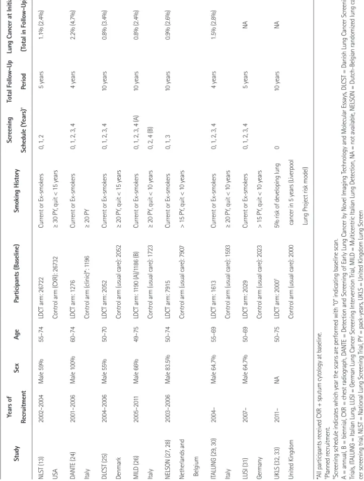
저선량 흉부CT를 이용한 폐암검진에 대한 RCT는 전세계적 으로 8개의 연구가 진행 중이거나 결과가 발표되었다(Table 1).
NLST의 연구 결과는 폐암특이사망률을 감소시켰지만, 유럽의 연구 결과는 부정적이다(13, 14, 24-33). NLST 연구는 55~
74세의 30갑년 이상의 흡연력을 가진 현재 흡연자 또는 금연한 지 15년 미만의 과거흡연자를 대상으로 하였는데, 덴마크에서 시행한 Danish Lung Cancer Screening Trials (이하 DLCST) 연 구나 이탈리아에서 시행한 Multicentric Italian Lung Detection (이하 MILD) 연구는 NLST 연구나 Detection and Screening of Early Lung Cancer by Novel Imaging Technology and Mo- lecular Essays (이하 DANTE) 연구에 비해 참여자의 평균 나이 나 흡연력이 낮은 편이었다(Table 1). 대조군은 NLST 연구는 흉 부X선이었고 유럽의 연구는 검사를 시행하지 않는 관찰이었다 (13, 24-26).
폐암검진의 이득
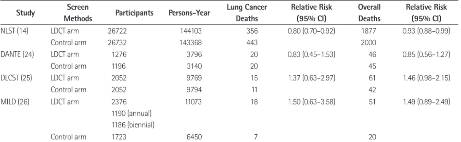
저선량 흉부CT를 이용한 폐암검진의 기대할 수 있는 이득은 폐암특이사망률 및 전체사망률의 감소이고 현재는 NLST 연구 만이 긍정적인 결과를 보였다(14). NLST 연구 결과에서 폐암 특이사망률에 대한 저선량 흉부CT군의 상대위험도를 평가하 였을 때 0.80(95% CI, 0.70~0.92)으로 유의하였고, 1명의 사 망을 예방하기 위해 필요한 검진수(Numbers Needed to be Screened to prevent 1 death; 이하 NNS)는 320(95% CI, 190~
840)이었다. 전체사망률에 대한 저선량 흉부CT군의 상대위험 도는 0.93(95% CI, 0.88~0.99)으로 유의하였고 NNS는 219 (95% CI, 112~5000)였다(14). 다른 세 연구 DLCST, MILD, DANTE에서는 저선량 흉부CT군과 대조군 간에 폐암특이사망 률 및 전체사망률에 유의한 차이가 없었다(Table 2).
유럽의 결과가 부정적인 이유에 대해서는 밝혀진 바는 없으나, NLST에 비해 연구 참여자 수가 적고(저선량 흉부CT군 1000~
2000명 vs. 26700명), 발견된 폐암의 수가 적으며(30~60 vs.
350~400), 추적 검사 기간이 짧았으며(34~58개월 vs. 78개월), 흡연력도 낮아(20갑년 vs. 30갑년) 상대적으로 위험도가 적었 기 때문일 가능성이 있다.
저선량 흉부CT의 위해
폐암검진으로 인한 위해는 높은 위양성률과 이차적인 진단과 정에서 부작용 발생과 불안감, 잠재적인 과진단(overdiagno-
Table 1. Randomized Controlled Trials of Lung Cancer Screening with Low-Dose CT StudyYears of RecruitmentSexAgeParticipants (Baseline)Smoking HistoryScreening Schedule (Years)‡Total Follow-Up Period
Lung Cancer at Initial (Total in Follow-Up) NLST (13) USA
2002–2004Male 59%55–74LDCT arm: 26722 Control arm (CXR): 26732 Current or Ex-smokers ≥ 30 PY, quit < 15 years
0, 1, 25 years1.1% (2.4%) DANTE (24) Italy
2001–2006Male 100%60–74LDCT arm: 1276 Control arm (clinic)*: 1196 Current or Ex-smokers ≥ 20 PY
0, 1, 2, 3, 44 years2.2% (4.7%) DLCST (25) Denmark
2004–2006Male 55%50–70LDCT arm: 2052 Control arm (usual care): 2052 Current or Ex-smokers ≥ 20 PY, quit < 15 years
0, 1, 2, 3, 410 years0.8% (3.4%) MILD (26) Italy
2005–2011Male 66%49–75LDCT arm: 1190 (A)/1186 (B) Control arm (usual care): 1723 Current or Ex-smokers ≥ 20 PY, quit < 10 years 0, 1, 2, 3, 4 (A) 0, 2, 4 (B)
10 years0.8% (2.4%) NELSON (27, 28) Netherlands and Belgium
2003–2006Male 83.5%50–74LDCT arm: 7915 Control arm (usual care): 7907 Current or Ex-smokers > 15 PY, quit < 10 years
0, 1, 310 years0.9% (2.6%) ITALUNG (29, 30) Italy
2004–Male 64.7%55–69LDCT arm: 1613 Control arm (usual care): 1593 Current or Ex-smokers ≥ 20 PY, quit < 10 years
0, 1, 2, 3, 44 years1.5% (2.8%) LUSI (31) Germany
2007–Male 64.7%50–69LDCT arm: 2029 Control arm (usual care): 2023 Current or Ex-smokers > 15 PY, quit < 10 years
0, 1, 2, 3, 45 yearsNA UKLS (32, 33) United Kingdom
2011–NA50–75LDCT arm: 2000† Control arm (usual care): 2000 5% risk of developing lung cancer in 5 years (Liverpool Lung Project risk model)
010 yearsNA *All participants received CXR + sputum cytology at baseline. †Planned recruitment. ‡Screening schedule indicates which year the scans are performed with “0” indicating baseline scan. A = annual, B = biennial, CXR = chest radiograph, DANTE = Detection and Screening of Early Lung Cancer by Novel Imaging Technology and Molecular Essays, DLCST = Danish Lung Cancer Screening Trials, ITALUNG = Italian Lung, LUSI = German Lung Cancer Screening Intervention Trial, MILD = Multicentric Italian Lung Detection, NA = not available, NELSON = Dutch-Belgian randomized lung can- cer screening trial, NLST = National Lung Screening Trial, PY = pack-years, UKLS = United Kingdom Lung Screen
sis), 그리고 방사선피폭(radiation exposure)에 의한 암발생 가 능성 등이 있다(34-48).
위양성(False Positive)
저선량 흉부CT의 양성률(비석회화 결절 발견율, positive rate)은 연구에 따라 6~53%로 CT 절편두께 및 검사대상에 따 라 다양하게 보고된다(34-36). NLST 연구에서 4 mm 이상의 결절을 양성으로 정의하였을 때, 결절이 24.2%의 수검자에서 발견되었다(37). 저선량 흉부CT는 흉부X선에 비해 약 3.5배 가량 결절이 더 많이 발견되지만 이들의 대부분은 양성(benign) 으로 위양성률(false positive rate)은 높다.
4~5 mm 이상의 결절을 양성(positive)이라 정의한 연구에
서는 위양성률이 약 59.4~96.4%였다(34-36). NELSON 연구 에서는 결절의 부피가 500 mm3 초과인 경우 혹은 50~500 mm3 인 경우 3개월 후 추적검사에서 용적이 25% 증가하였을 때를 양성이라 정의하였을 때 양성률이 6%였고 위양성률은 1.2%였 다(38). 발견된 결절을 진단하기 위해 추가 CT검사나 조직검사 로 인한 비용이 발생하고 시술과 관련된 부작용 및 불안감이 생 길 수 있다. 악성 가능성이 있다고 판단되는 경우에 결절에 대 한 침습진단검사를 시행하였고 이에 대한 양성예측도(positive predictive value; 이하 PPV)는 50~92%로 검사를 시행한 수검 자의 대부분은 폐암으로 진단된다. NLST에서는 결절이 발견된 경우 56.5%가 추가로 영상촬영을 하였고, 5.9%가 침습적 진단 을 받았다. 이에 의한 주요 합병증은 약 7.8%였다(37).
Table 2. Effects of Lung Cancer Screening with LDCT
Study Screen
Methods Participants Persons-Year Lung Cancer Deaths
Relative Risk (95% CI)
Overall Deaths
Relative Risk (95% CI)
NLST (14) LDCT arm 26722 144103 356 0.80 (0.70–0.92) 1877 0.93 (0.88–0.99)
Control arm 26732 143368 443 2000
DANTE (24) LDCT arm 1276 3796 20 0.83 (0.45–1.53) 46 0.85 (0.56–1.27)
Control arm 1196 3140 20 45
DLCST (25) LDCT arm 2052 9769 15 1.37 (0.63–2.97) 61 1.46 (0.98–2.15)
Control arm 2052 9794 11 42
MILD (26) LDCT arm 2376
1190 (annual) 1186 (biennial)
11073 18 1.50 (0.63–3.58) 51 1.49 (0.89–2.49)
Control arm 1723 6450 7 20
CI = confidence interval, DANTE = Detection and Screening of Early Lung Cancer by Novel Imaging Technology and Molecular Essays, DLCST = Danish Lung Cancer Screening Trials, LDCT = low-dose computed tomography, MILD = Multicentric Italian Lung Detection, NLST = National Lung Screening Trial Table 3. Lung CT Screening Reporting and Data System (39)
Category Findings Description Management
0. Incomplete Incomplete exam Additional LDCT
or comparisons
1. Negative No nodules/specific calcification No nodules and definitely benign
nodules
Annual LDCT
2. Benign appearance or behavior
Solid < 6 mm, new 4 mm Part solid < 6 mm
Non solid < 20 mm OR ≥ 20 mm unchanged or slow growing
Very low likelihood of becoming clinically active cancer due to size or lack of growth (stable > 3 mo)
Annual LDCT
3. Probably benign Solid 6–8 mm at baseline OR new 4–6 mm
Part solid ≥ 6 mm with solid < 6 mm OR new < 6 mm total Nonsolid ≥ 20 mm
Low likelihood of becoming clinically active cancer suggesting short term follow-up
6 month LDCT
4A. Suspicious Solid 8–15 mm at baseline OR growing < 8 mm OR new 6–8 mm Part solid ≥ 6 mm with solid 6–8 mm OR new growing < 4 mm total
Additional diagnostic testing and/or tissue sampling is advised
Tissue sampling OR 3 month LDCT, PET/CT when ≥ 8 mm solid 4B. Suspicious Solid ≥ 15 mm at baseline OR new or growing > 8 mm
Part solid with solid ≥ 8 mm OR new or growing ≥ 4 mm solid
Tissue sampling OR chest CT, PET/CT Modifier S, potentially clinically significant (non-lung cancer related); modifier C, diagnosed lung cancer.
LDCT = low-dose computed tomography, PET/CT = positron emission tomography/CT
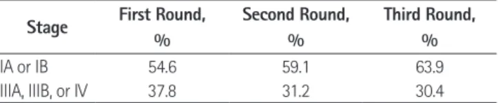
는 회차에 따른 말기 폐암의 감소가 약간 있다(Table 4)(43).
NLST 연구의 경우 6.5년 동안 폐암은 저선량 흉부CT군 26722 명에서 1060명(4.0%)이, 흉부X선군 26732명에서 941명(3.5%) 이 발생하여 저선량 흉부CT군에서 폐암발생이 119명 더 많았 다. NLST에서 발견된 폐암을 토대로 추정하였을 때 저선량 흉 부CT로 발견된 폐암의 과진단 가능성은 약 18.5%(95% CI, 5.4~30.6%)이며, 비소세포폐암의 약 22.5%(95% CI, 9.7~
34.3%), 세기관지폐포암의 78.9%(95% CI, 62.2~93.5%)가 과진단의 가능성이 있다. 한 명의 폐암으로 인한 사망을 방지하 기 위해서 320명이 선별검사를 시행해야 하는데 이 중 과진단 추정수는 1.38개이다(44). Veronesi 등(45)은 연구에서는 용적 배가시간이 400일이 넘는 경우 과진단의 가능성이 있는 것으로 보고하였다. 그러나, 현시점에서 과진단은 추정치일 뿐 해결되 지 않는 문제로 남아 있다.
방사선 피폭(Radiation Exposure)
저선량 흉부CT의 유효방사선량은 검사 건당 약 0.6~1.5 mSv 로 NLST 연구에서 3년의 기간 동안 검진과 진단을 위한 검사 까지 고려할 때 대략 일인당 방사선량은 8 mSv 정도였다(14).
발견된 병변을 진단하기 위해 노출되는 방사선량은 흉부CT는 약 ~8 mSv, positron emission tomography-CT는 약 ~14 mSv 이다. 현재까지 저선량 흉부CT검사로 인한 폐암 발생 위험 정 도는 정확히 알려져 있지 않으며 방사선 위해는 원폭생존자 및 방사선종사자 노출에 대한 연구에 근거하며, 대략 NLST 연구 의 고위험군에서 방사선에 의한 암사망이 2500명당 약 1명 있 을 것으로 예측된다(34, 46). Brenner (47)의 보고에 의하면 흡연자와 과거 흡연자인 50~75세의 미국 인구의 약 50%가 매년 검진을 받는다면 방사선과 연관된 폐암 발생은 약 1.8%(95% CI, 0.5~5.5%) 증가할 것으로 예측된다. Cancer Intervention and Surveillance Modeling Network Working Gr- oup에서 55~80세의 고위험군(30갑년, 금연 15년 이내)을 대 상으로 분석한 모델로 계산한 방사선노출과 관련된 폐암사망 추정치는 약 0.8%이다(인구 10만 명당 24명)(47). 최근 기기 의 발달로 방사선량을 현저히 낮출 수 있어, 저선량 흉부CT 촬 영에 의한 방사선 노출의 위험은 현재 추정하는 것보다는 낮을 것으로 예측되며, 방사선 노출에 의한 암발생은 10~20년이 지 미국영상의학회(American College of Radiology; 이하 ACR)
에서 저선량 흉부CT에서 발견된 결절을 표준화된 관리를 하기 위하여 현재까지 발표된 폐암검진에서 발견된 결절의 결과를 토대로 하여, 유방암 검진의 판독(Bi-RADS)과 유사한 Lung CT Screening Reporting and Data System (이하 Lung-RADS)을 제안하였다(Table 3)(39). Lung-RADS에서는 고형 결절의 경 우는 기준을 4 mm에서 6 mm로 하였고, 비고형(간유리음영) 결절의 경우는 기준을 5 mm에서 2 cm로 증가시켜 수행력을 향상시켰다. NLST에서 발견된 결절을 다시 후향적으로 분석한 최근 연구에 따르면, Lung-RADS에 따라 결절을 추적 검사하 게 되면 악성결절 발견하는 데 있어 민감도 감소 없이 NLST에 서 발견된 결절 양성률(positive rate)을 26.6%에서 10%로 감 소시킬 수 있었다. 결절 양성률을 첫 검사에서 13.7%, 그 후에 5.9%로 감소시킬 수 있었고, 또한, 4 mm 기준이었을 때 3.8%
였던 악성에 대한 양성예측도(PPV)도 첫 검사에서 6.9%, 그 후 검사에서 10.9%로 향상시킬 수 있었다(40, 41).
NELSON 연구에서 발견된 결절로 Lung-RADS와 용적을 이 용한 결절 관리 프로토콜을 시뮬레이션하여 성적을 비교 평가 한 논문에서 처음 발견된 결절에 대하여 용적(양성: 300 mm3 이상, 중간: 100~300 mm3, 음성: 100 mm3 미만), 컴퓨터 측 정 용적에 역산한 직경(양성: 10 mm 이상, 중간: 5~10 mm, 음성: 5 mm 미만), Lung-RADS (양성: 8 mm 이상, 중간: 4~
8 mm, 음성: 4 mm 미만)를 비교하였을 때, 민감도는 각각 90.9%, 92.4%, 90.9%, 특이도는 94.9%, 90.0%, 87.2%, 양 성예측도는 14.4%, 7.9%, 6.2%, 음성예측도는 모두 99.9%
로, 용적을 사용하였을 경우 민감도, 특이도를 높이고, 양성예 측도를 높일 수 있다고 하였고, 컴퓨터 측정 용적에서 역산한 직경이 수기로 측정한 직경보다 높은 수행률을 보였다(42). 이 연구에 따르면 부피가 100 mm3 미만 혹은 직경 5 mm 미만 크 기의 결절과 추적 검사하였을 때 용적배가시간이 600일 이상인 결절은 폐암 가능성이 낮았고(각각 0.4%, 0.8%), 부피가 300 mm3 이상 혹은 직경이 10 mm 이상인 결절과 용적배가시간이 400일 미만인 결절은 폐암가능성이 높았다(각각 16.9%, 9.9%).
과진단(Overdiagnosis)
수검자의 생명과 관련 없는 종양을 진단하는 것을 과진단이 라고 하며 이로 인하여 불필요한 치료 및 이환율 증가, 추적 검 사, 비용, 불안감이 발생한다(34-36). NLST를 제외한 다른 3 개의 RCT 결과에서 저선량 흉부CT군에서 대조군에 비해 초 기 폐암이 더 발견되었으나 말기 폐암의 수는 감소하지 않아서 잠재적인 과진단의 가능성을 암시하고 있으나, NLST 연구에서
Table 4. Lung Cancer Stage Detected in NLST According to Screening Rounds (37, 43)
Stage First Round,
%
Second Round,
%
Third Round,
%
IA or IB 54.6 59.1 63.9
IIIA, IIIB, or IV 37.8 31.2 30.4
NLST = National Lung Screening Trial
난 후에야 나타나는 결과로 고위험군의 경우 방사선 위해에 의 한 폐암사망보다는 폐암검진을 시행하는 것이 사망에 대한 이 득이 있을 것으로 생각된다. 그러나, 여러 모델에 의한 연구에서 비흡연자와 42세 이하의 연령에서는 이득보다 잠재적 위험이 더 클 것을 시사하였다(48).
저선량 흉부CT 검진의 권고대상 및 검진 주기
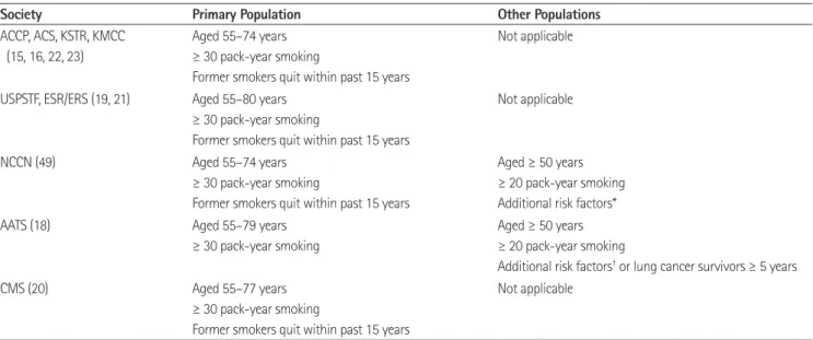
미국 여러 학회와 대한흉부영상의학회와 폐암검진제정위원 회에서 NLST 연구에 근거하여 30갑년 이상의 현재흡연자 또 는 금연한지 15년 이내의 과거흡연자로 55~74세의 고위험군 을 대상으로 저선량 흉부CT를 이용한 폐암검진을 시행할 것을 권고한다. 수검자는 추가적인 진단과 근치적 치료를 견딜 수 있 는 체력조건을 필수적으로 갖추고 있어야 한다(15-23).
미국종합암네트워크(National Comprehensive Cancer Net- work)와 미국흉부외과학회(American Association for Tho- racic Surgery)에서는 50~54세이면서, 20갑년 이상의 흡연력 이 있고 라돈 피폭, 폐암 가족력, 만성폐쇄성폐질환(chronic obstructive pulmonary disease) 또는 폐섬유증(pulmonary fi- brosis)에 해당하는 폐질환 병력, 위험 직업력(실리카, 카드뮴, 석면, 비소, 베릴리움, 크롬, 디젤배기가스, 니켈 등 폐암 발암물 질에 피폭되는 작업장) 또는 암병력(폐암에서 완치되었던 경우, 림프종, 두경부암, 식도암 등 흡연과 관련 있는 암병력자) 중
하나 이상의 요인을 가진 사람도 폐암검진의 대상으로 언급하 였으나 현 시점에서 이에 대한 근거는 없다(18, 49). USPSTF에 서는 시뮬레이션 모델을 토대로 55~80세로 권고하였다(Table 5)(50).
최근 CMS에서 고위험군에 해당하는 수검자에 대해 보험을 적용하기로 하였고, 30갑년 이상의 현재흡연자 또는 금연한지 15년 이내의 55~77세의 고위험군을 대상으로 폐암검진을 권 고하며 저선량 흉부CT를 시행하기 전에 의료기관을 방문하여 이득과 위해에 관해 충분한 상담을 거친 후 공동의사결정을 하 고 자격 있는 의료인의 처방을 받은 후 저선량 흉부CT를 시행 할 수 있도록 하고 자료는 반드시 국가에 등록하도록 하였다 (21). 연령을 77세로 한 것은 NLST가 74세부터 3년을 시행하 였기 때문이다.
검진의 주기와 지속시기는 아직 정확하게 밝혀진 바는 없지 만 USPSTF에서 검진대상과 시행주기를 다양하게 변경하여 평 생동안 저선량 흉부CT를 지속적으로 시행할 때 효과와 위해 의 크기를 보는 시뮬레이션 연구에서 최적의 조건은 30갑년 이 상의 흡연력이 있는 55~80세의 흡연자와 금연한지 15년 이내 의 과거흡연자를 대상으로 일년 주기로 시행할 때였다(49). 따 라서, 현재로서는 1년 주기로 시행하는 것을 권장한다. 폐암검 진을 시행하기를 원하는 고위험군에 해당하지 않는 사람에 대 해서는 저위험군에서는 검진의 이득과 위해의 균형이 부정적이 고 잠재적인 위해가 있으며, 현재는 이득이 있다는 증거가 불충
Table 5. Populations Recommended Lung Cancer Screening with Low-Dose CT
Society Primary Population Other Populations
ACCP, ACS, KSTR, KMCC (15, 16, 22, 23)
Aged 55–74 years
≥ 30 pack-year smoking
Former smokers quit within past 15 years
Not applicable
USPSTF, ESR/ERS (19, 21) Aged 55–80 years
≥ 30 pack-year smoking
Former smokers quit within past 15 years
Not applicable
NCCN (49) Aged 55–74 years
≥ 30 pack-year smoking
Former smokers quit within past 15 years
Aged ≥ 50 years
≥ 20 pack-year smoking Additional risk factors*
AATS (18) Aged 55–79 years
≥ 30 pack-year smoking
Aged ≥ 50 years
≥ 20 pack-year smoking
Additional risk factors† or lung cancer survivors ≥ 5 years
CMS (20) Aged 55–77 years
≥ 30 pack-year smoking
Former smokers quit within past 15 years
Not applicable
*Cancer history, lung disease history, family history of lung cancer, radon exposure, and occupational exposure.
†Chronic obstructive pulmonary disease, environmental and occupational exposures, any prior cancer or thoracic radiation, and genetic or family history.
AATS = American Association for Thoracic Surgery, ACCP = American College of Chest Physicians, ACS = American Cancer Society, CMS = Centers for Medicare & Medicaid Service, ESR/ERS = European Society of Radiology/European Respiratory Society, KMCC = Korean multisociety collaborative commit- tee, KSTR = Korean Society of Thoracic Radiology, NCCN = National Comprehensive Cancer Network, USPSTF = U.S. Preventive Service Task Services
분하다는 것을 설명해야 한다. 앞으로는 수검자의 위험도 평가 도구가 좀 더 개선되어 개별화된 위험도에 따라 검진을 시행하 는 것이 도움이 될 것이다.
검사의 질 관리 및 판독
폐암검진이 효과적이기 위해서는 폐암 위험도가 높고 위해가 상대적으로 적은 고위험군을 대상으로 NLST 연구의 기준에 부합하는 우수한 의료기관에서 시행할 때 사망률을 감소시키는 효과가 있을 것으로 생각된다. 이를 위해서는 저선량 흉부CT 촬영 및 판독에 있어서 결절의 발견과 관리가 표준화 되어야 하 고 질 관리가 중요하다. 폐암검진의 정확도를 높이기 위한 진단 기준을 마련하고, 검진의 위해를 감소시키는 방안을 마련하는 것이 중요한 과제이며 특히 저선량 흉부CT에서 발견되는 폐결 절의 적절한 양성(positive) 기준을 제시하고, 추적 검사 또는 추가적인 진단 검사에 대한 가이드라인을 마련하는 것이 필요하 다(23). 최근 ACR에서 저선량 흉부CT에서 발견된 결절의 표 준화된 관리를 위하여 현재까지 발표된 폐암검진에서 발견된 결절의 결과를 토대로 하여, 유방암 검진의 판독(Bi-RADS)과 유사한 Lung-RADS를 제안하였고(Table 3), 대한흉부영상의학 회에서도 폐암검진판독에 대한 권고안을 발표한 바 있고 이를 개정 중에 있다(22, 39).
최근 여러 연구에서, 컴퓨터를 이용하여 3차원적으로 결절의 용적배가시간을 측정하는 것이 수기로 측정하는 것보다 위양성 을 감소시키는 데 도움이 된다는 보고가 있고(38, 42), 소프트웨 어들이 많이 개발되어 대학병원이나 연구 기관을 중심으로 결절 에 대한 3차원 영상이 시행되고 있기는 하지만, 대규모로 검진 이 시행되었을 때 이에 대한 보급과 인력 부분 등을 고려할 때 3차원적 용적 측정의 실효성은 미지수이다(22).
우리나라 폐암검진권고안제정위원회의 권고에 따르면, 폐암 검진을 위한 저선량 흉부CT는 16채널 이상의 다중검출기를 사 용하도록 하고, 스캔의 절편두께는 2.5 mm 이하로 하며, volume CT dose index는 표준체중인 수검자에서 1.5 mGy 이하의 수준 을 권장하였다. 폐암검진을 위한 저선량 흉부CT는 16채널 이 상의 다중검출기를 보유한 병원 또는 검사기관에서 자격이 갖 추어진 영상의학과 전문의가 상근하는 경우 시행하도록 하였 다. 이상소견이 발견되는 경우는 다학제 진료가 가능한 종합병 원으로 수검자를 의뢰하여야 하며, 의뢰 받는 기관은 최소한 NLST를 시행했던 기관에 부합하는 자격을 갖추어야 한다고 하였다. 폐암검진을 위한 저선량 흉부CT를 판독하는 의사는 영상의학과 전문의여야 하며, 최근 3년간 연간 최소 300건 이 상의 흉부CT 촬영감독 및 판독경력이 있어야 하고, 또한 흉부
영상의학 전문가집단에 의해 시행하는 적절한 CT 영상획득방 법과 영상의 질, 다양한 흉부병변의 예와 적절한 판독방법에 대 한 교육을 이수하여야 한다고 하였고 이는 NLST와 미국 다른 학회에서도 요구하는 조건이다(22, 23).
결론
우리나라는 결핵 유병률이 높아 위양성 병변이 서구에 비해 높을 가능성이 있으며, 저선량 흉부CT에 대한 판독 경험이 축 적된 전문가와 적절한 검사의 질이 확보된 여건에서 검진을 시 행하는 것이 바람직할 것이다. 현재 유럽을 중심으로 진행 중인 유사 연구들의 결과가 발표되면 폐암검진 권고에 어떤 영향을 줄지 현시점에서 알기는 어려우나 유럽의 모든 연구를 다 합쳐 도 NLST 연구 대상자 수보다 적으므로 유럽의 결과가 큰 영향 을 미치지 못할 가능성도 있다. NLST 연구는 우수 연구기관에 서 상대적으로 사회경제적 여건이 높은 수검자를 대상으로 하 였으므로, 실제 다른 군으로 확대되었을 때는 어떤 결과를 가져 올지 현시점에서 예측하기는 어렵다. 미국 CMS에서 보험 적용 이 되는 수검자들의 결과가 이를 반영할 수 있을 것이다. 현시 점에서는 NLST 연구에 근거하여 우리나라 권고안에서도 30갑 년 이상의 흡연력이 있는 현재 및 금연한지 15년 이내의 과거 흡 연자로 55~74세인 고위험군을 대상으로 저선량 흉부CT를 매 년 시행할 것을 권고한다. 저선량 흉부CT 촬영 및 판독에 있어 서 결절의 발견과 관리가 표준화되어야 하고 질 관리가 매우 중요하다. 그리고, 폐암검진을 시행하는 것뿐 아니라 흡연자들 에게 금연에 대한 교육을 시행하고 금연을 적극적으로 유도하 여야 한다.
REFERENCES
1. International Agency for Research on Cancer. GLOBOCAN 2012: Estimated cancer incidence, mortality and prevalence worldwide in 2012. Available from: http://globocan.iarc.fr 2. Jung KW, Won YJ, Kong HJ, Oh CM, Cho H, Lee DH, et al.
Cancer statistics in Korea: incidence, mortality, survival, and prevalence in 2012. Cancer Res Treat 2015;47:127-141 3. Surveillance, Epidemiology, and End Results Program. SEER
Stat Fact Sheets: Lung and Bronchus Cancer. Available from:
http://seer.cancer.gov/statfacts/html/lungb.html
4. Manser RL, Irving LB, Stone C, Byrnes G, Abramson M, Campbell D. Screening for lung cancer. Cochrane Database Syst Rev 2004:CD001991
5. Oken MM, Hocking WG, Kvale PA, Andriole GL, Buys SS, Church TR, et al. Screening by chest radiograph and lung cancer mortality: the Prostate, Lung, Colorectal, and Ovari- an (PLCO) randomized trial. JAMA 2011;306:1865-1873 6. Tammemagi CM, Pinsky PF, Caporaso NE, Kvale PA, Hocking
WG, Church TR, et al. Lung cancer risk prediction: Prostate, Lung, Colorectal And Ovarian Cancer Screening Trial mod- els and validation. J Natl Cancer Inst 2011;103:1058-1068 7. Henschke CI, McCauley DI, Yankelevitz DF, Naidich DP, Mc-
Guinness G, Miettinen OS, et al. Early Lung Cancer Action Project: overall design and findings from baseline screen- ing. Lancet 1999;354:99-105
8. Sobue T, Moriyama N, Kaneko M, Kusumoto M, Kobayashi T, Tsuchiya R, et al. Screening for lung cancer with low-dose helical computed tomography: anti-lung cancer association project. J Clin Oncol 2002;20:911-920
9. Sone S, Nakayama T, Honda T, Tsushima K, Li F, Haniuda M, et al. Long-term follow-up study of a population-based 1996-1998 mass screening programme for lung cancer us- ing mobile low-dose spiral computed tomography. Lung Cancer 2007;58:329-341
10. Swensen SJ, Jett JR, Hartman TE, Midthun DE, Mandrekar SJ, Hillman SL, et al. CT screening for lung cancer: five-year prospective experience. Radiology 2005;235:259-265 11. International Early Lung Cancer Action Program Investi-
gators, Henschke CI, Yankelevitz DF, Libby DM, Pasmantier MW, Smith JP, et al. Survival of patients with stage I lung cancer detected on CT screening. N Engl J Med 2006;355:
1763-1771
12. Humphrey LL, Teutsch S, Johnson M; U.S. Preventive Ser- vices Task Force. Lung cancer screening with sputum cyto- logic examination, chest radiography, and computed to- mography: an update for the U.S. Preventive Services Task Force. Ann Intern Med 2004;140:740-753
13. National Lung Screening Trial Research Team, Aberle DR, Berg CD, Black WC, Church TR, Fagerstrom RM, et al. The National Lung Screening Trial: overview and study design.
Radiology 2011;258:243-253
14. National Lung Screening Trial Research Team, Aberle DR, Adams AM, Berg CD, Black WC, Clapp JD, et al. Reduced lung-cancer mortality with low-dose computed tomo- graphic screening. N Engl J Med 2011;365:395-409
15. Detterbeck FC, Mazzone PJ, Naidich DP, Bach PB. Screen- ing for lung cancer: diagnosis and management of lung cancer, 3rd ed: American College of Chest Physicians evi- dence-based clinical practice guidelines. Chest 2013;143(5 Suppl):e78S-e92S
16. Wender R, Fontham ET, Barrera E Jr, Colditz GA, Church TR, Ettinger DS, et al. American Cancer Society lung cancer screening guidelines. CA Cancer J Clin 2013;63:107-117 17. Field JK, Smith RA, Aberle DR, Oudkerk M, Baldwin DR,
Yankelevitz D, et al. International Association for the Study of Lung Cancer Computed Tomography Screening Work- shop 2011 report. J Thorac Oncol 2012;7:10-19
18. Jaklitsch MT, Jacobson FL, Austin JH, Field JK, Jett JR, Kes- havjee S, et al. The American Association for Thoracic Sur- gery guidelines for lung cancer screening using low-dose computed tomography scans for lung cancer survivors and other high-risk groups. J Thorac Cardiovasc Surg 2012;144:
33-38
19. Moyer VA; U.S. Preventive Services Task Force. Screening for lung cancer: U.S. Preventive Services Task Force recom- mendation statement. Ann Intern Med 2014;160:330-338 20. Centers for Medicare & Medicaid Services. Decision Memo
for Screening for Lung Cancer with Low Dose Computed Tomography (LDCT) (CAG-00439N). Available from: http://
www.cms.gov
21. Kauczor HU, Bonomo L, Gaga M, Nackaerts K, Peled N, Prokop M, et al. ESR/ERS white paper on lung cancer screen- ing. Eur Radiol 2015;25:2519-2531
22. Lee HJ, Kim JH, Kim YK, Park CM, Yi CA, Jeong YJ. Korean Society of Thoracic Radiology Guideline for lung cancer screening with low-dose CT. J Korean Soc Radiol 2012;67:
349-365
23. Jang SH, Sheen S, Kim HY, Yim HW, Park BY, Kim JW, et al.
The Korean guideline for lung cancer screening. J Korean Med Assoc 2015;58:291-301
24. Infante M, Cavuto S, Lutman FR, Brambilla G, Chiesa G, Ceresoli G, et al. A randomized study of lung cancer screen- ing with spiral computed tomography: three-year results from the DANTE trial. Am J Respir Crit Care Med 2009;180:
445-453
25. Saghir Z, Dirksen A, Ashraf H, Bach KS, Brodersen J, Clem- entsen PF, et al. CT screening for lung cancer brings forward
early disease. The randomised Danish Lung Cancer Screen- ing Trial: status after five annual screening rounds with low-dose CT. Thorax 2012;67:296-301
26. Pastorino U, Rossi M, Rosato V, Marchianò A, Sverzellati N, Morosi C, et al. Annual or biennial CT screening versus ob- servation in heavy smokers: 5-year results of the MILD trial.
Eur J Cancer Prev 2012;21:308-315
27. van Iersel CA, de Koning HJ, Draisma G, Mali WP, Scholten ET, Nackaerts K, et al. Risk-based selection from the general population in a screening trial: selection criteria, recruit- ment and power for the Dutch-Belgian randomised lung cancer multi-slice CT screening trial (NELSON). Int J Cancer 2007;120:868-874
28. Baecke E, de Koning HJ, Otto SJ, van Iersel CA, van Klaveren RJ. Limited contamination in the Dutch-Belgian random- ized lung cancer screening trial (NELSON). Lung Cancer 2010;
69:66-70
29. Lopes Pegna A, Picozzi G, Mascalchi M, Maria Carozzi F, Carrozzi L, Comin C, et al. Design, recruitment and baseline results of the ITALUNG trial for lung cancer screening with low-dose CT. Lung Cancer 2009;64:34-40
30. Lopes Pegna A, Picozzi G, Falaschi F, Carrozzi L, Falchini M, Carozzi FM, et al. Four-year results of low-dose CT screen- ing and nodule management in the ITALUNG trial. J Thorac Oncol 2013;8:866-875
31. Becker N, Motsch E, Gross ML, Eigentopf A, Heussel CP, Di- enemann H, et al. Randomized study on early detection of lung cancer with MSCT in Germany: study design and re- sults of the first screening round. J Cancer Res Clin Oncol 2012;138:1475-1486
32. Field JK, Baldwin D, Brain K, Devaraj A, Eisen T, Duffy SW, et al. CT screening for lung cancer in the UK: position state- ment by UKLS investigators following the NLST report.
Thorax 2011;66:736-737
33. NIHR Evaluation, Trials and Studies. Health technology as- sessment programme. Available from: http://www.hta.
ac.uk/2382
34. Bach PB, Mirkin JN, Oliver TK, Azzoli CG, Berry DA, Brawley OW, et al. Benefits and harms of CT screening for lung can- cer: a systematic review. JAMA 2012;307:2418-2429 35. Prokop M. Lung cancer screening: the radiologist’s per-
spective. Semin Respir Crit Care Med 2014;35:91-98
36. Humphrey LL, Deffebach M, Pappas M, Baumann C, Artis K, Mitchell JP, et al. Screening for lung cancer with low-dose computed tomography: a systematic review to update the US Preventive services task force recommendation. Ann In- tern Med 2013;159:411-420
37. National Lung Screening Trial Research Team, Church TR, Black WC, Aberle DR, Berg CD, Clingan KL, et al. Results of initial low-dose computed tomographic screening for lung cancer. N Engl J Med 2013;368:1980-1991
38. Horeweg N, van der Aalst CM, Vliegenthart R, Zhao Y, Xie X, Scholten ET, et al. Volumetric computed tomography screening for lung cancer: three rounds of the NELSON tri- al. Eur Respir J 2013;42:1659-1667
39. American College of Radiology, ACR [Internet]. Available from: http://www.acr.org/~/media/ACR/Documents/PDF/
QualitySafety/Resources/LungRADS/AssessmentCategories.
40. McKee BJ, Regis SM, McKee AB, Flacke S, Wald C. Perfor- mance of ACR Lung-RADS in a clinical CT lung screening program. J Am Coll Radiol 2015;12:273-276
41. Pinsky PF, Gierada DS, Black W, Munden R, Nath H, Aberle D, et al. Performance of Lung-RADS in the National Lung Screening Trial: a retrospective assessment. Ann Intern Med 2015;162:485-491
42. Horeweg N, van Rosmalen J, Heuvelmans MA, van der Aalst CM, Vliegenthart R, Scholten ET, et al. Lung cancer proba- bility in patients with CT-detected pulmonary nodules: a prespecified analysis of data from the NELSON trial of low- dose CT screening. Lancet Oncol 2014;15:1332-1341 43. Aberle DR, DeMello S, Berg CD, Black WC, Brewer B, Church
TR, et al. Results of the two incidence screenings in the National Lung Screening Trial. N Engl J Med 2013;369:
920-931
44. Patz EF Jr, Pinsky P, Gatsonis C, Sicks JD, Kramer BS, Tam- memägi MC, et al. Overdiagnosis in low-dose computed to- mography screening for lung cancer. JAMA Intern Med 2014;
174:269-274
45. Veronesi G, Maisonneuve P, Bellomi M, Rampinelli C, Durli I, Bertolotti R, et al. Estimating overdiagnosis in low-dose computed tomography screening for lung cancer: a cohort study. Ann Intern Med 2012;157:776-784
46. Albert JM. Radiation risk from CT: implications for cancer
screening. AJR Am J Roentgenol 2013;201:W81-W87 47. Brenner DJ. Radiation risks potentially associated with low-
dose CT screening of adult smokers for lung cancer. Radi- ology 2004;231:440-445
48. Berrington de González A, Kim KP, Berg CD. Low-dose lung computed tomography screening before age 55: estimates of the mortality reduction required to outweigh the radi- ation-induced cancer risk. J Med Screen 2008;15:153-158 49. National Comprehensive Cancer Network. NCCN Clinical
Practice Guidelines in Oncology. Lung Cancer Screening v 1.2014. Available from: http://www.nccn.org/profession- als/physician_gls/f_guidelines.asp
50. de Koning HJ, Meza R, Plevritis SK, ten Haaf K, Munshi VN, Jeon J, et al. Benefits and harms of computed tomography lung cancer screening strategies: a comparative modeling study for the U.S. Preventive Services Task Force. Ann Intern Med 2014;160:311-320
폐암검진: 최신지견
김혜영*
폐암은 전세계적으로뿐 아니라 우리나라에서도 암사망의 주요 원인이다. 최근 미국에서 고위험군을 대상으로 시행한 국 가폐암검진연구(National Lung Screening Trial)에서 저선량 흉부CT는 흉부X선과 비교하여 폐암특이사망률을 20% 감 소시켰고, 이 연구를 근거로 미국 여러 학회와 우리나라 폐암검진권고안제정위원회에서 고위험군에서 저선량 흉부CT로 폐암검진을 매년 시행할 것을 권고하고 있다. 대부분의 학회에서 고위험군을 55~74세의 30갑년 이상의 현재 흡연자 또 는 금연한지 15년 이내의 과거흡연자에서 정의한다. 고위험군에서는 저선량 흉부CT의 이득이 위해보다 높은 것으로 생각 되며, 위해로는 높은 위양성 소견, 과진단, 방사선 관련 사망, 위양성 소견에 대한 침습적 검사로 인해 발생하는 합병증이 있다. 저선량 흉부CT는 양질의 병원에서 시행되어야 하고 숙련된 영상의학과 의사가 판독해야 한다. 미국영상의학회에서는 영상소견과 결합하여 표준화된 권고안으로 폐암검진판독양식(Lung cancer CT screening Reporting and Data Systems) 을 제안하였다. 흡연자에게는 금연에 대한 교육 및 금연을 적극적으로 유도하여야 한다.
국립암센터 영상의학과