J Korean Soc Radiol 2015;73(5):307-311 http://dx.doi.org/10.3348/jksr.2015.73.5.307
INTRODUCTION
Osteofibrous dysplasia (OFD) is a benign fibro-osseous lesion found in long bones, frequently affecting the tibia of children (1, 2). Congenital OFD is rare, and only a few cases have been re- ported. A diagnosis of congenital OFD is difficult, due to its rar- ity and the absence of characteristic radiographic features. We, herein, present the case of a female infant with congenital OFD that presented with aggressive imaging features.
CASE REPORT
A 7-day-old female infant was admitted to our hospital for an evaluation of swelling in the right lower leg. She had no signifi- cant birth complications. Radiographs demonstrated an expans- ile osteolytic lesion with inner irregular sclerosis, involving the cortex and medullary cavity of the proximal meta-diaphysis of the right tibia. The margin of the lesion was irregular, multi-lob-
ulated, but relatively well-defined. The cortical disruption of the proximal tibia suggested a pathologic fracture, and a thin linear periosteal reaction at its distal part was noted (Fig. 1). Magnetic resonance (MR) images showed a mass replacing the entire med- ullary space, which revealed heterogeneous hyperintense signal intensity on the T2-weighted image and intermediate signal in- tensity on the T1-weighted image, as well as heterogeneous en- hancement on the Gd-enhanced T1-weighted image. A disrupt- ed periosteal reaction continuous to the pathologic fracture and prominent signal change in the extraosseous soft tissue were not- ed, and the transition between the extraosseous component and the normal muscles was wide and ill-defined (Fig. 2). The dif- ferential diagnoses of this lesion comprised a number of con- genital diseases (including malignancy), and a subsequent biop- sy was recommended. A surgical-excision biopsy on a portion of the lesion was performed, and the histological result showed fibrous stromas with immature bony trabeculae and osteoblas- tic rim. Subsequently, the final diagnosis of congenital OFD
Congenital Osteofibrous Dysplasia, Involving the Tibia of a Neonate
신생아 경골에 생긴 선천성 골섬유성 이형성증Sang Yoon Kim, MD
1,2, Sang Hoon Lee, MD
1*
1Department of Radiology and Research Institute of Radiology, University of Ulsan College of Medicine, Asan Medical Center, Seoul, Korea
2Department of Radiology, Dankook University Hospital, Cheonan, Korea
Osteofibrous dysplasia (OFD) is a benign fibro-osseous lesion found in long bones, and congenital OFD in neonates is very rare. The diagnosis of OFD in neonates is dif- ficult, and it is sometimes misidentified as any of a number of other congenital tu- mors or tumor-like lesions, in which case biopsies are often necessary. After a histo- logical confirmation of OFD, non-surgical or delayed surgical treatment is generally recommended. We present image findings from the radiographs and magnetic reso- nance images in the case of a 7-day-old female infant with pathologically con- firmed congenital OFD.
Index terms Congenital
Osteofibrous Dysplasia Tibia
Received June 2, 2015 Revised July 3, 2015 Accepted July 18, 2015
*Corresponding author: Sang Hoon Lee, MD Department of Radiology and Research Institute of Radiology, University of Ulsan College of Medicine, Asan Medical Center, 88 Olympic-ro 43-gil, Songpa-gu, Seoul 05505, Korea.
Tel. 82-2-3010-3983 Fax. 82-2-476-0090 E-mail: [email protected]
This is an Open Access article distributed under the terms of the Creative Commons Attribution Non-Commercial License (http://creativecommons.org/licenses/by-nc/3.0) which permits unrestricted non-commercial use, distri- bution, and reproduction in any medium, provided the original work is properly cited.
was made (Fig. 3). Follow-up radiographs (after 3 weeks) showed a callus formation at the pathologic fracture, and a bowing de- formity of the proximal tibia. On this image an osteolytic lesion, that was not detected on initial radiograph, was additionally found in the distal dia-metaphysis of the fibula. Although the lesion was also presumed to be OFD, MR images for this lesion
were not obtained (Fig. 1).
DISCUSSION
The term OFD, which was first described by Campanacci in 1976, appeared formerly in literature under “ossifying fibroma”-
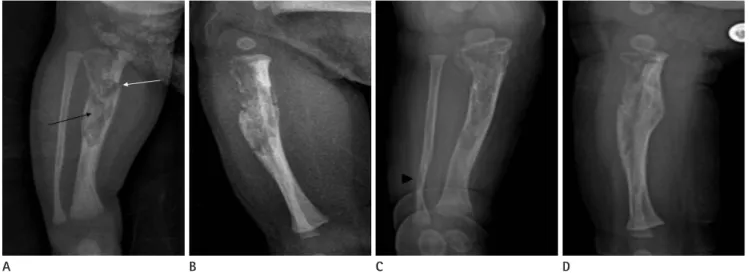
Fig. 1. Radiographs.
A, B. The initial AP and lateral image demonstrate an expansile osteolytic lesion (black arrow) with inner irregular sclerosis, involving the cortex and medullary cavity of the proximal meta-diaphysis of the right tibia. The margin of the lesion is irregular, multi-lobulated, but relatively well- defined. The cortical disruption of the proximal tibia suggests a pathologic fracture (white arrow), and a thin linear periosteal reaction at its distal part is noted.
C, D. The follow-up AP and lateral image (after 3 weeks) show a callus formation at the pathologic fracture, and a bowing deformity of the prox- imal tibia. An osteolytic lesion (arrowhead), not detected on the initial radiograph, is seen in the distal dia-metaphysis of the fibula.
A B C D
Fig. 2. MRI of the right lower leg.
A. A sagittal T2-weighted MR image shows an expansile mass replacing the entire medullary space, which reveals heterogeneous hyperintense signal intensity (white arrow). A disrupted periosteal reaction, continuous to the pathologic fracture, is noted (black arrow).
B-D. The axial T2-weighted (B), T1-weighted (C), and enhanced T1-weighted (D) MR images show prominent signal change with heterogeneous enhancement in extraosseous soft tissue (star). The transition between the extraosseous component and the normal muscles is wide and ill-defined.
A B C D
subsequent authors have preferred the term OFD (2, 3). The age range of patients typically affected by OFD is seven days to 22 years, with neonatal cases being very rare. Consistent with other infantile bone lesions, the diagnosis of congenital OFD is especially difficult. The clinical presentation is usually marked by swelling or bowing of the leg with pain (3).
The pathogenesis of OFD is not well-known. It has been pos- tulated that OFD results from excessive resorption of bone, with a subsequent fibrous repair of the defect (4). The histological ap- pearance of OFD is characterized by a fibrous matrix with nu- merous irregularly shaped trabeculae of woven bone that are of- ten rimmed by osteoblasts. Histologically, the main differential diagnoses are differentiated adamantinoma and fibrous dyspla- sia. Aside from its preferential involvement with the tibia, OFD has many overlapping clinical, morphologic and pathological similarities with differentiated adamantinoma; some authors have hypothesized that OFD and adamantinoma may simply be different stages of the same disease, but the relationship be- tween the two remains controversial (2, 5). Fibrous dysplasia typically originates in the intramedullary space rather than in the cortex, and bowing of the involved bone is rarely combined.
Histologically, fibrous dysplasia has more cellular and less-ma- ture fibrous tissue than OFD. In fibrous dysplasia, osteoblastic rimming of bone trabeculae may be composed of lamellar rath- er than of woven bone (3).
The common radiographic finding of OFD in children is well- defined eccentric osteolytic lesions involving the metaphysis of
long bones, with consequent cortical expansion and bowing of the bone in the antero-posterior direction. On the MR image, OFD reveals intermediate to high signal intensity on the T2- weighted image and intermediate signal intensity on the T1- weighted image, which may be similar to other tumors with fi- broblastic stroma. Superimposed hemorrhagic, cystic, myxoid change and cartilaginous differentiation can contribute to het- erogeneous signal intensity on the T2-weighted image. Cortical breakage and perilesional soft tissue extension can be com- bined, which is mostly associated with pathologic fracture (5).
According to previous literature, it seems that congenital OFD can more frequently be associated with pathologic fracture and pseudoarthrosis and, reveal more prominent perilesional reac- tive change (2, 6). Thus, although OFD is a benign, slowly-pro- gressive lesion, sometimes it can mimic other progressive tumors (or tumor-like lesions) such as Langerhans-cell histiocytosis (LCH), myofibromatosis, neurofibromatosis, infection-related lesions, and malignant tumors, especially in neonates (1, 3). Most of these are difficult to differentiate by their clinical or radio- graphic appearance, in which case biopsy is often necessary.
LCH, most commonly occurring in children, demonstrates a broad spectrum of clinical and radiologic features that can mimic those of infection, benign and malignant tumors. During the early phase, there is a more-aggressive pattern of osteolysis re- flective of the biologic activity. Localized LCH commonly oc- curs in patients of 5–15 years of age; younger patients under 2 years old are usually afflicted with disseminated multi-system involvement, which is known as Letterer-Siwe disease (7). As for congenital infantile myofibromatosis, it is a rare, potentially OFD-mimicking disorder. Common findings of bone lesions are well-defined lytic lesions with, or without, sclerotic borders.
The typical clinical course of the solitary form is initial rapid growth followed by spontaneous regression within the first two years. However, infantile myofibromatosis more commonly presents as the multi-centric type (8). Neurofibromatosis has been reported in 40–80% of patients suffering from congenital pseudoarthrosis of the tibia, whose radiographic appearance can mimic congenital OFD. However, neurofibromatosis mainly involves the periostium of the long bones; the medullary canal, unlike in congenital OFD, usually is narrowed by a thickening of the cortical bone (9). As for infection, neonatal osteomyelitis is characterized by destructive lesions with abundant periosteal Fig. 3. The histological result shows fibrous stromas (star) with imma-
ture bony trabeculae (white arrow) and osteoblastic rim (black arrow) (hematoxylin and eosin, × 20).
reaction in the metaphysis. It usually shows prompt articular in- volvement and various clinical signs, such as fever, leukocytosis and joint swelling (10). Finally, as an example of a malignant tu- mor, Ewing’s sarcoma should be considered. In infants, Ewing’s sarcoma (due to its rarity) is often misdiagnosed as either a frac- ture or an infection. The typical radiographic appearance of Ew- ing’s sarcoma in the long bones is a permeative lesion with a la- mellar “onion-skin” periosteal reaction.
The treatment of OFD is controversial. OFD usually shows a slow progression and stabilizing after skeletal maturity; howev- er, spontaneous regression of congenital OFD has been described in a few cases. Jobke et al. (1) reported a case with spontaneous post-biopsy regression, positing a potential for self-healing and remodeling at very young ages, and speculated that biopsy-in- curred alteration of the lesion comparable to the effect of a spon- taneous fracture through it can initiate healing in parallel with physiological bone maturation and diaphyseal growth. Though some authors have advocated radical surgery, others have em- phasized that surgery should be delayed as long as possible, and restricted to extensive lesions or to cases of combined patho- logical fracture (2).
In conclusion, congenital OFD is a benign, slowly-progres- sive lesion frequently affecting the tibia. Sometimes, it can pres- ent with aggressive imaging features such as complete intramed- ullary involvement, pathologic fracture with prominent perile- sional reactive change and even pseudoarthrosis. Although OFD is very rare in neonates, it should be considered in differential diagnoses of congenital tumor or tumor-like lesions in the low- er limbs.
REFERENCES
1. Jobke B, Bohndorf K, Vieth V, Werner M. Congenital os- teofibrous dysplasia Campanacci: spontaneous postbioptic regression. J Pediatr Hematol Oncol 2014;36:249-252 2. Zamzam MM. Congenital osteofibrous dysplasia of the
tibia, associated with pseudoarthrosis of the ipsilateral fibula. Saudi Med J 2008;29:1507-1509
3. Smith NM, Byard RW, Foster B, Morris L, Clark B, Bourne AJ. Congenital ossifying fibroma (osteofibrous dysplasia) of the tibia--a case report. Pediatr Radiol 1991;21:449-451 4. Hahn SB, Kang ES, Jahng JS, Park BM, Choi JC. Ossifying
fibroma. Yonsei Med J 1991;32:347-355
5. Jung JY, Jee WH, Hong SH, Kang HS, Chung HW, Ryu KN, et al. MR findings of the osteofibrous dysplasia. Korean J Radiol 2014;15:114-122
6. Teo HE, Peh WC, Akhilesh M, Tan SB, Ishida T. Congenital osteofibrous dysplasia associated with pseudoarthrosis of the tibia and fibula. Skeletal Radiol 2007;36 Suppl 1:S7-S14 7. Stull MA, Kransdorf MJ, Devaney KO. Langerhans cell his-
tiocytosis of bone. Radiographics 1992;12:801-823 8. Ang P, Tay YK, Walford NQ. Infantile myofibromatosis: a case
report and review of the literature. Cutis 2004;73:229-231 9. Maffulli N, Fixsen JA. Pseudoarthrosis of the ulna in neurofi-
bromatosis. A report of four cases. Arch Orthop Trauma Surg 1991;110:204-207
10. Kozlowski K, Beluffi G, Cohen DH, Padovani J, Tamaela L, Azouz M, et al. Primary bone tumours in infants. Short liter- ature review and report of 10 cases. Pediatr Radiol 1985;
15:359-367
신생아 경골에 생긴 선천성 골섬유성 이형성증
김상윤
1,2· 이상훈
1*
골섬유성 이형성증은 장골에 발생하는 양성 골섬유성 병변이며, 신생아의 선천성 골섬유성 이형성증은 매우 드물다. 선천 성 골섬유성 이형성증은 임상 및 영상 소견만으로는 다른 선천성 종양성 또는 비종양성 병변들과 감별이 어려우며, 따라서 조직 생검이 필요한 경우가 많다. 조직학적 확진 후에는 비수술적으로 치료를 하거나, 충분한 경과 관찰 후에 수술적 치료 를 하는 것이 권장된다. 저자들은 생후 7일된 여아의 경골에서 조직학적으로 확진된 선천성 골섬유성 이형성증의 단순촬 영 및 자기공명영상 소견을 보고하고자 한다.
1울산대학교 의과대학 서울아산병원 영상의학과, 2단국대학교병원 영상의학과