Amelogenesis imperfecta is characterized by inherited defects in the enamel that can be qualitative or quantita
tive. The primary dentition, permanent dentition, or both can be affected by this disorder. It may be associated with morphological or biochemical changes elsewhere in the body.1 The kidneys are the most commonly affected organ, and may present with disorders such as nephrocalcinosis, nephrolithiasis, and other functional abnormalities.2 The first case was reported by MacGibbon in 1972;3 multiple cases have been subsequently diagnosed and the condition has been referred to as enamel renal syndrome, amelogen
esis imperfecta and nephrocalcinosis, amelogenesis im
perfecta and gingival fibromatosis syndrome, or enamel renal gingival syndrome.4,5 According to a recent review article, only 16 cases have been reported with the com
plete oral, dental, and renal phenotype that is associated with enamel renal syndrome.4 Although the prevalence of
amelogenesis imperfecta is known, the prevalence of ena
mel renal syndrome has not been assessed, with the ex
ception of one study that emphasized its rarity.6 Affected patients generally suffer from functional and aesthetic pro
blems, leading to an inferior quality of life.7 The severity of the associated renal disorders has been described as ranging from no complications to chronic renal failure.8,9
Case Report
A 21yearold male patient presented with a chief com
plaint of small upper and lower permanent teeth. Accord
ing to the history reported by the patient, the size of his teeth never changed with age. No contributing family or medical history was identified. The patient did not report any abnormal oral habits. The findings of his general phy
sical examination were normal.
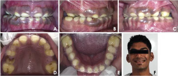
An intraoral soft tissue examination showed the pres
ence of brown pigmented gingiva with a normal contour (Fig. 1A). A hard tissue examination revealed retained de
ciduous teeth and permanent teeth with atypical findings, as well as multiple decayed teeth(Figs. 1AE). Many of Dhvani Bhesania , Ankit Arora *, Sonali Kapoor
1Department of Conservative Dentistry and Endodontics, Manubhai Patel Dental College, Maharaja Krishnakumarsinhji Bhavnagar University, Vadodara, India
AbstRACt
Numerous cases of enamel renal syndrome have been previously reported. Various terms, such as enamel renal syndrome, amelogenesis imperfecta and gingival fibromatosis syndrome, and enamelrenalgingival syndrome, have been used for patients presenting with the dental phenotype characteristic of this condition, nephrocalcinosis or nephrolithiasis, and gingival findings. This report describes a case of amelogenesis imperfecta of the enamel agenesis variety with nephrolithiasis in a 21yearold male patient who complained of small teeth. The imaging modalities employed were conventional radiography, conebeam computed tomography, and renal sonography. Such cases are first encountered by dentists, as other organ or metabolic diseases are generally hidden. Hence, cases of amelogenesis imperfecta should be subjected to advanced diagnostic modalities, incorporating both dental and medi cal criteria, in order to facilitate comprehensive longterm management.(Imaging Sci Dent 2015; 45: 181-5) KEy woRds: Amelogenesis Imperfecta; ConeBeam Computed Tomography; Dental Enamel; Kidney Disease
Copyright ⓒ 2015 by Korean Academy of Oral and Maxillofacial Radiology
This is an Open Access article distributed under the terms of the Creative Commons Attribution NonCommercial License(http://creativecommons.org/licenses/bync/3.0) which permits unrestricted noncommercial use, distribution, and reproduction in any medium, provided the original work is properly cited.
Imaging Science in Dentistry·pISSN 22337822 eISSN 22337830 Received December 10, 2014; Revised April 7, 2015; Accepted May 15, 2015
*Correspondence to : Dr. Ankit Arora
Department of Conservative Dentistry and Endodontics, Manubhai Patel Dental Col
lege, Vishwa Jyoti Ashram, Near Vishwamitri Bridge, Munjmahuda Road, Vadodara, Gujarat Pin: 390011, India
Tel) 919033975629, Fax) 912652330362, Email) [email protected]
the permanent teeth were missing, and the erupted teeth were widely spaced, with a yellowish brown discoloration (Fig. 1A). The enamel layer was absent clinically and posteruption wearing of the teeth was evident(Figs. 1D
F). The upper central incisors exhibited a semilunar shape and the molars had flat occlusal surfaces(Figs. 1A, D, and E). Severe attrition resulted in a collapsed bite and reduced vertical dimensions during occlusion(Figs. 1A and E).
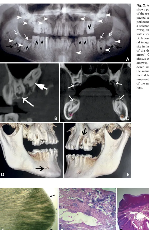
The panoramic radiography and conebeam computed tomography(CBCT) images revealed multiple findings.
All of the teeth were affected, especially the posterior teeth(Fig. 2A). Multiple calcification nodules were seen in the pulp chambers of the primary and permanent teeth;
these nodules were needleshaped in the incisors and round in the posterior teeth(Figs. 2A and B). The differ
ential radiodensity expected between the dentine and the enamel was absent(Fig. 2B). Pericoronal radiolucencies delineated by sclerotic margins around nonerupted teeth were significant(Figs. 2A and B). The impacted teeth pre
sented with complete root formation, exhibiting dilacera
tions and curvature(Fig. 2A). The dental follicles around the unerupted teeth appeared to be hyperplastic(Figs. 2A and B). Extensive crown resorption with partial replace
ment of the resorbed dentin by globular calcified struc
tures was observed(Fig. 2C).
The impacted permanent canines, second molars, and third molars were observed to have fully formed roots(Fig.
2A). An accessory mental foramen was seen on the right side of the mandible and extensive bone loss was observ
ed around the mesiobuccal root of the left maxillary first molar in the volumerendered CBCT images(Figs. 2D and E). A provisional diagnosis of amelogenesis imper
fecta was made based on the clinical and radiographic findings. The histopathological examination of an extract
ed deciduous canine using light microscopy confirmed the absence of enamel and the absence of scalloping of the dentinoenamel junction.(Fig. 3A). Hence, based on the Witkop classification,10 the final diagnosis was the enamel agenesis(type 1G) subtype of hypoplastictype amelogen
esis imperfecta, which is an autosomal recessive disorder.
The histopathological analysis of specimens obtained during treatment also confirmed the presence of calcified pulpal tissue(Fig. 3B) and dense collagen bundles with abundant vasculature in the gingival tissue(Fig. 3C).
As this type of amelogenesis imperfecta can be associ
ated with other organ and metabolic abnormalities, further investigations were carried out. An ultrasound examina
tion of his kidneys demonstrated multiple hyperechoic foci in the medulla of both the kidneys, suggesting bilate
ral nephrolithiasis(Figs. 4A and B). A 24hour urine spec
trophotometric analysis revealed very low citrate levels (18.9mg/24 hours; reference range, 116924mg/24 hours).
Other biochemical and haematological findings were nor
mal(alkaline phosphatase, 128U/L; urea, 12.84mg/dL, blood urea nitrogen, 6mg/dL; creatinine, 1.33mg/dL,
Fig. 1. A. A frontal view shows pigmented gingiva, the absence of enamel with spacing between the teeth, and a semilunar appearance of the maxillary central incisors. A right lateral view (B), left lateral view (C), and maxillary and mandibular occlusal views(D and E) show the presence of decayed teeth, molars with a flat occlusal surface, multiple deciduous teeth, and multiple missing permanent teeth. F. An extraoral photograph shows the evidence of wear of anterior teeth.
A B C
D E F
rows), and complete root formation with curvatures(long black arrows).
B. A conebeam CT(CBCT) sagit
tal image shows uniform radioden
sity in the crown(long white arrow) of the dental follicle(long white arrow). C. A CBCT coronal image shows crown resorption(white arrows). D. A CBCT volumeren
dered image of the right side of the mandible shows an accessory mental foramen. E. A CBCT vol
umerendered image of the left side of the maxilla shows severe bone loss.
C A
B C
D E
Fig. 3. A. A photomicrograph of a ground section shows the absence of enamel and scalloping of the dentinoenamel junction(black ar
rows). B. A photomicrograph of the pulpal tissue shows connective tissue with numerous basophilic calcifications(H&E stain, 40×). C. A photomicrograph of the gingival tissue shows parakeratinised epithelium and dense collagen bundles in the lamina propria with fibroblasts and blood vessels(H&E stain, 40×).
A B C
blood urea nitrogen to creatinine ratio, 4.51; estimated glomerular filtration rate, >60mL/min/1.73m; sodium, 136mmol/L, potassium, 4.0mmol/L; chloride, 98mmol/L;
bicarbonate, 19mmol/L; calcium, 9.4mg/dL; vitamin D3, 34.63ng/mL; urine albumin, 32.1mg/L). A physical, mi
croscopic, and chemical examination of a urine sample did not reveal any abnormalities(urine creatinine, 17.51 mg/dL; urine albumincreatinine ratio, 183.32mg/g; 24 hours calculated citrate in urine, 18.9mg/24 hours; urine oxalate, 14.27mg/24 hours, urinary calcium, 98.7mg/24 hours; parathyroid hormone, 19.70pg/mL).
Thus, based on previous findings,4 the diagnosis was modified to enamel renal syndrome with associated amel
ogenesis imperfecta, nephrolithiasis, and hypocitraturia.
discussion
A general dental practitioner may come across many cases of amelogenesis imperfecta; however, it is difficult to ensure an accurate diagnosis due to its complex clas
sification and the likely absence of relevant data from family members.11 The clinical features of amelogenesis imperfecta vary depending on the type.1113 In the hypo
plastic type of amelogenesis imperfect, the teeth exhibit a chalky white to dark brown colour, the occlusal surfaces and incisal edges are generally abraded, and occasionally, the complete loss of the enamel is observed. In the hy
pocalcified form of amelogenesis imperfecta, the enamel exhibits a cheesy consistency and can be easily removed with a sharp explorer. The hypomaturative type of amelo
genesis imperfecta is characterized by normal enamel thickness with the appearance of white opaque areas on the incisal surfaces. Another important fact is the associa
tion of amelogenesis imperfecta with other features, com
prising a systemic disease or syndrome.11 One such entity is known as enamel renal syndrome.4 De la DureMolla et al.4 have established an oral diagnosis criteria which includes clinical, radiographic, and microscopic features characteristic of this syndrome, along with the presence of renal abnormalities. Almost all the findings in our case fit these criteria except for few mentioned below. Radio
graphically, an abnormal pathway of eruption was not evident in the second molars, and thickening of the max
illary or nasal sinus lining was not observed. Microscop
ically, the cellular cementum layer characteristic of this syndrome could not be seen. The diagnosis of enamel renal syndrome with associated amelogenesis imperfec
ta, nephrolithiasis and hypocitraturia was made in this case, as most of the criteria were fulfilled, nephrolithiasis was evident in the ultrasound, and hypocitraturia was ob
served in the urine analysis.
A renal ultrasound is now recommended in cases with such oral findings.14 Abnormalities in the interstitial ma
trix have been hypothesized to be the causative factor of dystrophic calcification in the kidney and of enamel ab
normalities.15 Low urinary citrate levels indicate that stone formation is more likely, because urinary citrate inhibits stone formation by forming soluble complexes with calci
um.16 Other relevant findings in such cases are localized aggressive periodontitis, gingival fibromatosis, and soft tissue calcifications, as described by Kantaputra et al.5,17 Hence, they coined the term enamelrenalgingival syn
drome. In the present case, localized periodontitis was ev
ident, but the clinical findings did not justify a diagnosis of gingival fibromatosis and histopathological analysis did not reveal any soft tissue calcifications. However, the
Fig. 4. A and B. Ultrasound images of the left and right kidneys, respectively, show multiple calcified deposits(white arrows).
A B
Enamel renal syndrome is a rare autosomal disorder that has been proven to be associated with mutations in the FAM 20A gene.4,5,17 However, the mutation test was not feasible in the present case.
Detecting an accessory mental foramen on the right side of the mandible was relevant, because it is a rare finding.19 It is important to be aware of variations of the mental fora
men in order to achieve successful anaesthesia that avoids nerve injury during periodontal or maxillofacial surgeries and dental implant placement.20 It was observed that vol
umerendered images were more useful in detecting ac
cessory foramina and localized aggressive periodontitis.
Based on the findings of ultrasonography and other di
agnostic modalities, the patient was informed that he had a higher risk of renal stone formation and was referred to a nephrologist for the necessary preventive treatment.
Long term followup is imperative for patients with this condition, as some patients may eventually suffer from serious renal dysfunction.9
In conclusion, an adequate knowledge of diseases that involve both dental and medical factors is indispensable for making the correct diagnosis and ensuring comprehen
sive treatment. Volumerendered images from CBCT scans can be very valuable in detecting certain abnormalities.
Acknowledgements
We thank Dr. Rashmi Gubbi, Dr. Arpan Shah and all other staff members of the Department of Oral and Maxil
lofacial Pathology and Microbiology for their support. We would like to extend our sincere gratitude to Dr. Satish Shah and Dr. Sheel Shah for helping with the imaging modalities.
References
1. Aldred MJ, Savarirayan R, Crawford PJ. Amelogenesis imper
fect: a classification and catalogue for the 21st century. Oral Dis 2003; 9: 1923.
2. Poornima P, Katkade S, Mohamed RN, Mallikarjuna R.
Amelogenesis imperfecta with bilateral nephrocalcinosis.
BMJ Case Rep 2013; 2013: bcr2013009370.
3. MacGibbon D. Generalized enamel hypoplasia and renal dys
function. Aust Dent J 1972; 17: 613.
4. de la DureMolla M, Quentric M, Yamaguti PM, Acevedo AC,
A 2014; 164A: 19.
6. Chosack A, Eidelman E, Wisotski I, Cohen T. Amelogenesis imperfecta among Israeli Jews and the description of a new type of local hypoplastic autosomal recessive amelogenesis imperfecta. Oral Surg Oral Med Oral Pathol 1979; 47: 14856.
7. Hashem A, Kelly A, O’Connell B, O’Sullivan M. Impact of moderate and severe hypodontia and amelogenesis imperfec
ta on quality of life and selfesteem of adult patients. J Dent 2013; 41: 68994.
8. Jaureguiberry G, De la DureMolla M, Parry D, Quentric M, Himmerkus N, Koike T, et al. Nephrocalcinosis(enamel renal syndrome) caused by autosomal recessive FAM20A muta
tions. Nephron Physiol 2012; 122: 16.
9. Elizabeth J, Lakshmi Priya E, Umadevi KM, Ranganathan K.
Amelogenesis imperfecta with renal disease a report of two cases. J Oral Pathol Med 2007; 36: 6258.
10. Witkop CJ Jr. Amelogenesis imperfecta, dentinogenesis im
perfecta and dentin dysplasia revisited: problems in classifica
tion. J Oral Pathol 1988; 17: 54753.
11. Crawford PJ, Aldred M, BlochZupan A. Amelogenesis im
perfecta. Orphanet J Rare Dis 2007; 2: 17.
12. Chamarthi V, Varma BR, Jayanthi M. Amelogenesis imper
fecta: a clinician’s challenge. J Indian Soc Pedod Prev Dent 2012; 30: 703.
13. Chaudhary M, Dixit S, Singh A, Kunte S. Amelogenesis im
perfecta: report of a case and review of literature. J Oral Max
illofac Pathol 2009; 13: 707.
14. Hunter L, Addy LD, Knox J, Drage N. Is amelogenesis imper
fect an indication for renal examination? Int J Paediatr Dent 2007; 17: 625.
15. Lubinsky M, Angle C, Marsh PW, Witkop CJ Jr. Syndrome of amelogenesis imperfecta, nephrocalcinosis, impaired renal concentration, and possible abnormality of calcium metabo
lism. Am J Med Genet 1985; 20: 23343.
16. Del Valle EE, Spivacow FR, Negri AL. Citrate and renal stones. Medicina(B Aires) 2013; 73: 3638.
17. Kantaputra PN, Bongkochwilawan C, Kaewgahya M, Ohaz
ama A, Kayserili H, Erdem AP, et al. EnamelRenalGingival syndrome, hypodontia, and a novel FAM20A mutation. Am J Med Genet A 2014; 164A: 21248.
18. MartelliJúnior H, Bonan PR, Dos Santos LA, Santos SM, Cavalcanti MG, Coletta RD. Case reports of a new syndrome associating gingival fibromatosis and dental abnormalities in a consanguineous family. J Periodontol 2008; 79: 128796.
19. Udhaya K, Saraladevi KV, Sridhar J. The morphometric anal
ysis of the mental foramen in adult dry human mandibles: a study on the South Indian population. J Clin Diagn Res 2013;
7: 154751.
20. Pancer B, GaraicoaPazmiño C, Bashutski JD. Accessory mandibular foramen during dental implant placement: case re
port and review of literature. Implant Dent 2014; 23: 11624.