155 155
THE EWHA MEDICAL JOURNAL THE EWHA MEDICAL JOURNAL
Halo Nevus Arising from Congenital Melanocytic Nevus Featuring an Early Onset Vitiligo
Osung Kwon, Yongwoo Choi, Hyun Chung, Joonsoo Park
Department of Dermatology, Catholic University of Daegu School of Medicine, Daegu, Korea
Introduction
Halo nevus is characterized by a round to oval shaped depig- mented patch surrounding a precedent skin lesion. Involvement of congenital melanocytic nevus at the center of a halo nevus is reported to be a minor aspect and the patient presenting simultaneous vitiligo is extremely rare [1]. Although the etiol- ogy concerning both congenital halo nevus and vitiligo further requires more validity, pathological and laboratory evaluation confirming massive infiltration of CD8 T cells with antibodies directed to melanocytes conjecture similar pathogenesis between the two entities [2]. The causal relationship between the halo and vitiligo loads significant relevance in terms of pathogenesis and disease progression. Herein, the authors present a case of congenital melanocytic nevus along with vitiligo presented con- currently in an adjacent and distant manner.
Case
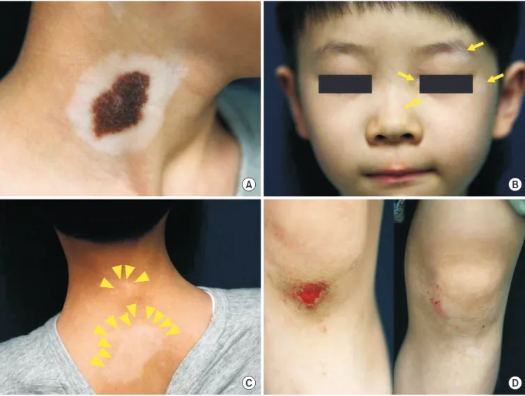
A 6-year-old male presented complaining of recent occur- rence of whitish patches on face, both knees and the posterior neck area. A 6 cm-sized hairy and brownish patch surrounded by a whitish patch along the pigmented lesion was noticeable on the neck in addition to the complaint. The patient was born with a congenital melanocytic nevus and the size of the ne- vus grew in a parallel manner to the height and a depigmented patch surrounding the lesion was developed 3 years in prior to the visit (Fig. 1A). There was no specific family history of skin cancer or significant medical condition. In addition, physi- cal evaluation revealed multiple whitish patches along the face where vision corrective braces were placed and the wound bear- ing knee area, featuring Koebner phenomenon (Fig. 1B-D).
Wood light inspection revealed accentuation at the depigmented.
Case Report
Ewha Med J 2017;40(4):155-158
https://doi.org/10.12771/emj.2017.40.4.155 eISSN 2234-2591
Halo nevus and vitiligo are known to be associated with immunologic defect that result in typical skin lesions. Random shapes and sizes of whitish patches, depending on the type, are featured in vitiligo. Halo, on the other hand, presents by surrounding the previ- ous pigmented lesion leaving a whitish-halo-like appearance. The mechanisms under- lying these entities remain to be elucidated. Various immunological responses along with biomechanical activities suggest causal relationship between the two diseases. A 6-year-old male patient was recently presented with multiple whitish patches on the various parts of the body in a Koebner phenomenon manner. A noticeable hairy con- genital melanocytic nevus surrounded a well-demarcated halo of depigmentation was also observed. Clinical and pathological findings were conclusive of as halo nevus with multiple concurrent vitiligo. The pathogenic relationship between the two entities must be underlined since the nature of disease progression is associated and the respective management may also be altered accordingly. (Ewha Med J 2017;40(4):155-158)
Received June 27, 2017 Revised September 5, 2017 Accepted September 15, 2017 Corresponding author Joonsoo Park
Department of Dermatology, Catholic University of Daegu School of Medicine, 33 Duryugongwon-ro 17-gil, Nam-gu, Daegu 42472, Korea
Tel: 82-53-650-4161, Fax: 82-53-650-4891 E-mail: ashkwon@naver.com
Key Words
Nevus, halo; Congenital mealnocytic nevus;
Vitiligo
This is an Open Access article distributed under the terms of the Creative Commons Attribution Non-Commercial License (http://creativecommons.org/licenses/by-nc/4.0) which permits unrestricted non-commercial use, distribution, and reproduction in any medium, provided the original work is properly cited.
156 THE EWHA MEDICAL JOURNAL Kwon O, et al
Skin biopsy was performed on the center of the congenital melanocytic nevus and the scanning view revealed heavy infil- tration of nevus cells at the upper dermis (Fig. 2A). Middle to lower dermis presented groups of nevus cluster with no definite atypia (Fig. 2B). In addition, skin biopsy was performed on the surrounding whitish area and revealed significant reduction in melanocytes and melanin pigmentation (Fig. 2C). Higher magnification revealed mild infiltration of the lymphocytes (Fig.
2D). Based on the clinical and pathological findings the patient was diagnosed with halo nevus surrounding congenital melano- cytic nevus concurrent with vitiligo. The patient was prescribed
with tacrolimus 0.03% based ointment and excimer therapy on the whitish patches was delivered.
Discussion
Vitiligo and halo nevus are two common T-cell-mediated autoimmune disorders [3]. Halo nevus is clinically defined as whitish patch surrounding the preceding pigmented lesion and is also known as leukoderma acquisitum centrifugum, Sutton’
s nevus, leukopigmentary nevus, perinevoid vitiligo, perinevoid leukoderma [4]. The prevalence of halo nevus was suspected to
A B
C D
Fig. 1. Clinical images at initial visit. (A) A 6 cm-sized patch with central pigmented hairy lesion surrounded by a whitish halo phenomenon on right neck. The whitish halo surrounding the congenital central lesion was developed 3 years in prior to visit. (B) Multiple variegated-shaped whitish patches on left periorbital area (arrows) where the corrective vision braces was in touch. Lesions had developed 1 year in prior to initial visit. (C) Multiple irregularly shaped whitish patches on posterior nape and upper back area (arrow heads). Lesions had developed 1 year in prior to initial visit. (D) Whitish patches around wound area of the right knee (right) and left knee (left). Lesions had developed 1 year in prior to initial visit.
157
THE EWHA MEDICAL JOURNAL Concurrent Vitiligo in Congenital Halo Nevus
be 1% in general population and halo nevus accompanied with vitiligo was especially pronounced in children [5]. Moreover, concurrent halo nevus was reported to range from 0.5% to 1.4% in all age groups of vitiligo patients [6]. The majority of the halo nevus arises on acquired nevus followed by congenital melanocytic nevus, melanoma, neurofibroma, seborrheic kera- tosis and lichen planus. Although, no exact chronological study has been elucidated, halo nevus arising in congenital melano- cytic nevus composites 4.7% of all halo nevus and is common under the age of 20 [7]. In addition, the presence of concurrent congenital melanocytic nevus was suspected to be involved with significantly higher occurrence of halo nevus, lower age onset of
vitiligo and higher risk for Koebner phenomenon compared to the control group that was without congenital melanocytic nevus [4].
Histologic findings confirm the two diseases as variant type of one another. Barona et al. [8] stated that the presence of a halo nevus indicates a risk factor of vitiligo progression and a prospective observational study suggest that non-segmental vitiligo-associated halo nevus affects age of onset and depig- mentation pattern, and has a more frequent familial background of autoimmunity [9]. Moreover, patients with multiple non-seg- mented vitiligo with congenital melanocytic nevus had a higher frequency of halo nevi at distance, which suggests a relationship
A B
C D
Fig. 2. (A) Histopathological image from the central pigmented area of the right neck presenting heavy infiltration of nevus cells at the upper dermis (H&E, ×20). (B) Higher magnification reveals groups of nevus cluster with no definite atypia at the middle to lower dermis (H&E, ×100).
(C) Skin biopsy was performed on the whitish surrounding and revealed significant reduction in melanocytes and melanin pigmentation (H&E,
×20). (D) Higher magnification revealed mild infiltration of the lymphocytes (H&E, ×100).
158 THE EWHA MEDICAL JOURNAL Kwon O, et al
between vitiligo and halo nevus [9,10]. This relation was even more significant in halo nevus from congenital melanocytic ne- vus with higher presentation of vitiligo. Numerous research state that congenital melanocytic nevus can be considered as a pos- sible trigger leading to an earlier development of vitiligo [4].
Although autoimmunity has been suggested to be involved in both diseases, the relationship between vitiligo and halo nevus is not fully understood. CD8+ T lymphocytic infiltration remains as the key role of the disease progression resulting in destruction of melanocytes in both diseases [2,11]. Interferon (IFN)-γ is con- sidered as the key regulative cytokine and induction of IFN-γ leads to up-regulation of chemokine receptors known as CCR5 and CXCR3 [12]. Oxidative stress has also been demonstrated to be enhanced in pathological melanocytes and closely related to melanocyte apoptosis and autoimmune destruction. Oxidative stress triggered by H2O2 accelerates chemokine productions for the recruitment of cytotoxic T cells in lesions of the diseases [3,13]. The clinical similarity presenting both Koebner phenom- enons also further states the link.
The presented case features pathologic and clinical similar- ity of the halo nevus and vitiligo. The immunologic similarity, however, is insufficient as to the order of occurrence differs.
Based on the immunologic susceptibility, it has been postulated that the antibodies to congenital melanocytic nevus may pre- cede and affect normal melanocytes to cause lesions consistent that of vitiligo [14]. Microstructural transformation and cellular injury progresses into multiple whitish patches after occurrence of halo nevus. van Geel et al. [4] conjectured improvement in skin lesion after early surgical excision and Workman et al. [15]
observed initial improvement after excision of a halo nevus aris- ing in congenital melanocytic nevus. However, elucidation must be supported through sufficient cases and studies. There are currently only three cases reported of halo nevus around con- genital melanocytic nevus with concurrent vitiligo in the Korean literature. The present case is to underline the significance of this entity and to alert early management to prevent further complications knowingly, vitiligo.
Informed Consent
Regarding provision of pictorial images, the informed consent
was received from parents of the patient in presented Fig. 1B.
References
1. Taieb A, Picardo M. Clinical practice: vitiligo. N Engl J Med 2009;
360:160-169.
2. van den Boorn JG, Konijnenberg D, Dellemijn TA, van der Veen JP, Bos JD, Melief CJ, et al. Autoimmune destruction of skin me- lanocytes by perilesional T cells from vitiligo patients. J Invest Dermatol 2009;129:2220-2232.
3. Yang Y, Li S, Zhu G, Zhang Q, Wang G, Gao T, et al. A similar lo- cal immune and oxidative stress phenotype in vitiligo and halo nevus. J Dermatol Sci 2017;87:50-59.
4. van Geel N, Van Poucke L, Van de Maele B, Speeckaert R. Rel- evance of congenital melanocytic naevi in vitiligo. Br J Dermatol 2015;172:1052-1057.
5. Handa S, Dogra S. Epidemiology of childhood vitiligo: a study of 625 patients from north India. Pediatr Dermatol 2003;20:207- 210.
6. Handa S, Kaur I. Vitiligo: clinical findings in 1436 patients. J Der- matol 1999;26:653-657.
7. Suh KY, Bolognia JL. Signature nevi. J Am Acad Dermatol 2009;
60:508-514.
8. Barona MI, Arrunategui A, Falabella R, Alzate A. An epidemio- logic case-control study in a population with vitiligo. J Am Acad Dermatol 1995;33:621-625.
9. Ezzedine K, Diallo A, Leaute-Labreze C, Seneschal J, Mossalayi D, AlGhamdi K, et al. Halo nevi association in nonsegmental vitiligo affects age at onset and depigmentation pattern. Arch Dermatol 2012;148:497-502.
10. Nicolaidou E, Antoniou C, Miniati A, Lagogianni E, Matekovits A, Stratigos A, et al. Childhood- and later-onset vitiligo have diverse epidemiologic and clinical characteristics. J Am Acad Dermatol 2012;66:954-958.
11. Zeff RA, Freitag A, Grin CM, Grant-Kels JM. The immune re- sponse in halo nevi. J Am Acad Dermatol 1997;37:620-624.
12. Ogg GS, Rod Dunbar P, Romero P, Chen JL, Cerundolo V. High frequency of skin-homing melanocyte-specific cytotoxic T lymphocytes in autoimmune vitiligo. J Exp Med 1998;188:1203- 1208.
13. Li S, Zhu G, Yang Y, Guo S, Dai W, Wang G, et al. Oxidative stress- induced chemokine production mediates CD8(+) T cell skin trafficking in vitiligo. J Investig Dermatol Symp Proc 2015;17:32- 33.
14. Yu HS, Kao CH, Yu CL. Coexistence and relationship of antikera- tinocyte and antimelanocyte antibodies in patients with non- segmental-type vitiligo. J Invest Dermatol 1993;100:823-828.
15. Workman M, Sawan K, El Amm C. Resolution and recurrence of vitiligo following excision of congenital melanocytic nevus. Pedi- atr Dermatol 2013;30:e166-e168.