www.e-enm.org
61
Endocrinol Metab 2013;28:61-64
http://dx.doi.org/10.3803/EnM.2013.28.1.61 pISSN 2093-596X · eISSN 2093-5978
Case Report
Anaplastic Thyroid Carcinoma Following Radioactive Iodine Therapy for Graves’ Disease
Sun Hwa Kim¹, Hee Young Kim¹, Kwang Yoon Jung², Dong Seop Choi¹, Sin Gon Kim¹
¹Division of Endocrinology and Metabolism, Department of Internal Medicine, ²Department of Otolaryngology, Head and Neck Surgery, Korea University College of Medicine, Seoul, Korea
Radioactive iodine (RAI) therapy has been used as a treatment option for Graves’ disease, and it has been widely accepted to be safe. On the other hand, some evidence suggests that RAI therapy is possibly associated with a small increased risk of thyroid cancer. Herein, we report a rare case of anaplastic thyroid carcinoma (ATC) associated with Graves’ disease, following RAI treat- ment. A 42-year-old woman had been diagnosed with Graves’ disease and although she was treated with an antithyroid drug, she remained in a hyperthyroid state, which led to two RAI treatments. More than 10 years later, the patient revisited our clinic due to hoarseness, dysphagia, and dyspnea, which had lasted for 2 months. Neck computed tomography suggested thyroid carcinoma and a lymph node biopsy showed metastatic papillary carcinoma. The patient underwent total thyroidectomy and was finally di- agnosed as having an ATC. It is not clear if the occurrence of ATC reported here was influenced by the RAI therapy or alterna- tively, it may only represent the delayed recognition of a rare change in the natural history of Graves’ disease. Nevertheless, this report is worthwhile since it presents a very rare case of ATC that occurred eleven years after the RAI therapy for Graves’ disease.
Keywords: Thyroid cancer, anaplastic; Graves disease; Radioactive iodine therapy
INTRODUCTION
The treatment options for Graves’ disease consist of antithy- roid drugs, radioactive iodine (RAI) therapy, and surgery. RAI therapy has been used for the treatment of Graves’ disease for more than seven decades, and in most studies, it has not been associated with an increased risk of cancer [1]. On the other hand, several studies have proposed the potential increase in thyroid malignancies with RAI treatment [2-4].
Here, we describe a very rare case of anaplastic thyroid car- cinoma (ATC) associated with Graves’ disease, following RAI treatment.
CASE REPORT
A 42-year-old woman was referred to our clinic in 1998, be- cause of palpitations, hand tremor, and laboratory findings of hyperthyroidism (serum thyrotropin [thyroid-stimulating hor- mone, TSH] 0.05 µIU/mL [reference range, 0.17 to 4.05], free thyroxine 4.01 ng/dL [reference range, 0.79 to 1.86], and posi- tive TSH receptor antibody at 201.7% [percent binding inhibi- tion index, radioreceptor assay, reference range; ±15.0%]), and had been diagnosed as Graves’ disease. A technetium-99m thyroid scan showed enlarged and homogeneously increased uptake, and ultrasonography revealed a diffuse goiter without nodules. The patient received 200 mg/day of propylthiouracil
Received: 27 August 2012, Accepted: 12 November 2012 Corresponding author: Sin Gon Kim
Division of Endocrinology and Metabolism, Department of Internal Medicine, Korea University College of Medicine, 73 Inchon-ro, Seongbuk-gu, Seoul 136-705, Korea
Tel: +82-2-920-5890, Fax: +82-2-953-9355, E-mail: [email protected]
Copyright © 2013 Korean Endocrine Society
This is an Open Access article distributed under the terms of the Creative Com- mons Attribution Non-Commercial License (http://creativecommons.org/
licenses/by-nc/3.0/) which permits unrestricted non-commercial use, distribu- tion, and reproduction in any medium, provided the original work is properly cited.
Kim SH, et al.
62
www.e-enm.org Copyright © 2013 Korean Endocrine Societyand β-adrenergic blocking agents for 1 year. However, due to recurrent hyperthyroidism, she received RAI therapy twice with an administered dose 370 MBq (10 mCi), 296 MBq (8 mCi), each. Two years later, she received thyroid hormone re- placement due to subsequent hypothyroidism at a local clinic.
In September 2010, over 10 years later, she revisited us with complaints of hoarseness, dysphagia, and dyspnea, which had been presented for 2 months. An anterior neck mass, which was hard and fixed, and some lymph nodes (LNs) around the mass were palpable. Her chest radiography showed a tracheal narrowing and deviation to the right without active lung lesion.
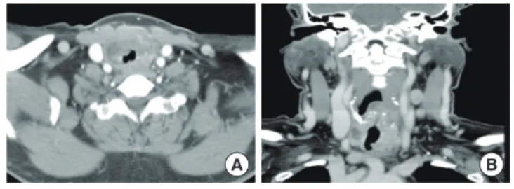
On computed tomography (CT) of the neck, the thyroidal mass invaded the tracheal cartilage and the right tracheoesophageal groove (Fig. 1). In fludeoxyglucose positron emission tomog- raphy-CT, an intense hypermetabolic lesion was noted in the thyroid gland and LNs on her left neck without distant metas- tasis. LN biopsy was performed and microscopically showed metastatic papillary carcinoma (Fig. 2A). In September 2010, a total thyroidectomy with selective neck dissection, tracheal sleeve resection, and end-to-end anastomosis were performed.
The surgical specimen from the thyroid was diagnosed as ana- plastic carcinoma (Fig. 2B). The final pathologic staging was T4bN1bM0. Subsequently, she had concurrent cisplatin-based
chemoradiotherapy for her neck and superior mediastinum (total 6,600 cGy/33 fractions).
After 4 months of follow-up, despite the aggressive treat- ment, the disease progressed. She developed shortness of breath, dyspnea, and pain in her left arm. On neck CT, a new focal necrotic mass on the left side of her neck was noted, sug- gesting local recurrence. The mass extended up to the tracheal lumen and the patient died of sudden uncontrolled bleeding in the airway.
DISCUSSION
The patient in this case received RAI therapy twice for recur- rent hyperthyroidism. After more than 10 years, she developed respiratory symptoms and was finally diagnosed with ATC.
This case raises questions about the causal relationship be- tween RAI therapy for Graves’ disease and the subsequent oc- currence of thyroid cancer. Although it has been widely ac- cepted that RAI is safe, some studies have suggested that RAI therapy is possibly associated with a small increased risk of thyroid cancer [2-4]. DeGroot [5] reported that the RAI treat- ment can induce an increase of thyroid antibodies, such as TSH receptor antibody, and it was suggested that thyroid stimulat- ing antibodies play an important role in thyroid carcinogenesis [6,7]. These antibodies also can stimulate angiogenesis, which contributes to thyroid tumor cell growth, by activating vascu- lar endothelial growth factor [8]. Sturgis [9] reported cytomor- phologic alterations in thyroid follicular epithelium, which is associated with RAI therapy. Before treatment, Graves’ dis- ease typically lack significant atypia, while after RAI therapy, nuclear atypia, and progressive cellular metaplasia could be seen microscopically. Furthermore, it was prominent in those who remain in a hyperthyroid state despite RAI treatment.
In addition to our case, two previously reported cases of ATC after RAI therapy for documented Graves’ disease are summarized in Table 1. Baker [10] reported a 64-year-old woman with ATC 12 years after RAI therapy. Because thyroid toxicity persisted after one course of RAI therapy, the patient received a second therapy in 4 months. Gossage et al. [11] re- ported a 46-year-old woman who had her first RAI therapy, when aged only 23 years. Although she underwent partial thy- roidectomy prior to the RAI therapy, hyperthyroidism recurred and she received several subsequent doses of radioiodine. Fi- nally, 20 years later, she developed ATC. An analysis of these cases is noteworthy, because since the interval to cancer diag- nosis was over 10 years in all cases, it supports the importance Fig. 1. Computed tomography (CT) findings of anaplastic thyroid
carcinoma. Neck CT shows an exophytic thyroidal mass with ex- tracapsular invasion, tracheal deviation, and narrowing.
A B
Fig. 2. The histological findings. (A) The histologic section of lymph node shows clear nuclei and formation of papillary struc- ture (H&E stain, ×200). (B) The surgical specimen shows atypi- cal spindle cells with irregular nuclei; a feature of anaplastic thy- roid carcinoma (H&E stain, ×200).
A B
Anaplastic Thyroid Carcinoma after Radioactive Iodine Therapy
Copyright © 2013 Korean Endocrine Society www.e-enm.org
63
of long-term vigilance in patients receiving RAI therapy. In addition, these cases had something in common in terms of the occurrence of ATC after repetitive, small doses of RAI thera- py. The Cooperative Thyrotoxicosis Therapy Follow-up Study showed that thyroid neoplasms developed in children treated with lower, rather than higher, doses of RAI therapy [4], which is compatible with our finding. Therefore, it can be hypothe- sized that multiple courses of RAI therapy, with small doses, may play a role in carcinogenesis, especially in the poorly dif- ferentiated type. In particular, Ogawa et al. [12] demonstrated that 131I treatment inactivates the tumor suppressor gene that is frequently found in ATC rather than in the differentiated type.
However, to discuss the causal relationship between RAI ther- apy and the occurrence of cancer, the number of reports is too small, therefore a population-based study is required and this is a limitation of our report.
In addition to the RAI therapy, we cannot exclude that Graves’ disease itself might also increase the risk of cancer.
Although our patient had no thyroid nodule by ultrasonogra- phy, the palpable thyroid nodules found in Graves’ patients have a 16.9% malignancy rate, which is higher than that of the general population [13]. Moreover, the finding that thyroid au- toantibody stimulates the malignant transformation of the thy- roid gland supports this suggestion.
It is not clear that the occurrence of ATC reported here was influenced by RAI therapy or, alternatively, it may only repre- sent the delayed recognition of a rare change in the natural history of Graves’ disease. Nevertheless, this report is worth- while since it presents a very rare case of ATC that occurred 11 years after RAI therapy for Graves’ disease.
CONFLICTS OF INTEREST
No potential conflict of interest relevant to this article was re- ported.
REFERENCES
1. Lucignani G. Long-term risks in hyperthyroid patients treated with radioiodine: is there anything new? Eur J Nucl Med Mol Imaging 2007;34:1504-9.
2. Dobyns BM, Sheline GE, Workman JB, Tompkins EA, McConahey WM, Becker DV. Malignant and benign neo- plasms of the thyroid in patients treated for hyperthyroid- ism: a report of the cooperative thyrotoxicosis therapy fol- low-up study. J Clin Endocrinol Metab 1974;38:976-98.
3. Franklyn JA, Maisonneuve P, Sheppard M, Betteridge J, Boyle P. Cancer incidence and mortality after radioiodine treatment for hyperthyroidism: a population-based cohort study. Lancet 1999;353:2111-5.
4. Rivkees SA, Dinauer C. An optimal treatment for pediatric Graves’ disease is radioiodine. J Clin Endocrinol Metab 2007;92:797-800.
5. DeGroot LJ. Radioiodine and the immune system. Thyroid 1997;7:259-64.
6. Fujikawa M, Okamura K, Sato K, Asano T, Yamasaki K, Hirata T, Ohta M, Mizokami T, Kuroda T, Fujishima M.
Anaplastic transformation of a papillary carcinoma of the thyroid in a patient with Graves’ disease with varied activ- ity of thyrotropin receptor antibodies. Thyroid 1998;8:53-8.
7. Majima T, Komatsu Y, Doi K, Shigemoto M, Takagi C, Fukao A, Kojima M, Tamaki H, Ito J, Nakao K. Anaplastic thyroid carcinoma associated with Graves’ disease. Endocr J 2005;52:551-7.
8. Hoffmann S, Hofbauer LC, Scharrenbach V, Wunderlich A, Hassan I, Lingelbach S, Zielke A. Thyrotropin (TSH)-in- duced production of vascular endothelial growth factor in thyroid cancer cells in vitro: evaluation of TSH signal transduction and of angiogenesis-stimulating growth fac- tors. J Clin Endocrinol Metab 2004;89:6139-45.
9. Sturgis CD. Radioactive iodine-associated cytomorpho- logic alterations in thyroid follicular epithelium: is recog- nition possible in fine-needle aspiration specimens? Diagn Cytopathol 1999;21:207-10.
10. Baker HW. Anaplastic thyroid cancer twelve years after radioiodine therapy. Cancer 1969;23:885-90.
11. Gossage AA, Neal FE, Ross CM, Talbot CH, Blake GM, Table 1. Cases of Anaplastic Thyroid Carcinomas Following
Radioiodine Therapy for Graves’ Disease
Author Sex
receiving Age radioiodine,
yr
Dose, mCi
Age at cancer diagnosis,
yr
Interval to cancer diagnosis,
yr
Baker [10] F 52 9 64 12
52 7
Gossage et al. [11] F 23 11.6 43 20
25 3.8
26 3.2
27 3.2
32 3.7
This study F 43 10 54 11
44 8
Kim SH, et al.
64
www.e-enm.org Copyright © 2013 Korean Endocrine SocietyMunro DS. Cases of carcinoma of thyroid following io- dine-131 therapy for hyperthyroidism. Oncology 1984;41:
8-12.
12. Ogawa M, Hori H, Hirayama M, Kobayashi M, Shiraishi T, Watanabe Y, Komada Y. Anaplastic transformation from
papillary thyroid carcinoma with increased serum CA19-9.
Pediatr Blood Cancer 2005;45:64-7.
13. Belfiore A, Russo D, Vigneri R, Filetti S. Graves’ disease, thyroid nodules and thyroid cancer. Clin Endocrinol (Oxf) 2001;55:711-8.