66 https://e-jcvi.org
An 80-year-old gentleman with history of coronary artery bypass surgery, repaired abdominal artery aneurysm and repaired bilateral iliac arteries aneurysms was admitted for subacute right lower limb ischemia from partially thrombosed right popliteal artery aneurysm. Before his scheduled surgery an echocardiogram was ordered to complete work-up for possible cardiac cause of thromboembolism. Echocardiogram showed a big rounded atrial mass just proximal to the tricuspid valve at the right atrioventricular junction (Movie 1 and Figure 1). The mass was non-obstructive but appeared to be mobile. This finding triggered a CT angiogram of the thorax. It revealed a large calcified and thrombosed right coronary artery aneurysm compressing the right atrium externally (Movie 2 and Figure 2). He underwent a successful femoral-popliteal bypass surgery and recovered well post-operatively.
SUPPLEMENTARY MATERIALS
Movie 1
Echocardiogram in four-chamber views showing relationship of the right atrial mass at the level of the right atrioventricular junction and tricuspid valve.
Click here to view J Cardiovasc Imaging. 2019 Jan;27(1):66-67
https://doi.org/10.4250/jcvi.2019.27.e6 pISSN 2586-7210·eISSN 2586-7296
Images in Cardiovascular Disease
Received: Sep 17, 2018 Revised: Oct 30, 2018 Accepted: Nov 20, 2018 Address for Correspondence:
Yi Chuan Tham, MD
Cardiothoracic Surgery Department, National Heart Center Singapore, 5 Hospital Drive, Singapore 169609.
E-mail: [email protected] Copyright © 2019 Korean Society of Echocardiography
This is an Open Access article distributed under the terms of the Creative Commons Attribution Non-Commercial License (https://
creativecommons.org/licenses/by-nc/4.0/) which permits unrestricted non-commercial use, distribution, and reproduction in any medium, provided the original work is properly cited.
ORCID iDs Yi Chuan Tham
https://orcid.org/0000-0002-2540-6993 Kok Hooi Yap
https://orcid.org/0000-0002-4730-7830 Conflict of Interest
The authors have no financial conflicts of interest.
Yi Chuan Tham , MD1, Kok Hooi Yap , MBChB1, and Jack Kian Ch'ng, MBBCh2
1Cardiothoracic Surgery Department, National Heart Center Singapore, Singapore
2Vacular Surgery Department, Singapore General Hospital, Singapore
Large Thrombosed Right Coronary Artery Aneurysm Mimicking Right Atrial Mass
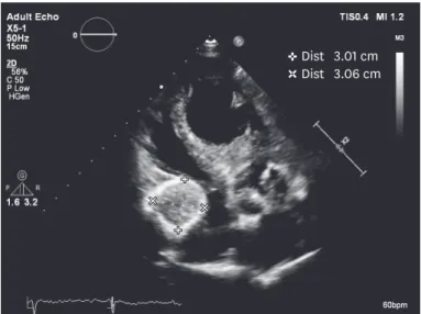
Dist 3.01 cm Dist 3.06 cm
Figure 1. Echocardiogram still image showing calcified and rounded right atrial mass.
Movie 2
CT angiogram showing a large calcified and thrombosed right coronary artery aneurysm mimicking right atrial mass that we saw from transthoracic echocardiogram.
Click here to view
67 https://e-jcvi.org https://doi.org/10.4250/jcvi.2019.27.e6
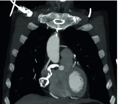
Coronary Artery Aneurysm Mimicking Atrial Mass
Figure 2. CT angiogram showing coronal images of the heavily calcified and thrombosed right coronary aneurysm.