ABSTRACT
Purpose: The purpose of this study was to assess the presence of contralateral occult malignant foci (OMF) among patients who confined unilateral papillary thyroid carcinomas (PTCs).
Methods: From January 2010 to December 2012, we retrospectively evaluated 714 patients who received total thyroidectomy with central lymph node (LN) dissection after being diagnosed as having unilaterally confined PTC. By dividing the patients into 2 groups according to the presence or absence of contralateral OMF, the relationship between OMF and clinicopathological factors such as age, sex, tumor size, multifocality, chronic lymphocytic/Hashimoto's thyroiditis, contralateral lobe benign nodule, extrathyroidal extension (ETE), and central LN metastasis.
Results: When the patients were subdivided based on primary tumor size, OMF in the contralateral lobe were more frequently found as the tumor sizes increased, with statistical significance (P=0.012). In the patients with multifocal thyroid cancer in the lobe that had primary tumor, OMF was observed in the other lobe regardless of the number of primary malignant nodules, and the difference was statistically significant (P=0.001).
Conclusion: The primary tumor size or multifocality is a risk factor that can predict the presence of contralateral lobe occult cancers. We suggest that patient needs to carefully observe the remaining contralateral lobe, taking into consideration the size and multifocality of the primary tumor, when performing unilateral thyroid lobectomy.
Keywords: Papillary thyroid carcinoma; Contralateral occult malignant foci
INTRODUCTION
Papillary thyroid carcinoma (PTC) is the most common type of thyroid cancers. Its incidence rate is recently growing worldwide because of rapid development of imaging instruments and diagnostic techniques such as fine-needle aspiration biopsy (FNAB) (1-3). In addition, most PTCs are known as an indolent mild cancer with favorable prognosis, having a survival rate of 30 years in >90% of cases (4).
The first-line treatment of PTC is thyroidectomy (5). When only unilateral lobectomy was performed, thyroid cancer in the contralateral lobe should be excluded before surgery.
Received: Dec 12, 2017 Revised: Feb 21, 2018 Accepted: Mar 30, 2018 Correspondence to Tae Kwun Ha
Department of Surgery, Inje University Busan Paik Hospital, Inje University College of Medicine, 75 Bokji-ro, Busanjin-gu, Busan 47392, Korea.
E-mail: [email protected]
Copyright © 2018. Korean Association of Thyroid and Endocrine Surgeons; KATES This is an Open Access article distributed under the terms of the Creative Commons Attribution Non-Commercial License (https://
creativecommons.org/licenses/by-nc/4.0/).
ORCID iDs Sang Wook Jo
https://orcid.org/0000-0003-4946-3519 Ha Kyoung Park
https://orcid.org/0000-0002-7610-8590 Tae Kwun Ha
https://orcid.org/0000-0001-7980-3700 Author Contributions
Conecptualiztion: Ha Kyoung Park, Tae Kwun Ha; Data curation: Sang Wook Jo; Formal analysis: Sang Wook Jo; Investigation: Sang Wook Jo; Methodology: Sang Wook Jo, Tae Kwun Ha; Resources: Sang Wook Jo, Tae Kwun Ha; Software: Ha Kyoung Park, Tae Kwun Ha;
Supervision: Ha Kyoung Park, Tae Kwun Ha;
Validation: Ha Kyoung Park, Tae Kwun Ha;
Writing - original draft: Sang Wook Jo, Ha Kyoung Park, Tae Kwun Ha; Writing - review&
editing: Sang Wook Jo, Ha Kyoung Park, Tae Kwun Ha.
Sang Wook Jo , Ha Kyoung Park , Tae Kwun Ha
Department of Surgery, Inje University Busan Paik Hospital, Inje University College of Medicine, Busan, Korea
Prediction of Contralateral Occult Malignant Nodule in Patients with Unilaterally Confined Papillary Thyroid Carcinomas
Original Article
Conflict of Interest
No potential conflict of interest relevant to this article was reported.
However, occult malignant foci (OMF) in the contralateral lobe not found before surgery (6). Preoperatively undiagnosed OMF in contralateral thyroid lobes can cause postoperative remnant disease not only may lead to an additional surgery, but also can be associated with regional and distant metastasis (7).
The primary objective of this study was to assess the presence of contralateral OMF in patients with unilateral confined PTC.
METHODS
We retrospectively evaluated 714 patients who received total thyroidectomy with central lymph node (LN) dissection after being diagnosed as having unilaterally confined PTC, between January 2010 and December 2012. Preoperative ultrasonography (US) and US-guided FNAB was performed for all the patients. Neck computed tomography (CT) was further performed if an extrathyroidal extension (ETE) is suspected from US findings, or central or lateral LN metastasis was present.
The patient group of 2010 to 2012, according to 2009 American Thyroid Association (ATA) Guideline (5) or 2010 Korean Thyroid Association (KTA) Management Guidelines (8,9) went through total thyroidectomy for thyroid cancer larger than 1 cm or <1 cm with multifocality, ETE (included gross ETE and micro/minimal/minor ETE by American Joint Committee on Cancer (AJCC) Cancer Staging Manual, 7th edition (10), central LN metastasis (only level 6, AJCC 7th edition), a personal history of radiation therapy to the head and neck, or familial thyroid carcinoma (5). Additionally, when gross finding and operator's empirical decision at the operation field favored it, total thyroidectomy was done.
The contralateral OMF was defined as foci found in the pathological examination after surgery, but not diagnosed based on preoperative radiological evaluation and FNAB results.
By dividing the patients into 2 groups according to the presence or absence of contralateral OMF, the relationship between OMF and clinicopathological factors such as age, sex, tumor size, multifocality (more than 2 malignant nodules in unilateral lobe), chronic lymphocytic/
Hashimoto's thyroiditis, contralateral lobe benign nodule, ETE and central LN metastasis were analyzed by using SPSS 18.0 (SPSS Inc., Chicago, IL, USA). The relationship between the predictive factors and OMF was analyzed by using χ2 test. A multivariate analysis was performed by using multivariate logistic regression analysis. Data were considered statistically significant when the significance level was <0.05
RESULTS
Among the 714 patients included in the present study, 637 were women and 77 were men. The mean age was 47.5 years (range, 8–83 years) at the time of surgery.
After performing total thyroidectomy, contralateral OMF was found in 61 patients (8.5%) on pathological examination. The mean size of the tumors in 61 patients with contralateral malignant nodules was 0.2 cm (range, 0.1–0.3 cm). Furthermore, all of the OMF were papillary thyroid microcarcinoma (PTMC).
The mean size of the primary malignant tumors was 1.25 cm (range, 0.1–7 cm). The mean size of the primary malignant tumors with OMF was 1.43 cm (range, 0.4–4 cm). Of the patients, 280 (39.2%) had multifocal PTCs with ≥ malignant 2 nodules in one lobe, with a maximum of 4 nodules found. ETE of tissues surrounding the thyroid, and metastasis to the central neck LN occurred in 321 (45%) and 370 (51.8%), respectively. Chronic lymphocytic/Hashimoto's thyroiditis was found in 148 patients (20.7%; Table 1).
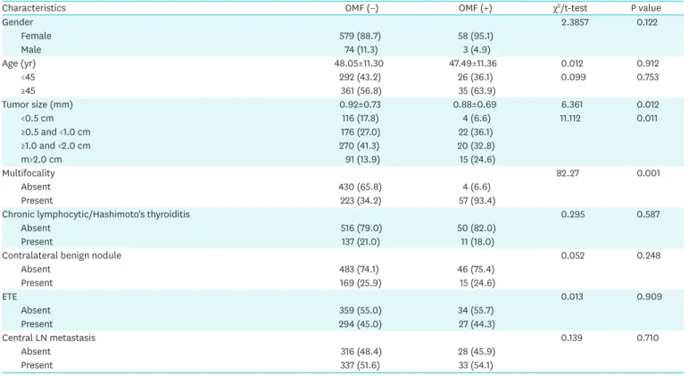
When the patients were divided into 2 groups depending on the presence or absence of OMF, the mean (standard deviation) ages of the patients in the 2 groups were 48.05±11.30 and 47.49±11.36, respectively. OMF were more frequently found in the group aged >45 years, although the difference was not statistically significant (P=0.753). The sex distribution was 58 women and 3 men (P=0.122). When the patients were subdivided based on primary tumor size, OMF in the contralateral lobe were more frequently found as the tumor sizes increased, with statistical significance (P=0.012). In the patients with multifocal thyroid cancer in the lobe that had primary tumor, OMF was observed in the other lobe regardless of the number of primary malignant nodules, and the difference was statistically significant (P=0.001).
However, accompanying chronic lymphocytic/Hashimoto's thyroiditis, contralateral lobe benign nodule, ETE or central LN metastasis had no significant relationship with the presence or absence of OMF (Table 2).
Multivariate logistic regression analysis was performed for primary tumor size and multifocality, which were statistically significant on univariable analysis. OMF in the other lobe were more frequently found as the primary tumor sizes (P=0.027) and multifocality (P<0.001) increased (Table 3).
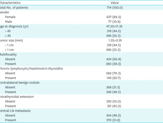
Table 1. Demographic characteristics of 714 patients with papillary thyroid carcinoma
Characteristics Value
Total No. of patients 714 (100.0)
Gender
Female 637 (89.2)
Male 77 (10.8)
Age at diagnosis (yr) 47.50±11.35
< 45 318 (44.5)
≥ 45 396 (55.5)
Tumor size (mm) 1.25±0.91
< 1 cm 318 (44.5)
≥ 1 cm 396 (55.5)
Multifocality
Absent 434 (60.8)
Present 280 (39.2)
Chronic lymphocytic/Hashimoto's thyroiditis
Absent 566 (79.3)
Present 148 (20.7)
Contralateral benign nodule
Absent 368 (51.5)
Present 346 (48.5)
Extrathyroidal extension
Absent 393 (55.0)
Present 321 (45.0)
Central LN metastasis
Absent 344 (48.2)
Present 370 (51.8)
Values are shown as number (%) or mean±standard deviation.
LN = lymph node.
DISCUSSION
Decisions regarding the extent of thyroid surgery are influenced by several factors (11).
Older age (>45 years), contralateral thyroid nodules may be criteria for recommending a bilateral procedure either because of plans for radioactive iodine (RAI) therapy, to facilitate follow-up strategies, or to address suspicions of bilateral disease (12-15). However, in this study, age (<45 years, ≥45 years), contralateral lobe benign nodules had no significant (P=0.248) relationship with the presence or absence of OMF (Table 2). The 2017 AJCC Cancer Staging Manual, 8th edition (16) suggest the age at diagnosis cutoff used for staging was increased from 45 years to 55 years. The significant clinical benefit of preventing upstaging based only on age of diagnosis between 45 and 55 years in patients who otherwise would be considered low-risk (stage I or II). Other investigators have endorsed using an age cutoff of 55 years as the optimal single time point for papillary thyroid cancer prognostic models (17-21).
Kasai and Sakamoto (22) reported that the invasiveness differed depending on the primary tumor size. 2015 ATA Guidelines (23) suggest that if the primary thyroid carcinoma is >4 Table 2. Clinicopathologic factors in relation to OMF of contralateral lobe in 714 patients with papillary thyroid carcinoma
Characteristics OMF (−) OMF (+) χ2/t-test P value
Gender 2.3857 0.122
Female 579 (88.7) 58 (95.1)
Male 74 (11.3) 3 (4.9)
Age (yr) 48.05±11.30 47.49±11.36 0.012 0.912
<45 292 (43.2) 26 (36.1) 0.099 0.753
≥45 361 (56.8) 35 (63.9)
Tumor size (mm) 0.92±0.73 0.88±0.69 6.361 0.012
<0.5 cm 116 (17.8) 4 (6.6) 11.112 0.011
≥0.5 and <1.0 cm 176 (27.0) 22 (36.1)
≥1.0 and <2.0 cm 270 (41.3) 20 (32.8)
m>2.0 cm 91 (13.9) 15 (24.6)
Multifocality 82.27 0.001
Absent 430 (65.8) 4 (6.6)
Present 223 (34.2) 57 (93.4)
Chronic lymphocytic/Hashimoto's thyroiditis 0.295 0.587
Absent 516 (79.0) 50 (82.0)
Present 137 (21.0) 11 (18.0)
Contralateral benign nodule 0.052 0.248
Absent 483 (74.1) 46 (75.4)
Present 169 (25.9) 15 (24.6)
ETE 0.013 0.909
Absent 359 (55.0) 34 (55.7)
Present 294 (45.0) 27 (44.3)
Central LN metastasis 0.139 0.710
Absent 316 (48.4) 28 (45.9)
Present 337 (51.6) 33 (54.1)
Values are shown as number (%) or mean±standard deviation.
OMF = occult malignant foci; m = mass; ETE = extrathyroidal extension; LN = lymph node.
Table 3. Multivariate analysis of clinicopathologic factors associated with OMF in patients with unilateral papillary thyroid carcinoma
Characteristics B SE Sig. Exp (B) 95% CI
Lower Upper
Tumor size 0.317 0.144 0.027 1.374 1.036 1.821
Multifocality 0.852 0.116 <0.001 2.345 1.868 2.943
Constant −4.434 0.370 0.000 0.012
OMF = occult malignant foci; B = coefficient of regression; SE = standard error; Sig. = significance probability; Exp (B) = odds ratio; CI = confidence interval.
cm, near-total or total thyroidectomy is necessary if the overall strategy is to include RAI therapy post-operatively (2015 ATA). for tumors that are between 1 and 4 cm in size, either a bilateral thyroidectomy (total or near-total) or a unilateral procedure (thyroid lobectomy) may be suitable as treatment plan (2015 ATA). Based on our study, the mean size of the primary malignant nodules with OMF was 1.43 cm (range, 0.4–4 cm). When the patients were subdivided based on primary tumor size, OMF in the contralateral lobe were more frequently found as the tumor sizes increased, with statistical significance (P=0.012).
2016 KTA Management Guidelines (24) and 2017 The Korean Association of Thyroid and Endocrine Surgeons (KATES) Guidelines (25) suggest that the initial surgical procedure of gross extrathyroidal extension should include a near-total or total thyroidectomy (24,25).
In this study, ETE had no significant (P=0.909) relationship with the presence or absence of OMF (Table 2). The limitation of this study is our data included grossETE and micro/minimal/
minor ETE. Pathologically, the thyroid has an incomplete capsule. Because, the thyroid gland may contain adipose tissue and skeletal muscle under normal circumstances. According to the College of American Pathologists, defining (minimal) ETE may be problematic and subjective (26). Microscopic ETE is not an independent prognostic factor for persistent/
recurrent disease. The disease-free survival is equivalent in patients with microscopic ETE and those with completely intrathyroidal tumors (26-30). The 2017 AJCC Cancer Staging Manual 8th edition suggested minor ETE was removed from the definition of T3 disease. As a result, minor ETE does not affect either T category or overall stage.
In the present study, the primary tumor multifocality is a risk factor that can predict the presence of contralateral lobe occult cancers. Kim et al. (31) published that multifocality is a unique factor that can predict the incidence of contralateral cancer after unilateral surgery of thyroid cancer. In addition, the presence of PTMC in the contralateral lobe is related with the multifocality of primary cancer (32,33). Park et al. (34) and Giannini et al. (35) reported that each multifocal cancer occurs independently by studying BRAF mutation rate. Sugg et al. (36) thought that individual tumors are associated with multifocality via a distinct pattern of RET/PTC rearrangement. According to Wang et al. (37), if bilateral PTCs are observed, the disease would have already progressed, disease-free survival duration would be shorter, different biological characteristics would be observed, and the stage would be more advanced than that of unilateral PTC. Patients with indeterminate nodules who have bilateral nodular disease, those with significant medical comorbidities, or those who prefer to undergo bilateral thyroidectomy to avoid the possibility of requiring a future surgery on the contralateral lobe, may undergo total or near-total thyroidectomy, assuming completion thyroidectomy would be recommended if the indeterminate nodule proved malignant following lobectomy. including the estimated pre-surgical likelihood of malignancy based upon clinical risk factors (>4 cm, family history, and/or radiation history) (38-41). However, not only the growing rate and prognosis of OMF after thyroid lobectomy, but also the usefulness of routine total thyroidectomy for detection of thyroid cancer of <0.5 cm in size are controversial. In PTMC patients, delayed surgery was not associated with higher risk of structural recurrent/persistent disease compared to immediate surgery. These findings support the notion that surgical treatment can be safely delayed in patients with PTMC under close monitoring (42). Ito et al. (43) proposed active surveillance is the optimal first line of management for all adult patients with low-risk papillary microcarcinomas.
In this retrospective study, the incidence of contralateral OMF among 714 patients who received total thyroidectomy after being diagnosed as having unilateral PTC was 61 patients
(8.5%), which is lower than that reported in a previous study (15.6%–27%) (44,45). In the our study, the mean size of the contralateral OMF was 0.2 cm (range, 0.1–0.3 cm) and all of the OMF were PTMCs. As small PTMC grows slowly, has favorable prognosis, and shows a pattern like that of benign lesion (46), active monitoring and observation are necessary when unilateral lobectomy is performed by considering primary tumor size and multifocality.
Among the clinicopathological factors related to the presence of contralateral OMF before surgery, age, sex, chronic lymphocytic/Hashimoto's thyroiditis, benign nodule, ETE, and central LN metastasis were not significant predictive factors, but the primary tumor size and multifocality had a significant relationship with the incidence of contralateral OMF (Table 3).
However, in this study, the mean size of the contralateral OMF was very small 0.2 cm (range, 0.1–0.3 cm) and all of the OMF were PTMCs. So if the lobectomy was performed already, meticulous follow-up is necessary about remnant lobe than completion thyroidectomy.
ACKNOWLEDGMENTS
Thank you Professor Bae Sung Kwon for his medical and statistical advice.
REFERENCES
1. Kim EK, Park CS, Chung WY, Oh KK, Kim DI, Lee JT, et al. New sonographic criteria for recommending fine-needle aspiration biopsy of nonpalpable solid nodules of the thyroid. AJR Am J Roentgenol 2002;178:687-91.
PUBMED | CROSSREF
2. Hong YJ, Son EJ, Kim EK, Kwak JY, Hong SW, Chang HS. Positive predictive value of sonographic features of solid thyroid nodule. Clin Imaging 2010;34:127-33.
PUBMED | CROSSREF
3. Siegel R, Ma J, Zou A, Ahmedin J. Cancer statistics, 2014. CA Cancer J Clin 2014;64:9-29.
PUBMED | CROSSREF
4. Markovina S, Grigsby PW, Schwarz JK, DeWees T, Moley JF, Siegel BA, et al. Treatment approach, surveillance, and outcome of well-differentiated thyroid cancer in childhood and adolescence. Thyroid 2014;24:1121-6.
PUBMED | CROSSREF
5. American Thyroid Association (ATA) Guidelines Taskforce on Thyroid Nodules and Differentiated Thyroid CancerCooper DS, Doherty GM, Haugen BR, Kloos RT, Lee SL, et al. Revised American Thyroid Association management guidelines for patients with thyroid nodules and differentiated thyroid cancer.
Thyroid 2009;19:1167-214.
PUBMED | CROSSREF
6. Pasieka JL, Thompson NW, McLeod MK, Burney RE, Macha M. The incidence of bilateral well- differentiated thyroid cancer found at completion thyroidectomy. World J Surg 1992;16:711-6.
PUBMED | CROSSREF
7. So YK, Kim MW, Son YI. Multifocality and bilaterality of papillary thyroid microcarcinoma. Clin Exp Otorhinolaryngol 2015;8:174-8.
PUBMED | CROSSREF
8. Yi KH, Park YJ, Koong SS, Kim JH, Na DG, Ryu JS, et al. Revised Korean Thyroid Association management guidelines for patients with thyroid nodules and thyroid cancer. Endocrinol Metab 2010;25:270-97.
CROSSREF
9. Yi KH, Park YJ, Koong SS, Kim JH, Na DG, Ryu JS, et al. Revised Korean Thyroid Association management guidelines for patients with thyroid nodules and thyroid cancer. Korean J Otorhinolaryngol-Head Neck Surg 2011;54:8-36.
CROSSREF
10. Edge SB; American Joint Committee on Cancer. AJCC Cancer Staging Manual. 7th ed. New York (NY):
Springer; 2010.
11. Stojadinovic A, Peoples GE, Libutti SK, Henry LR, Eberhardt J, Howard RS, et al. Development of a clinical decision model for thyroid nodules. BMC Surg 2009;9:12.
PUBMED | CROSSREF
12. Hay ID, Bergstralh EJ, Goellner JR, Ebersold JR, Grant CS. Predicting outcome in papillary thyroid carcinoma: development of a reliable prognostic scoring system in a cohort of 1779 patients surgically treated at one institution during 1940 through 1989. Surgery 1993;114:1050-7.
PUBMED
13. Hay ID, Thompson GB, Grant CS, Bergstralh EJ, Dvorak CE, Gorman CA, et al. Papillary thyroid carcinoma managed at the Mayo Clinic during six decades (1940–1999): temporal trends in initial therapy and long-term outcome in 2444 consecutively treated patients. World J Surg 2002;26:879-85.
PUBMED | CROSSREF
14. Matsuzu K, Sugino K, Masudo K, Nagahama M, Kitagawa W, Shibuya H, et al. Thyroid lobectomy for papillary thyroid cancer: long-term follow-up study of 1,088 cases. World J Surg 2014;38:68-79.
PUBMED | CROSSREF
15. Nixon IJ, Ganly I, Patel SG, Palmer FL, Whitcher MM, Tuttle RM, et al. Thyroid lobectomy for treatment of well differentiated intrathyroid malignancy. Surgery 2012;151:571-9.
PUBMED | CROSSREF
16. Amin MB, Edge SB; American Joint Committee on Cancer. AJCC Cancer Staging Manual. 8th ed.
Switzerland: Springer; 2017.
17. Hendrickson-Rebizant J, Sigvaldason H, Nason RW, Pathak KA. Identifying the most appropriate age threshold for TNM stage grouping of well-differentiated thyroid cancer. Eur J Surg Oncol 2015;41:1028-32.
PUBMED | CROSSREF
18. Ito Y, Fukushima M, Tomoda C, Inoue H, Kihara M, Higashiyama T, et al. Prognosis of patients with papillary thyroid carcinoma having clinically apparent metastasis to the lateral compartment. Endocr J 2009;56:759-66.
PUBMED | CROSSREF
19. Ito Y, Ichihara K, Masuoka H, Fukushima M, Inoue H, Kihara M, et al. Establishment of an intraoperative staging system (iStage) by improving UICC TNM classification system for papillary thyroid carcinoma.
World J Surg 2010;34:2570-80.
PUBMED | CROSSREF
20. Kim SJ, Myong JP, Suh H, Lee KE, Youn YK. Optimal cutoff age for predicting mortality associated with differentiated thyroid cancer. PLoS One 2015;10:e0130848.
PUBMED | CROSSREF
21. Mazurat A, Torroni A, Hendrickson-Rebizant J, Benning H, Nason RW, Pathak KA. The age factor in survival of a population cohort of well-differentiated thyroid cancer. Endocr Connect 2013;2:154-60.
PUBMED | CROSSREF
22. Kasai N, Sakamoto A. New subgrouping of small thyroid carcinomas. Cancer 1987;60:1767-70.
PUBMED | CROSSREF
23. Haugen BR, Alexander EK, Bible KC, Doherty GM, Mandel SJ, Nikiforov YE, et al. 2015 American Thyroid Association management guidelines for adult patients with thyroid nodules and differentiated thyroid cancer: the American Thyroid Association Guidelines Task Force on thyroid nodules and differentiated thyroid cancer. Thyroid 2016;26:1-133.
PUBMED | CROSSREF
24. Yi KH, Lee EK, Kang HC, Koh Y, Kim SW, Kim IJ, et al. 2016 revised Korean Thyroid Association
management guidelines for patients with thyroid nodules and thyroid cancer. Int J Thyroidol 2016;9:59-126.
CROSSREF
25. Park JW, Chung KW, Yun JS, Kwon H, Kim HY, Nam KH, et al. Surgical treatment guidelines for patients with differentiated thyroid cancer: the Korean Association of Thyroid and Endocrine surgeons (KATES) Guidelines Taskforce. Korean J Endocr Surg 2017;17:1-18.
CROSSREF
26. Arora N, Turbendian HK, Scognamiglio T, Wagner PL, Goldsmith SJ, Zarnegar R, et al. Extrathyroidal extension is not all equal: implications of macroscopic versus microscopic extent in papillary thyroid carcinoma. Surgery 2008;144:942-7.
PUBMED | CROSSREF
27. Leboulleux S, Rubino C, Baudin E, Caillou B, Hartl DM, Bidart JM, et al. Prognostic factors for persistent or recurrent disease of papillary thyroid carcinoma with neck lymph node metastases and/or tumor extension beyond the thyroid capsule at initial diagnosis. J Clin Endocrinol Metab 2005;90:5723-9.
PUBMED | CROSSREF
28. Radowsky JS, Howard RS, Burch HB, Stojadinovic A. Impact of degree of extrathyroidal extension of disease on papillary thyroid cancer outcome. Thyroid 2014;24:241-4.
PUBMED | CROSSREF
29. Shin JH, Ha TK, Park HK, Ahn MS, Kim KH, Bae KB, et al. Implication of minimal extrathyroidal extension as a prognostic factor in papillary thyroid carcinoma. Int J Surg 2013;11:944-7.
PUBMED | CROSSREF
30. Woo CG, Sung CO, Choi YM, Kim WG, Kim TY, Shong YK, et al. Clinicopathological significance of minimal extrathyroid extension in solitary papillary thyroid carcinomas. Ann Surg Oncol 2015;22 Suppl 3:S728-33.
PUBMED | CROSSREF
31. Kim ES, Kim TY, Koh JM, Kim YI, Hong SJ, Kim WB, et al. Completion thyroidectomy in patients with thyroid cancer who initially underwent unilateral operation. Clin Endocrinol (Oxf ) 2004;61:145-8.
PUBMED | CROSSREF
32. Koo BS, Lim HS, Lim YC, Yoon YH, Kim YM, Park YH, et al. Occult contralateral carcinoma in patients with unilateral papillary thyroid microcarcinoma. Ann Surg Oncol 2010;17:1101-5.
PUBMED | CROSSREF
33. Zhou YL, Zhang W, Gao EL, Dai XX, Yang H, Zhang XH, et al. Preoperative BRAF mutation is predictive of occult contralateral carcinoma in patients with unilateral papillary thyroid microcarcinoma. Asian Pac J Cancer Prev 2012;13:1267-72.
PUBMED | CROSSREF
34. Park SY, Park YJ, Lee HS, Choi SH, Choe G, Jang HC, et al. Analysis of differential BRAF V600E mutational status in multifocal papillary thyroid carcinoma. Cancer 2006;107:1831-8.
PUBMED | CROSSREF
35. Giannini R, Ugolini C, Lupi C, Proietti A, Elisei R, Salvatore G, et al. The heterogeneous distribution of BRAF mutation supports the independent clonal origin of distinct tumor foci in multifocal papillary thyroid carcinoma. J Clin Endocrinol Metab 2007;92:3511-6.
PUBMED | CROSSREF
36. Sugg SL, Ezzat S, Rosen IB, Freeman JL, Asa SL. Distinct multiple RET/PTC gene rearrangement in multifocal papillary thyroid neoplasia. J Clin Endocrinol Metab 1998;83:4116-22.
PUBMED | CROSSREF
37. Wang W, Zhao W, Wang H, Teng X, Wang H, Chen X, et al. Poorer prognosis and higher prevalence of BRAF (V600E) mutation in synchronous bilateral papillary thyroid carcinoma. Ann Surg Oncol 2012;19:31-6.
PUBMED | CROSSREF
38. Tuttle RM, Lemar H, Burch HB. Clinical features associated with an increased risk of thyroid malignancy in patients with follicular neoplasia by fine-needle aspiration. Thyroid 1998;8:377-83.
PUBMED | CROSSREF
39. Goldstein RE, Netterville JL, Burkey B, Johnson JE. Implications of follicular neoplasms, atypia, and lesions suspicious for malignancy diagnosed by fine-needle aspiration of thyroid nodules. Ann Surg 2002;235:656-62.
PUBMED | CROSSREF
40. Schlinkert RT, van Heerden JA, Goellner JR, Gharib H, Smith SL, Rosales RF, et al. Factors that predict malignant thyroid lesions when fine-needle aspiration is “suspicious for follicular neoplasm”. Mayo Clin Proc 1997;72:913-6.
PUBMED | CROSSREF
41. Mehta RS, Carty SE, Ohori NP, Hodak SP, Coyne C, LeBeau SO, et al. Nodule size is an independent predictor of malignancy in mutation-negative nodules with follicular lesion of undetermined significance cytology. Surgery 2013;154:730-6.
PUBMED | CROSSREF
42. Jeon MJ, Kim WG, Kwon HM, Kim MJ, Park SY, Oh SY, et al. Clinical outcomes after delayed thyroid surgery in patients with papillary thyroid microcarcinoma. Eur J Endocrinol 2017;177:25-31.
PUBMED | CROSSREF
43. Ito Y, Miyauchi A, Oda H. Low-risk papillary microcarcinoma of the thyroid: a review of active surveillance trials. Eur J Surg Oncol 2018;44:307-15.
PUBMED | CROSSREF
44. Pitt SC, Sippel RS, Chen H. Contralateral papillary thyroid cancer: does size matter? Am J Surg 2009;197:342-7.
PUBMED | CROSSREF
45. Lee YC, Eun YG, Sohn YM, Rhee SY, Hong IK, Chon S, et al. Predictive factors for occult contralateral carcinoma in patients with unilateral papillary thyroid microcarcinoma by preoperative ultrasonographic and pathological features. World J Surg 2015;39:1736-41.
PUBMED | CROSSREF
46. Ito Y, Miyauchi A. Nonoperative management of low-risk differentiated thyroid carcinoma. Curr Opin Oncol 2015;27:15-20.
PUBMED | CROSSREF