YUJM 29(1):14-18, 2012
나선식 CT를 이용한 혐색소형 신세포암과 투명세포형 신세포암의 감별
김홍철, 조재호
영남대학교 의과대학 영상의학과학교실
Differentiation of Chromophobe Renal Cell Carcinoma and Clear Cell Renal Cell Carcinoma by Using Helical CT
Hong Chul Kim, Jae Ho Cho
Department of Diagnostic Radiology, College of Medicine, Yeungnam University, Daegu, Korea
Background: The purpose of this study was to differentiate chromophobe renal cell carcinoma and clear cell renal cell carcinoma on helical CT.
Methods: The CT images of 9 patients histopathologically proven to have chromophobe renal cell carcinoma and 20 patients with clear cell renal cell carcinoma were reviewed. The tumor sizes, margins, enhancement degrees and patterns, presence or absence of calcification, and tumor spread patterns (including perinephric changes, venous invasion, lymphadenopathy, and distant metastasis) were compared.
Results: All the chromophobe renal cell carcinomas showed well-demarcated margins. Thechromophobe renal cell carcinomas showed milder enhancements than the clear cell renal cell carcinomas. The sensitivity and specificity for differentiating the chromophobe renal cell carcinoma from the clear cell renal cell carcinoma were 100 and 88%, respectively, when 101 Hounsfield units was used as the cut-off value in the corticomedullary phase, and 95 and 100% when a less-than-three-time enhancement change was used as a cut-off value in the corticomedullary phase (p<0.05). The chromophobe renal cell carcinomas (67%) tended to show a homogeneous enhancement whereas the clear cell renal cell carcinomas (85%) usually showed a heteroge- neous enhancement (p<0.05). Statistical analysis revealed that the frequencies of the tumor spread pattern and calcification in the two subtypes didnot differ significantly (p>0.05).
Conclusion: The CT findings of the chromophobe renal cell carcinomascompared to those of the clear cell renal cell carcinomas showed that there were mild enhancements in the corticomedullary phase, homoge- neous enhancements, and well-demarcated margins.
Key Words: Clear cell renal cell carcinoma, Chromophobe renal cell carcinoma, Spiral CT
교신저자: 조재호, 705-717, 대구광역시 남구 현충로 170 영남대학교 의과대학 영상의학과학교실
Tel: (053) 620-3043, Fax: (053) 653-5484 E-mail: [email protected]
서 론
신세포암은 신 실질에서 발생하는 가장 흔한 원발성 악성종 양이며, 성인의 모든 악성 종양의 3%를 차지한다.1 2004년
개정된 신세포암의 WHO(World Health Organization, WHO) 분류에 따르면 투명세포형(clear cell renal cell carcinoma), 유 두형(papillary renal cell carcinoma), 혐색소형(chromophobe renal cell carcinoma), 벨리니관 암종(carcinoma of the collecting ducts of Bellini), 다방성 투명세포형(multilocular clear cell renal cell carcinoma), 수질암종(renal medullary carcinoma), Xp11 전좌를 동반한 암종(Xp11 translocation carcinomas), 신경아세포종
을 동반한 암종 (carcinoma associated with neuroblastoma), 점액성 세관 방추세포형(mucinous tubular and spindle cell carcinoma), 미분류형 (unclassified)으로 분류하고 있다.2 신 세포암의 분류는 예후와 밀접한 관련이 있는데, 투명세포형 이 전체 신세포암 중 70% 정도로 가장 흔한 아형이고, 5년 생존율은 55-60%인 반면,3 혐색소형은 전체신세포암 중 6- 11%를 차지하고, 5년 생존율은 90%로 예후가 가장 좋은 아형이다.4 따라서 치료 전 병기 및 예후 예측 그리고 치료계 획에 있어서 신세포암의 아형을 분류하는 것이 중요하다. 신 세포암의 전반적인 나선식 CT (Computed Tomography, CT) 소견에 대해서 여러 보고들이 있었는데, Jinzaki 등5이 3.5
cm 이하의 신장 종양의 이중 나선식 CT 감쇠를 보고하였고, 피질-수질기 영상에서 100 Hounsfield unit (HU) 이상으로 조영증강 되고, 혐색소형은 100 HU 이하로 조영증강을 보인 다고 하였고, Kim 등6의 연구에서는 투명세포형은 피질-수질 기 영상에서 84 HU 이상으로 강한 조영증강을 보이고, 혐색 소형 및 다른 아형들은 84 HU 이하로 약한 조영증강을 보인 다고 하였다. 또한 조영증강 양상 및 석회화 유무, 종양의 파급 양상이 감별에 중요하다고 하였다. 이와 같이 신세포암 의 전반적인 CT 소견은 잘 알려져 있으나, 각각의 세포형, 특히 혐색소형 신세포암의 나선식 CT 소견에 대한 보고는 아직 충분치 않다. 그러므로 가장 많은 아형인 투명세포형과 혐색소형 신세포암을 감별할 수 있는 CT 소견이 있는지를 알아보고자 하였다.
재료 및 방법
2002년 1월부터 2011년 6월까지 병리조직학적으로 확진 된 9명의 혐색소형 신세포암 환자와 2011년 6월을 기준으로 최근 병리조직학적으로 확진된 20명의 투명세포형 신세포 암 환자를 대상으로 하였다.
모든 예에서 수술 전 나선식 CT를 시행하였고, 조영증강 전 영상과 조영제를 조입한 후 피질-수질기의 영상을 얻었다.
영상을 후향적으로 분석하였고, 통상적인 종괴의 평가 기 준에 따라 CT 상 종양의 크기, 경계, 조영증강의 정도 및 양상, 석회화의 유무, 신 주위 공간으로의 파급 유무, 정맥 침범, 림프절로의 전이와, 원격 전이의 유무를 살펴보았다.
종양의 경계가 분명하고, 평활한 경우 (well demarcated and smooth margin), 분명하고 엽상인 경우(well demarcated and lobulated margin), 그리고 불분명한 경우(pooly demarcated margin)로 나누었다. 조영증강 정도는 조영증강 전과 피질-
수질기에서 종양의 고형성 부위 중 3군데의 감쇠를 측정하여 평균값을 구하였고, 조영증강 전과 피질-수질기의 감쇠를 비 교하였다. 조영증강 양상은 균질성 조영증강 (homogeneous enhancement)과 비균질성 조영증강(heterogeneous enhance- ment)으로 나누어 평가하였다. 신주위 공간으로의 파급 유무 는 Gerota 근막의 비후, 신주위 지방층의 소실 혹은 흐려짐으 로 평가하였다. 정맥으로의 종양 파급여부를 알기 위하여 신정맥이나 하대정맥의 내강에 비정상적인 음영의 충만 결 손이 있는지를 관찰하였고, 1 cm 이상으로 커진 림프절이나 원격 전이의 유무를 함께 알아보았다.
두 아형의 CT 소견을 비교하기 위해서 통계학적 분석은 카이 제곱 검정을 이용하여 종양의 크기, 경계, 조영증강의 정도 및 유형, 석회화의 유무, 신주위 공간으로의 파급 유무, 정맥 침범, 림프절로의 전이, 원격 전이의 유무를 비교하였다.
또한 조영증강 정도의 진단적 정확도를 평가하기 위해 recei- ver operating characteristic curve (ROC curve)를 사용하여 절 단값(cut-off value)을 판단하였다. 모든 통계 분석에는 PASW statistics 18.0 프로그램을 사용하였으며, 통계 유의 수준은 95% 신뢰구간에서 p 값이 0.05 미만일 경우로 정의하였다.
결 과
병리조직학적으로 확진된 9명의 혐색소형 신세포암 환자와 20명의 투명세포형 신세포암 환자가 대상이 되었고, 혐색소형 은 남자 5명, 여자 4명, 평균 연령 56 (41-70)세였고, 투명세포 형은 남자 13명, 여자 7명, 평균 연령 60 (40-78)세였다.
혐색소형 신세포암의 평균 직경은 3.6 (2-16) cm였고, 투명 세포형 신세포암은 4.6 (2-11) cm였다. 경계가 분명하고평활 한 경우는 혐색소형에서 7예(78%) (Fig. 1), 투명세포형에서 7예(35%) (Fig. 2), 분명하고 엽상인 경우는 혐색소형에서 2 예(22%), 투명세포형에서 11예(55%), 그리고 불분명한 경우 는 투명세포형에서 2예(10%) (Fig. 3)였고, 혐색소형에서는 없었다. 조영증강 정도를 평가하는 피질-수질기에서의 평균 감쇠는 혐색소형에서 89 (68-108) HU였고(Fig. 1), 투명세포 형에서 138 (103-157) HU였다(Fig. 2, 3). 또한 조영증강 전과 피질-수질기의 감쇠를 비교했을 때 혐색소형에서는 평균 2.4 배, 투명세포형에서는 4.7배의 조영증강을 보였다. 투명세포 형과 구별되는 혐색소형의 조영증강 정도를 ROC curve를 사용하여 절단값을 피질-수질기에서의 감쇠를 101 HU 이하 로 정했을 때 민감도 100%, 특이도 88%였고, 3배 이하의 조영증강 정도로 정했을 때 민감도 95%, 특이도 100%였다
Fig. 1. A 69-year-old man with chromophobe renal cell carci- noma. (A) Unenhanced CT scan obtained at the level of the lower pole of the right kidney shows a well-demarcated mass with a 5 cm diameter and 35 Hounsfield units. (B) In the contrast-enhan- ced CT scan obtained at the same level as in (A), the mass shows a homogeneous enhancement measuring 76 Hounsfield units.
Fig. 2. A 77-year-old woman with clear cell renal cell carcinoma.
(A) Unenhanced CT scan obtained at the level of the interpolar area of the right kidney shows a well-demarcated mass with a 3 cm diameter and 39 Hounsfield units. (B) In the contrast-enhan- ced CT scans obtained at the same level as in (A), the mass shows a heterogeneous enhancement measuring 146 Hounsfield units.
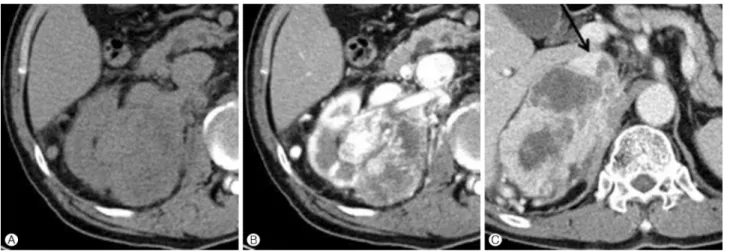
Fig. 3. A 73-year-old man with clear cell renal carcinoma. (A) Unenhanced CT scan obtained atthe level of the interpolar area of the right kidney shows a lobulated mass with an 11 cm diameter and 38 Hounsfield units and perirenal changes. (B) On the contrast-enhanced CT scan obtained at the same level as in (A), Mass shows a heterogeneous enhancement and 130 Hounsfield units. (C) Contrast-enhanced CT scan obtained in the portal venous phase 4 cm above (A) shows a tumor invasion (arrow) of the inferior vena cava.
(p<0.05). 조영증강 양상은 혐색소형은 6예 (67%)에서 균 질성 조영증강을 보였고 (Fig. 1), 투명세포형은 17예 (85%) 에서 비균질성 조영증강을 보였다 (Fig. 2, 3, p<0.05).
종양의 파급 여부는 투명세포형 신세포암인 경우 신주위 공간으로의 파급은 3예(15%), 정맥으로의 침범은 2예(10%), 림프절로의 전이는 1예(5%), 원격 전이는 2예(10%)로 관찰 되었지만, 혐색소형 신세포암의 경우는 신주위 공간, 정맥 침범이나 림프절로의 전이 혹은 원격 전이를 보인 예는 없었 고 (p>0.05), 석회화는 혐색소형에서는 2예 (22%), 투명세 포형에서는 6예 (30%)가 관찰되었고, 통계학적 유의성은 없 었다 (p>0.05). 이러한 연구결과는 Table 1과 같다.
고 찰
신세포암의 분류는 종양의 현미경적 양상과 유전정보에 근거하였고, 이 분류는 종양을 가진 환자의 예후와 종양의 행태에 잘 부합되고 있다.7 투명세포형이 전체 신세포암 중 70% 정도로 가장 많은 아형이고, 5년 생존율은 55-60%인 반면,3 혐색소형은 전체 신세포암중 6-11%를 차지하고, 5년 생존율은 90%로 예후가 가장 좋은 아형이다.4 혐색소형의 86%에서 T1이나 T2의 낮은 병기를 가지고, 10% 정도에서 피막을 통과해서 주위 지방조직을 침범할 수 있고, 약 4%에 서 신장 정맥을 침범한다고 한다.2 Cho 등8에 의하면 혐색소 형은 조직학적으로 높은 핵등급을 보이는 경우에도 다른 아
Table 1. Spiral CT findings of chromophobe renal cell carcinoma and clear cell renal cell carcinoma
Chromophobe renal cell carcinoma Clear cell renal cell carcinoma
Size (Range) 3 (2-12) cm 4.6 (2-11) cm
Margin
Well demarcated and smooth margin 78% 35%
Well demarcated and lobulated margin 22% 55%
Poorly demarcated margin 0% 10%
Precontrast phase (HU) 38 30
Corticmedullary phase (HU)* 89 138
Corticomedullary phase/precontrast phase (HU)* 2.4 4.7
Enhancement pattern*
Homogeneous enhancement 67% 15%
Heterogeneuos enhancement 33% 85%
Calcification 22% 30%
Perinephric change 0% 15%
Venous invasion 0% 10%
Lymphadenopathy 0% 5%
Distant metastasis 0% 10%
Values are mean.
HU: Hounsfield units.
*p<0.05.
형의 신세포암에 비해 상대적으로 낮은 병기와 좋은 예후를 가진다고 한다. 같은 병기일 때도 투명세포형 보다 혐색소형 신세포암의 5년 생존율이 더 높은 것으로 알려져 있다.7 Crotty 등9은 50예의 혐색소형 신세포암 중 86%는 Robson 병기 I이었고, 수술 후 추적 검사 상 47예(94%)는 이 병과는 무관한 이유로 사망하였으며, 종양의 재발 혹은 전이된 예는 없었다고 하였다. 혐색소형 신세포암은 이처럼투명세포형 과 비교하여 좋은 예후를 갖는다. 수술 전 신세포암의 아형을 평가하게 되면 수술의 범위를 결정하는데 도움을 주고 필요 없는 광범위한 절제를 피하여 수술 후 이환율과 사망률을 줄일 수 있으며, 예후 예측에도 도움을 준다.6
신세포암의 일반적인 CT 소견은 잘 알려져 있으나, 각각 의 세포형, 특히 혐색소형 신세포암의 나선식 CT 소견에 대 한 보고는 아직 충분치 않다. Fujimato 등10은 장경 5 cm 이하 의 신세포암 96개의 감쇠를 조영증강 역동적 CT상에서 알아 보았는데, 조영 초기의 CT에서 96예의 신세포암 중 72예에 서 고음영의 부분을 확인하였다. 이들 72예의 고음영의 부분 은 모두 투명세포형 신세포암이었고, 조직학적으로 폐포구 조를 가지고 있었다. 이 연구에 포함되었던 5예의 혐색소형 신세포암은 모두 조영증강 초기의 역동적 CT상 저음영으로
관찰되었다. Cho 등8은 혐색소형 신세포암으로 진단된 11예 의 나선식 CT 소견을 알아보았는데, 조영증강 전, 조영증강 초기 및 후기 나선식 CT상 신 실질보다 저음영이고 경계가 좋은 고형 종양이며, 핵등급이 높은 경우에도 상대적으로 병기가 낮고 예후가 좋았다. Kim 등6의 연구에서는 투명세포 형은 피질-수질기 영상에서 84 HU 이상으로 강한 조영증강 을 보이고 혐색소형 및 다른 아형들은 84 HU 이하로 약한 조영증강을 보인다고 하였다. 또한 투명세포형인 경우는 비 균질성 조영증강을 보이고, 혐색소형인 경우는 균질성 조영 증강을 보이며, 석회화 유무 및 종양의 파급 양상이 감별에 중요하다고 하였다.
본 연구에서도 조영증강 정도와 양상이 두 아형의 감별에 가장 중요한 지표였다. 혐색소형 신세포암은 투명세포형과 비교하여 약한 조영증강을 보였는데, 피질-수질기에서 감쇠 는 101 HU 이하, 조영증강 전과 피질-수질기를 비교했을 때 3배 이하의 조영증강을 보였다. 이는 병리조직학적으로 볼 때 상대적으로 혐색소형 신세포암의 혈관성이 적은 것과관 계가 있다.8 또한 혐색소형은 균질성 조영증강을 보이는데, 이는내부 괴사 및 출혈성 변화가 없고, 고형성 성장과 관련이 있다. 대조적으로 투명세포형 신세포암은 비균질성 조영증
강을 보이고, 이는 내부 괴사 및 출혈성 변화, 낭성 변화와 관련이 있다.6 석회화는 투명세포형 보다 유두형과 혐색소형 에서 더 흔히 관찰된다고 알려져 있으나, 본 연구에서는 통계 학적 의미가 없었다. 혐색소형은 신주위 공간, 정맥 침범이나 림프절로의 전이 혹은 원격 전이가 없었는데, 이는 종괴가 천천히 자라고 덜 침습적인 것을 의미한다. 또한 본 연구에서 투명세포형의 신주위 공간, 정맥 침범이나 림프절로의 전이 혹은 원격 전이가 이전 논문들과 비교하여 빈도가 다소 낮았 는데, 이는 최근 CT의 이용 증가로 조기에 발견되기 때문이 라고 생각된다. 본 연구에서 제한점이 있었는데, 가장 큰 제 한점은 혐색소형 신세포암의 환자 수가 CT 소견을 분석하기 에 적었는데, 이는 낮은 유병률과 관련이 있는 것으로 생각된 다. 또한 9명 중 3명은 외부 병원에서 CT를 시행하였고, 조영 제를 주입 후 피질-수질기의 영상 획득 시간이 본원과 다소 차이가 있었다.
요약하면 혐색소형 신세포암은 모두 분명한 경계를 보이 고, 투명세포형과 비교해서 약한 조영증강을 보였는데, 피질 -수질기에서 감쇠는 101 HU 이하, 조영증강 전과 피질-수질 기를 비교했을 때 3배 이하의 조영증강을 보였으며, 조영양 상은 67%에서 균질성 조영증강을 보였다 (p<0.05). 투명세 포형 신세포암과 대조적으로 혐색소형 신세포암의 경우는 신주위 공간, 정맥 침범이나 림프절로의 전이 혹은 원격 전이 를 보인 예는 없었으며 (p>0.05), 석회화의 유무는 혐색소형 에서 2예 (22%), 투명세포형에서 6예 (30%)가 관찰되어 통 계학적 유의성은 없었다 (p>0.05).
결 론
결론적으로 혐색소형 신세포암을 투명세포형 신세포암으 로 부터 감별 진단하는데 유용한 CT 소견은 피질-수질기에 서 약한 조영증강과 균질한 조영증강 그리고 분명한 경계를
가지는 것이다. 또한 신주위 공간, 정맥이나 림프절로의 침 범 혹은 원격 전이가 없는 소견은 보조적인 역할을 한다고 생각된다.
참고문헌
1. Fernández López-Peláez MS, GarcíaGómez JM, Ortíz Vico F, Roldán Ramos J. Tumor thrombosis of the left renal vein and inferior vena cava secondary to renal cell carcinoma. findings with ultrasonography, Echo-Doppler, and computerized tomo- graphy. Actas Urol Esp 2000;24:664-8.
2. Eble JN, Sauter G, Epstein JI, Sesterhenn IA, editors. Patho- logy and genetics of tumors of the urinary system and male genital organs. Lyon: IARC Press; 2004.
3. Bonsib SM. Risk and prognosis in renal neoplasms. a path- ologist’s prospective. Urol Clin North Am 1999;26:643-60.
4. Megumi Y, Nishimura K. Chromophobe cell renal carcinoma.
Urol Int 1998;61:172-4.
5. Jinzaki M, Tanimoto A, Mukai M, Ikeda E, Kobayashi S, Yuasa Y, et al. Double-phase helical CT of small renal paren- chymal neoplasms: correlation with pathologic findings and tumor angiogenesis. J Comput Assist Tomogr 2000;24:835- 42.
6. Kim JK, Kim TK, Ahn HJ, Kim CS, Kim KR, Cho KS. Diffe- rentiation of subtypes of renal cell carcinoma on helical CT scans. Am J Roentgenol 2002;178:1499-506.
7. Reuter VE, Presti JC Jr. Contemporary approach to the classi- fication of renal epithelial tumors. Semin Oncol 2000;27:
124-37.
8. Cho KR, Park CM, Chung HH, Kim HR, Park BJ, Lee YH, et al. Spiral CT findings of chromophobe renal cell carcinoma:
correlation with pathologic featuresand prognosis. J Korean Radiol Soc 2002;46:57-62. Korean.
9. Crotty TB, Farrow GM, Lieber MM. Chromophobe cell renal carcinoma: clinicopathological features of 50 cases. J Urol 1995;154:964-7.
10. Fujimoto H, Wakao F, Moriyama N, Tobisu K, Sakamoto M, Kakizoe T. Alveolar architecture of clear cell renal carci- nomas (< or =5.0 cm) show high attenuation on dynamic CT scanning. Jpn J Clin Oncol 1999;29:198-203.