약학회 지 제 49 권 제 2 호 174〜 179 (2005)
Yakhak Hoeji
Vol. 49,No. 2藥 學 含 職
사 람 섬 유 아 세 포 에 서
UVB
조 사 에 대 한 능 소 화 추 출 물 의 항 산 화 효 과김진화# • 이범천 . Y o n g H e Z h a n g * • 표형배
한 불 화 장 품 기 술 연 구 소 , *Department of Pharmacology, Peking University (Received February 4,2005; Revised March 16, 2005)
Effect of C am psis g ran d iflo ra on Antioxidative Activity in UVB-irradiated Human Dermal Fibroblasts
Jin H w a K i m # , B u m C h u n L e e , Y o n g H e Z h a n g * a n d H y e o n g B a e P y o
Hanbul Cosmetics, R&D Center 72-7, Samsung-myun, Umsung-kun, Chungbuk 369-830, Korea
^Department O f Pharmacology, School of Basic Medical Science, Peking University, 38 Xueyuan Road, Beijing 100083, China
A bstract — The human skin is constantly exposed to environmental irritants such as ultraviolet, smoke, chemicals. Free radicals and reactive oxygen species (ROS) caused by these environmental facts play critical roles in cellular damage. These irritants are in themselves damaging to the skin structure but they also participate the immensely complex inflammatory reaction. The purpose of this study was to investigate the skin cell protective effect of
Campsis grandiflora
extract on the UVB-irradiated human dermal fibroblasts (HDFs). We tested free radical and superoxide scavenging effectin vitro. C. gran
diflora
extracts had potent radical scavenging effect by 82% at 100 |ig/m/, respectively. For testing intracellular ROS scavenging activity the cultured HDFs were analyzed by increase in DCF fluorescence upon exposure to UVB 20 mj/cm 2 after treatment of C.
grandiflora
extracts. The results showed that oxidation of CM-DCFDA was inhibited byC. grandiflora
extracts effectively and C.
grandiflora
extracts has a potent free radical scavenging activity in UVB-irradiated HDFs. In ROS imaging using confocal microscope we visualized DCF fluorescence in HDFs directly. In conclusion, our results suggest that C.grandiflora
can be effectively used for the prevention of UV-induced adverse skin reactions such as radical production, and skin cell damage.K eyw ords □
Campsis grandiflora,
fibroblast, anti-oxidation, free radical scavenging, UVB자외선
,
약물,
환경오염물질과 같은여러 가지 외부 요인및 그외의여러 가지 이유에 의해서 몸안에활성산소종의 양이 크게늘어날경우생명체는산화적 스트레스를받아세포사멸,
질병발생그리고세포의노화를일으키게된다.
즉,
활성산소종 의생성과세포의 항산화력 간의 균형이 산화적 스트레스를결 정하게되고,
산화적 스트레스는세포내 단백질,
지질 및D N A 에손상을입히게 된다.
일반적으로세포는항상성유지를위하 여자체적으로항산화제의 생산과산화에의한손상의복구 기작을가지지만
, u y
흡연,
공해물질,중금속등의물리,화학적인외부환경에의해세포내작용에 이상미초래하거나활성산소의 생성이방어계의용량을초과할정도로과생성될경우산화스트
#본 논문에 관한 문의는 저자에게로 (전화) 043-879-2282 (팩스) O43-88^ ^ 28 (E-mail) [email protected]
레스가야기되며
,
피부의 노화를촉진시키게 된다.
태양광선과산소는피부세포에 자유라디칼(free radical)과활 성산소종(reactive oxygen species, ROS)을
:
생성시켜 DN A의손 상과세포막지질의 과산화를유발한다.
또한콜라겐과엘라스 틴과같은세포외기질을파괴,또는비정상적인교차결합유발 에관여하는 효소(matrix metalloproteinase, M M Ps)의발현에 영향을주어,
결합조직을손상시킨다.1^
능소화는쌍떡잎식물능소화과의 낙엽성 덩굴식물로원산지는 중국이며
,
금등화라고부르기도하고,
한자이름으로'
자위’
'등라 화1가 있다.
서양에서는 능소화를 '차이니즈 트렘펫 클리퍼 (Chinese trum pet creeper)'라고부르기도한다.
정원수로많이 쓰이고한방에서는꽃을사용하며,
피부소양,
풍진발홍,
좌창,
및 어혈과 혈열로인한질병에효과가있다고한다.
Jin 등은능소 화의 잎으로부터 triterpenoids 성분을분리하여 혈소판억제작 용에대하여 연구하였으며,
그외에 iridoids와phenyl propanoid174
능소회추출물의 항산화 ^과 175
glycosides를분리한예가보고되었으나,7) 사■람피부세포에대한
자외선으로부터의보호효과께대한연구예는거의보고되지 않
^ 다.
본연구는능소화추출물의 항산화효과와자외선에 의해 유발 되는자유라디칼생성저해효과등을 검색하여 다양한생리활 성효과와피부세포의보호효과소재로서이용하고자하였다.
실험 방법
실험재료
본실험에서사용한능소화
iCampsis grandiflora
)는중국북경 대학에서 제공받아사용하였다. 능소화 100 g을분쇄하여 5 0 %E t O H 1000 m/로 환류시키면서 3시간씩 2회반복추출하고감
압농축, 동결 건조하여그분말을사용하였다.
세포내
free radical
확인을 위해5-(6-) chloromethyl-2',7'- dichloro-dihydro-fluoresceindiacetate(CM-H2D C FD A )^ Molecular Probe
사(Eugene, OR, USA
)에서구입하였고,
형광스펙트로포토U1
터(luminescence spectrophotometer, Perkin Elmer, UK)
및 flow cytometry(FACS Calibur-S System, Becton Dickinson, NJ, USA), Leica D M IRE2 inverted microscope(Leica, Germany)로 즉정하였다. Butylated hydroxytoluene(BHT), epigallocatechin-
3-gallate(EGCG)는 Sigma Chemical Co.(St. Louis, M O , USA)에서구입하였다
.
DPPH에 의한 자유라디칼 소거 효과
자유라디칼소거 효과측정은 0.1 m M , DPPH(2,2-Di(4-tert-
octylphenyl)-l-picrylhydrazyl radical)에실험원료를농도별로동
량을첨가하여 3 7 ° C에서 10분간반응시킨후 5 6 0 n m에서 흡광
도를측정하였다8)
Superoxide radical ; 소 효 고 !'
Xanthine, xanthine oxidase 반 응 에 서 형 성 된 superoxide
radical
를소거하는효과를SOD Test Wako
방법에의해측정하 였 다 .9) 0.05 M Na2C 0 3 buffer(pH 10.2) 에 3 m M xanthine, 3 m M EDTA, 0.72 m M NBT와능소화추출물을 가한후250C에 서10
§:간반응하였다.
이반응액에0.25 U/m/ xanthine oxidase
를 가하고 250C에서 25분' 동 안 반응 후 superoxide radical 소거효과를
565nm
에서흡광도를 측정하였다.
세포 배양(Cell culture)
신생아의 포피조직에서 분리한
human dermal fibroblast
(H D F)는 M odern Tissue Technology(MTT, Korea)로부터 구입 하였다.
구입한H DF
를Dulbecco's Modified Eagle's Media
(D M E M )/F12(3:1 ) 배지 에 10% fetal bovine serum (FBS), 1%penicillin-streptomycini:
첨가하여37°C, 5% C 0 2
조건하에 배 양하고trypsinization
으로계대배양한뒤6~10
세대세포를실 험에 이용하였다.
Free radical scavenging activity in H um an Dermal Fibroblasts(Microtiter plate assay)
섬 유 아 세 포 (H um an derm al fibroblasts, H D F s)를 96 well plate에 l x 105/mZ로분주하여약 8 0%의 c o n f l u e n c e 도달할때
까지 배양한다
.
자외선(UVB)
조사 전에배양 배지를 제거한후H CSS(HEPES-buffered control salt solution : 120 m M NaCl, 5 m M KC1, 1.6 m M M gCl2, 2.3 m M CaCl2, 15 m M Glucose,
20 mM HEPES, 10 m M NaOH
)로세척하여 배지 내serum
성 분을 제거하였다. 4
나M CM-H2DCFDA in HCSS with 0.1%
Pluronic F-127(Molecular Probe)를빛이 들어가지 않도록주의
하여 처리한후37°C
20
분빈움시킨후농도 별로 처리해준다.
37°C 30
분반응시킨 후20m j/cm 2 UVB(UVB G15T8E, Sankyo Denki, Japan
)를 조사하였다.
모든시료를 처리 후37°0
에서 반 응시켜 Lum inescenceSpectrophotometer
를 사용하여DCF
(Ex=488 nm; E m =525nm
)에 의한 세포내 형광값을 측정하였 다 비 2)
Flow cytometry assay
섬유아세포를
6 well plate
에lx 105/mZ
로분주하여 약80
%의confluency
에도달할 때까지 배양한후능소화추출물을 적정농도별로세포배양액에 처리하여
1
시간배양한다.
자외선(UVB)
조사전에 배양배지를제거한후
HEPES-buffered control salt solution(HCSS
)으로세척하여 배지내serum
성분을제거하였 다.
자외선조사를위하여 배지를제거하고세쏘를세척한후각dish
에PBS
를lm
/씩 채워자외선(UVB
)은 조사량을 증가시키 면서 조사하였다. Trypsin
을처리하여 세포를떼어낸후HCSS
로washing
하면서 세포를잘혼함하고500 rpm, 3
분원심 분리 후1
나M CM-H2DCFDA
처리하고빛을차단하여37°C 20
분반 응시킨 후유세포분류기를사용하여D CF(Ex=488nm ; E m = 525 nm
)에의한세포내 형광값을측정하였다.10-13)
ROS imaging using confocal microscope
섬유아세포를
cover slip
에lx io 5/mZ
로분주하여 배양후 능 소화추출물을농도별로희석하여 세포배양액에 처리하고1
시간동안 배양한다
.
자외선(UVB)
조사 전에 배양배지를 제거한후
HEPES-buffered control salt solution(HCSS
)으로세척하여배지 내
serum
성분을제거하였다.
자외선 조사를 위하여 배지를제거하고세포를세척한후각
dish
에PBS
를1 m
/씩채워 자외선
(UVB
)의 조사량을 중가시키면서 조사한 후l|xM CM-
DCFDA
를처리하고빛을차단하여37°C 20
분반응시킨후Leica
Vol. 49, No. 2, 2005
176 김진화 • 이범천 . Yong He Zhang • 표형배
C o n c e n tra tio n U g /m l)
Fig. 2 - Superoxide scavenging activity (NBT method). BHT was
used as a positive control. The activity indicated by percentage of increase in comparison with that of control.
The activity is significant (*p<0.05) and the values are mean±S.E. from 5 individual experiments.
우수한항산화효과를 나타내었으며
,
세포에 유해효과를나타내는 라디칼인
0 2-
소거효과도BHT
보다 우수하게 나타났다(Fig. 2).
Free rad ical scavenging a c tiv ity in H D F s
DCFH의산화로생성된DCF의형광값을측정한실험으로세
포내에 생성된 과산화수소나산화적 스트레스를 실험하였다
.
DCFH 는 hydroxyl radical, nitrogen oxide radical (NO2'), thiyl radical, bicarbonate radical anion 둥에 의해 산화된다. DCFH 는apoptosis 동안세포내산화적 스트레스를평가하는데사용되어
져왔으며
, H20
2나자외선에 의해 생성된라디칼및세포내신C. grandif lora 500 AgZml C. grandif lora 100 /ig/ml C. grandif lora 50 «a/ml C. g randif lora 10 Ao/ml
C ontrol + U VB 2 0 m J /c m 2 C ontrol Blank
0 50 100 150 200 250 300 350 400
DCF fluorescence (AU)
Fig. 3 -
Effect of
C. grandifloraextract on the production of intracellular ROS in human dermal fibroblasts. HDFs were incubated with 4 |jM CM-H2DCFDA for 20 min, and irradiated by UVB 20 mj/cm2. ROS generation was assessed by luminescence spectrophotometer. The values of DCF fluorescence is significant (*p<0.05) and the values are mean±S.E. from 5 individual experiments.
s u *
■ ■ ■ IM M M M M ih
*
H H I
DM IRE2 inverted microscope(Leica, Germany
)를 사용하여라디칼에의해녹색형광을나타내는세포를직접관찰하였으며,
background signal
을:
최소화하기위해20xH C X FLUOTA lens/
TCS-SP2 confocal system(Leica, Germany
)으로illumination
후3
초이내에 촬영하였다.
자료분석 및 통계처리
모든실험결과는평균±표준편차로표기하였고통계적유의성 은
Studenfs t-test
로하였으며p
값이0.05
미만일때통계적으 로유의하다고판단하였다.
결과 및 고찰
피부 유해 라디칼 소거 효과
활성산소는 체내 각종 세포들의 여러 대사과정에서 끊임없 이 생성되며,자외선 및외부환경에 의해서도 세포내에서 발생 된다
.
활성산소와지질이 반응하면 과산화지질이 생성되어 혈 관벽이나 세포막의 구조가파괴되어 조직이 손상된다.
이런산 화반응에서 활성산소에 의해피부노화가가속화되며,
활성산소 의프리라디칼에 대한피부보호효과를 측정하기 위해 프리라 디칼인D PPH
에 대한 라디칼소거효과와xanthine oxidase
가xanthine
과반응하여uric acid
로전환되고 이때 생성되는0 2' (superoxide radical
)를소거히는 효과를평가하였다.1446)
능소 화추출물의 농도별프리라디칼소거효과를측정한결과100
[ig/m l실험농도에서
82
%의 우수한프리라디칼소거효과를나타 내었다(Fig. 1).
또한,
능소화추출물은100|xg/m
/에서81%
의C o n c e n t r a tio n ( 仰 / m l)
Fig. 1 一
Free radical scavenging activity (DPPH method). BHT was used as a positive control. The activity indicated by percentage of increase in comparison with that of control.
The activity is significant (*p<0.05) and the values are mean±S.E. from 5 individual experiments.
{%
) A}j
1>
c0o CDu 'o)
u9
>r o wo
l
to
ojf:>RIoplxalQdns
/.
Pharm. Soc. Korea능소화4 # 물의 항산화 ^과 177
Concentration (pg/m l)
Fig. 4 - Free radical scavenging activity of
C. grandiflora
extract and EGCG after UV irradiation in human dermal fibroblasts (HDFs).C. grandiflora
extract reduced the UVB-induced fluorescence increase in the dose dependent manner.EGCG was used as a positive control.
호전달과관련된연구에 최근시용되고있다.1^ 형광스펙트로
포토미터를이용하여 세포가 well plate에부착되어 살아있는상
태에서의형광값을측정한결과, 세포가없는 C M - H 2D C F D A 용
액상태의 형광값이약 5 0 A U 정도의 값으로나타났으며,섬유
아세포( H D F s)를배양함으로써 세포자체의 생리작용에 의한기
본적인형광값이 1 0 0 - 1 3 0 A U 정도의 값으로높아졌으며, 자외
선( U V B 20nxf/cm 2) 조사결과 3 4 0 A U 정도로급격히 증가함을 알수있었다. 이러한조건에서 능소화추출물을농도별로처리 한결과자외선에 의해높아졌던 형광값이농도 의존적으로다 시감소하였으며 (Fig. 3),이 결과로자외선조사군과비교하여 프리라디칼 소거효과를 분석한 결과 1 0 n g /m/의농도에서는
E G C G에비해비교적낮은효과를나타내었으나, 능소화추출물
이 50 의농도에서 5 3 % 정도로 E G C G와비슷하게 우수한
프리라디칼소거 효과를나타내었다(Fig. 4).
a i - H
Fig. 5 - DCF fluorescence profile comparing non-UVB with UVB irradiated human dermal fibroblasts (HDFs) and the effect of
C. grandiflora
extract. This panel reports the generation of intracellular ROS after UVB exposure and the effect of the
C. grandiflora
extract 25 (...) and 50 jig/ml
(■ ). HDFs were incubated with theC. grandiflora
extract in the culture medium for 2 hrs. After washing twice the cells were irradiated by a UVB lamp (UVB 20 m j/
cm2) and treated with carboxymethyl-27'-dihydrodichloro- fluorescein diacetate (CM-H2DCFDA, 1 fxM). After 20 min the ROS generation was assessed by flow cytometry, using CM- H2DCFDA as probe.
Flow cytometry assay
배양세포각각에 대한상태와전체세포군에서 프리라디칼의
생성 분포를 측정하기 위해 flow cytom etry(FA CS CaUbur-S
System, Becton Dickinson, NJ, U S A)를시용하여 형광생성링을
측정한결과, 자외선조사전(一)과 비교하여 U V B 2 0 m J을조
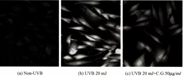
(a) N on-U V B (b ) U V B 20 m J (c) U V B 20 m J+ C .G .5 0 (ig /m /
Fig. 6 - Confocal microscopic observation of ROS by CM-H2DCFDA staining in cultured human dermal fibroblasts (HDFs). (a) C ontrol: HDFs on non-UVB, (b) exposed to 20 mj/cm2 UVB-irradiation, (c) pretreated with 50 |ig/m/
C. grandiflora
extract for 2 hrs, followed by treatment with 20 mj/cm2 UVB-irradiation, Magnification : x 400.60 50 40 30 20 10^
s
A l
>! o Oe U O U CDA COO W
(ojlDeJo
9a l u_
Vol. 49,No. 2, 2005
178 김진화 . 이범천 • Yong He Zhang • 표형배
사하였을경우세포내형광값이 급속히 중가하며피크
(
ᅳ)가오 른쪽으로이동하였으며,
능소화추출물25 ng/m/
처리 시(…)
자 외선에의해전체적인세포분포피크가높은형광을나타내던것 이컨트롤에가까운쪽으로이동하는것을확인할수있었다.
능 소화추출물5(Hig/m/
처리시(■ )에는 컨트롤과거의 비슷하게 이동하는것으로나타났다.
또한cell viability
를측정하여 실험 에사용된 세포의 상태도 정상적으로 살아있음을 확인하였다(Fig. 5).
ROS imaging using confocal microscopy
DCF
는녹색형광을나타내는것으로실제 세포가형광을띠고 있는상태를confocal microscope
로관찰하였다.
세포가살아있 는상태에서 형광을바로측정,
촬영해야하므로시간이나,
조건 이많은 영향을 미쳤으며,
현미경의 불빛으로도 형광물질의 세 기가강해지므로3
초이내에신속히 관찰 및촬영하였다.
실제 로세포관찰에서도자외선에 의해형광값이 강해짐을볼수 있 었으며,
능소화추출물에 의해 형광밝기가줄어드는것을관찰 할수있었다(Fig. 6).
피부에자외선에조사되면
0 2-, H20 2> OH
ᅵ와같은ROS
의생 성이 중가하여 피부손상을 유발시킨다. Superoxide dismutase (SOD), catalase(CAT), glutathione peroxidase(GSH-Px
)와같은ROS
소거효소들은자외선조사에 의해유발되는세포손상에 대 해보호효과가있다. Bestwick
등18>은과일이나야채와같은식 물에다량 함유된quercetin
이세포내에서ROS
에의해중가된DCF
형광값을 줄여주는효과가있음을보고하였으며,
세포생존률과
DNA
손상에 대한보호효과도확인하였다.
능소화꽃의성분으로는
apigenin
및기타배당체,폴라보노이드등이 함유되어있다고알려져 있으며
,19)
이러한성분들을함유하며 피부섬유아세포에서 자외선에 의한라디칼생성을소거시켜주는효과가 우수하게 나타나는것으로추측된다
.
결 론
능소화추출물의 유해라디칼소거효과실험 결과프리라디 칼및
superoxide
소거효과가우수하게 나타났으며,
세포내에 서ROS
에의해형광을띠는물질로전환되는CM-H2DCFDA
를이용하여
ROS
의양을측정한결과자외선에 의해중가된 세포내
ROS
의양이능소화추출물을처리함으로써50 ng/m/
농도에서
53%
이상의 우수한소거효과를나타내었으며,
공초점 현미경을이용하여 직접세포내에 나타나는
ROSSI
생성량을형광물질을이용하며 확인할수있었다
.
능소화추출물은자외선 및외부자극물질에 의해서 발생할수 있는피부손상에 대하여 효과적으로보호할수 있는우수한피 부세포보호소재로 적용될수 있을것으로사료된다
.
문 헌
1) Claude, S., Manabu, K., Laura, M . and Lester, R :
Antioxidantsmodulate acute solar ultraviolet radiation-induced
NF-kappa-B activation in a human keratinocyte cell lin e .Free Radical Biol & Med.
26,174 (1999).2) Naqui, A., Chance, B. and Cadenas, E. : Reactive oxygen intermediate in biochemistry.
Ann. Rev. Biochem.
55,137(1986).
3) Cadenas, E. : Biochemistry of oxygen toxicity.
Ann. Rev.
Biochem.
58,79 (1989).4) Davies, K. J. : Protein damage and degradation by oxygen radicals.
]. Biol. Chem.
262, 9895 (1987).5) Park, S. N. : Skin aging and antioxidant./.
Soc. Cos. Sci. Kor.
23, 75 (1997).
6) Yaar, M. and Gilchrest, B. A. : Aging versus photoaging:
postulated mechanisms and effectors.
J. Investing. Dermatol Symp. Proc.
3,47 (1998).7) Jin, J. L., Lee, Y. Y., Heo, J. E., Lee, S., Kim, J. M. and Yun- Choi, H. S .: Anti-platelet pentacyclic triterpenoids from leaves of
Campsis grandiflora. Arch. Pharm. Res.
27(4), 376 (2004).8) Blois, M. S. : Antioxidant determinations by the use of a stable free radical.
Nature
181,1199 (1958).9) Furuno, K.,Akasako, T. and Sugihara, N. : The contribution of
the pyrogallol moiety to the superoxide radical scavenging
activity of flavonoids.Boil. Pharm. Bull.
25,19 (2002).10) Seo, S. Y., Kim, E. Y , Kim, H. and Gwang, B. J. :
Neuroprotective effect of high glucose agains NMDA, free radical and oxygen-glucose deprivation through enhanced
mitochondrial potentials.]• Neurosci.
19(20),8849 (1999).11) Trayner, I. D., Rayner,A. R, Freeamn, G. E. and Farzaneh, E :
Quantitative multiwell myeloid differentiation assay using
dichlorodihydrofluorescein diacetate (H2DCFDA) or dihydro - rhodamine 123 (H2R123). /.Immunological Methods
186,275(1995).
12) Lee, B. C.’ Bae, J. T, Pyo, H. B., Choe, T. B.,Kim, S. W , Hwang, H. J. and Yun, J. W. : Biological activities of the
polysaccharides produced from submerged culture of the
edible BasidiomyceteGrifola Frondosa. Enzyme Microb.
Technol.
6274,1 (2003).13) Ryoo, Y. W, Suh, S. I., Mun, K. C.,Kim, B. C. and Lee, K. S. :
The effects of the melatonin on ultxaviolet-B irradiated
cultured dermal fibroblasts.J. Dermatol. Sci.
27, 162 (2001).14) Masaki, H., Sakaki, S., Atsumi, T. and Sakurai, H. : Activeoxygen scavenging activity of plant extracts.
Biol.
Pharm. Bull.
18, 162 (1995).15) Kuppusamy,R and Zweier, J. L. : Characterization of free radical generation by xanthine oxidase.
J. Biol. Chem.
264,9880 (1989).
J. Pharm. Soc. Korea
능소화추출물의 항산화 ^과 179
16) Ryoo, Y. W, Suh, S. I., Mun, K. C., Kim, B. C. and Lee, K. S. : The effects of the melatonin on ultraviolet-B irradiated cultured dermal fibroblasts.
J. Dermatol Science
27, 162(2001).
17) Tampo, Y., Kotamraju, S., Chitambar, C. R .,Kalivendi, S. V, Keszler’ A., Joseph, J. and Kalyanaraman, B .: Oxidative stress- induced iron signaling is responsible for peroxide-dependent
oxidation of dichlorodddihydrofluorescein in endothelial cells.
Circ. Res.
92,56 (2003).18) Bestwick, C. S. and M ilne, L. : Quercetin modifies reactive oxygen levels but exerts only partial protection against oxidative stress within HL-60 cells.
Biochim. Biophys. Acta
1528, 49 (2001).
19) 육 창 수 : 생 약 도 감 ,도 서 출 판 경 원 ,p. 486 (1997).
Vol. 49, No. 2,2005