척수원추에 발생한 혈관모세포종
- 증 례 보 고 -
고려대학교 의과대학 신경외과학교실
문수현·김세훈·권택현·박윤관·정흥섭·서중근
= Abstract =
Hemangioblastoma of the Conus Medullaris
---
- Case Report ----
Soo-Hyeon Moon, M.D., Se-Hoon Kim, M.D., Taek-Hyon Kwon, M.D., Youn-Kwan Park, M.D., Heung-Seob Chung, M.D., Jung Keun Suh, M.D.
Department of Neurosurgery, College of Medicine, Korea University, Seoul, Korea
ntramedullary spinal hemagioblastomas usually develope in cervical or thoracic region of spinal cord, but rarely in conus medullaris. We report a case of hemangioblastoma developed in conus medullaris. The 19-year-old male patient presented with slowly progressing low back pain and paresthesia of both legs. MRI and spinal angiography revealed a well-vascularized mass lesion in the conus medullaris with syrinx formation. Total excision of heman- gioblastoma was achieved via posterior approach. Postoperatively, patient’s walking difficulty was worsened transiently, but it was improved at discharge.
KEY WORDS:Conus medullaris・Hemagioblastoma・Spinal cord・Syrinx.
서 론
혈관모세포종은 내피세포(endothelial cell), 간질세포(st- romal cell), 혈관주위세포(pericyte)로 구성된 양성 종양으 로 그 기원은 혈관조직이라고 알려져 있으나7) 간질세포의 기 원에 대하여는 이견이 있다1)6)10)14). 혈관모세포종은 후두와 에 가장 빈발하나 신경계 어느 부위에서나 발생할 수 있다고 알려져 있다9). 그중 척수내 혈관모세포종은 척수내 종양의 3~8%를 차지하며 호발 부위는 경수와 흉수로 보고되고 있 다12). 하지만 본 례에서와 같이 척수원추에 발생하였다고 보 고된 례는 드물다9). 본 저자들은 척수원추에 발생한 혈관모 세포종 1례를 치험하고 문헌 고찰과 함께 보고하는 바이다.
증 례
19세 된 남자 환자로 내원 2개월 전부터 발생한 요통과 기
침을 하거나 허리를 구부릴 때 양측의 하지로 퍼지는 저린 감 각을 주소로 내원하였다. 과거력에 특이 사항은 없었으며 가 족력에도 별다른 이상은 없었다. 육안적 관찰상 기형이나 피 부에 이상 소견 등은 관찰되지 않았다. 신체 검사상 양측하 지에 약간의 근력 약화와 심부 건반사의 증가, 간대 경련이 관찰되었으며 Barbinski 증후는 없었다. 양측 하지에 약 60 도 정도의 하지 직거상 제한을 보였으나 감각 기능은 정상이 었다. 말초혈액검사, 혈액 생화학 검사, 뇨검사등 임상병리 검사에는 이상이 없었으며 심전도 검사도 정상이었다. 방사선 학적 검사 중 단순촬영에는 이상이 없었으나 흉-요추부 자기 공명 촬영상 척수원추에 1×2cm 크기의 타원형 종괴가 발 견되었으며 주위에 척수 공동증이 동반되어 있었다. 종괴는 조영이 잘되었고 종괴의 주위에 풍부한 혈관발달이 관찰되 었다. 척추혈관촬영에서 이상 증식된 혈관과 조영이 잘되는 종괴를 확인할 수 있었다(Fig. 1). 수술전 von Hippel-Lin- dau 증후군을 의심하고 시행한 안저 검사, 두부 컴퓨터 단층 촬영, 복부 초음파 검사, 24시간 뇨검사는 정상이었다.
IIII
수술은 복와위에서 정중앙 절개를 하고 흉추 11번 아후궁 절제술, 흉추 12번, 요추 1번 전 후궁 절제술을 시행하였다.
경막의 표면은 이상이 없었으나 종괴로 인해 약간 부풀려져 있었으며 경막을 절개하였을 때 척수원추가 확장되어 있었고 척수 표면으로 약간 돌출된 붉은 색의 종괴를 관찰할 수 있 었다. 정중앙 척수절개를 하고 종괴와 척수의 경계를 따라 전
종양 적출술을 시행하였으며 척수 공동증은 미미하여 별다 른 처치를 하지 않았다.
수술후 환자는 양측 하지의 근력은 정상을 보였으나 위치 감각이 저하되어 약간의 보행 장애를 보였다. 하지만 점차 호 전되어 자발적인 보행이 가능한 상태로 퇴원하였으며 외래 추적관찰 중이다.
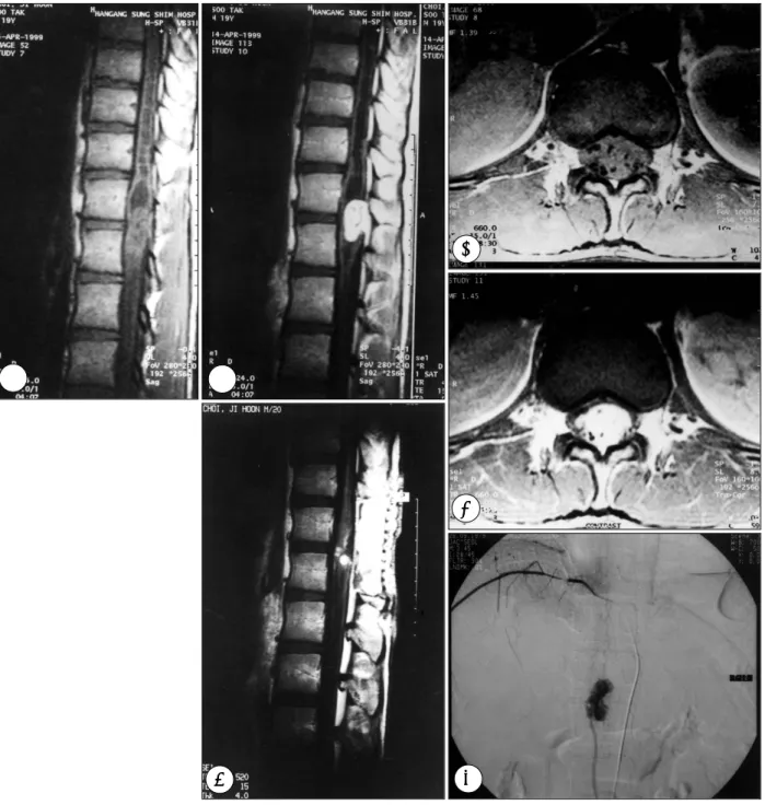
Fig. 1. A:T1-weighted MR image showing mass lesion and syrinx in the spinal cord. B:Gd-DTPA enhanced image showing remar- kably enhanced mass. C & D:Pre- and post-contrast enhanced axial MR images demonstrating a intramedullary mass with flow voids. E:Early venous phase of spinal angiography revealing tumor blush and draining vein. F:T1-weighted MR image obtained on postop 7 days showing complete removal of mass with small blood clot in the cornus medullaris and resolved syrinx.
AAA
A BBBB
F F F
F EEEE
C C C C
DDD D
조직학적 검사상 종양조직 내에 잘 발달된 혈관들과 증 식된 내피세포, 특징적인 간질세포들이 관찰되어 혈관모세포 종으로 확진되었다(Fig. 2).
고 찰
혈관모세포종은 1912년 Schultze가 처음 보고하였으며 혈 관모세포종(hemangioblastoma)이라는 병명은 1928년 Cu- shing과 Bailey가 동정맥 기형과 이를 구분하면서 사용하기 시작하였다9)16). 1943년 Wyburn-Mason이 47례를 보고한 뒤 로 Browne, Yasargil, Murota, Xu 등이 척수내 혈관모세포종 에 대하여 보고한 바 있다9)15)16). 호발 부위는 소뇌가 가장 많 고 다음으로 연수-경수 경계부위가 많다고 보고되고 있으며 그 비율은 6:4 정도라고 보고되고 있다11). 또한 척수에 발생 한 대부분의 경우는 경추와 흉추부에 발생하며 그 비율은 반 반 정도라고 보고되고 있다1)9). 본례와 같이 척수원추에 발생 한 혈관모세포종은 국내에는 보고된 바가 없으며 문헌 고찰상 검토가 가능하였던 외국 사례들1)9)15)16)에서는 63례 중 3례가 척수원추 부위에 발생하여 약 5%정도의 발생률이 추정된다.
혈관모세포종의 호발 연령은 없으나 남자에서 약간 더 호 발하며 3세 미만의 소아에서는 무척 드물다고 알려져 있다.
von Hippel-Lindau 증후군 환자의 척수내 혈관모세포종의 발생률은 약 5%정도이며 또한 척수에 발생한 혈관모세포종 의 15~25%가 von Hippel-Lindau 증후군과 관련되어 있 다고 한다11). von Hippel-Lindau 증후군과 관련된 염색체는 3번 염색체의 3p 25~26 부위라고 알려져 있으며12) VHL 종양억제 유전자(tumor suppressor gene)의 돌연변이가 관찰된다는 보고도 있다14). 따라서 혈관모세포종으로 진단 을 받은 경우 von Hippel-Lindau 증후군의 가능성을 염두
에 두고 안저검사, 복부 초음파 검사, 염색체 검사 등을 포함 한 전반적인 신체검사가 필요하다.
조직학적으로 보면 혈관모세포종은 혈관조직(angiogenic mesenchyme)7)에서 기원하는 종양으로 알려져 있으며 혈 관을 이루는 내피세포와 약간의 혈관주위세포, 및 간질세포 로 구성되어 있다. 그중 간질세포는 내피세포 기원이라는 의 견6)과 성상세포 기원이라는 의견5)이 있으며 혈관주위세포 가 섬유조직세포형 분화(fibrohistiocytic differentiation)되 어 만들어진다는 주장10)도 있으나 아직 그 기원에 대하여는 확정되지는 못한 상태이며4) 간질세포 내에 혈관내피 성장인 자(VEGF:vascular endothelial growth factor)와 태반성 성장인자(P1GF:placental growth factor)의 mRNA 증가 가 관찰되고 내피세포에 혈관내피 성장인자 수용체-1, -2, 태반성 성장인자 수용체 알파, 베타의 농도가 증가하였다고 알려져 있어 아마도 간질세포가 측분비(paracrine)의 형태로 혈관형성(angiogenesis)을 유발하는 것이 유력한 발생기전 으로 알려져 있다1)14).
척수내 혈관모세포종의 증상은 주로 추체외로 증상이 많으 나 드물지 않게 방사통을 호소하는 경우가 많다고 알려져 있 으며 그 이유로는 후근 진입부(dorsal root entry zone)를 따라 종양이 발생하는 경우가 많기 때문이다. 또한 척수공동 증이 동반되는 경우가 많아 종양의 위치와 환자의 증상이 서 로 맞지 않는 경우가 많아 진단에 주의를 요한다15).
가장 좋은 진단 방법으로는 MRI가 추천되며 척수혈관조영 술은 감별진단 및 수술의 계획에 많은 도움을 준다. 척수 MRI 소견으로는 T1 강조 영상에서 척수와 같은 정도의 신호강도 로 구형, 또는 타원형의 종괴를 보이고 종괴의 주위, 또는 내 부에 풍부하게 발달된 혈관의 신호강도가 나타나며 종괴의 주위로 척수공동증이 잘 동반되고 gadollinium의 투여로 조
Fig. 2. A:Microscopic photograph showing prominent vascular channels, endothelial cells and lipid-laden stromal cells, which are typical of hemangioblastoma(H & E, ×100). B:Immunohistochemical stain of CD34 showing positive for endothelial cells forming vascular channels(×100).
AA
AA BBBB
영증강이 잘되어 진단에는 별다른 어려움이 없다. 따라서 종 양의 진단과 추적관찰에 gadollinium을 이용한 MRI가 가장 유용한 방법으로 알려져 있다9). 본례에서도 전형적인 MRI 소견이 관찰되어 진단에 별다른 어려움이 없었다. 척수혈관조 영술은 수술전 가능하면 시행하는 것이 좋다고 하며 술전 색 전술을 시행하여 수술시 과다출혈을 예방하는 것이 추천되 나13) 일부에서는 색전술로 인한 척수의 허혈손상이 발생할 수 있어 주의하여야한다는 보고8)도 있다.
감별진단을 요하는 경우는 척수공동증, 상의세포종, 혈관기 형 등이 있다. 종양이 동반되지 않은 척수공동증과 혈관아세 포종으로 인한 척수공동증은 gadollinium을 이용한 MRI촬 영을 통해 종양을 발견함으로써 가능하며 상의세포종은 T1 강조 영상에서 혈관이 관찰되지 않는 경우가 많고 조영증강 이 잘 되지 않는 경우가 많아 감별이 가능하며 혈관기형은 척 수공동증이 동반되는 경우가 드물며 조영증강되는 종괴가 없 어 감별이 가능하다고 한다15).
치료는 종양과 척수간의 경계가 비교적 명확하고 종양 내 에 혈관발달이 풍부하여 piecemeal pattern의 수술방법은 피 하여야하며 en bloc 의 형태로 완전 제거하는 방법이 추천된 다. 수술성공의 관건은 배액정맥(drain vein)을 최후까지 보 존하고 수술초기에 영양동맥(feeding artery)을 차단하는 것 이라고 강조되고 있다15). 방사선 치료는 종양이 비교적 방사 선 내성이 강하여 추천되지는 않으나 일부 von Hippel-Li- ndau 증후군에서 관찰되는 다발성 혈관모세포종은 방사선 수 술로 좋은 결과를 보였다는 보고2)가 있어 과다 후궁 절제술로 인한 척추불안정성이 예상될 때 도움이 될 것이라고 판단된다.
혈관모세포종의 일반적인 재발 율은 약 25%정도로 알려 져 있으며 재발의 위험인자는 30세 이전 발병, von Hippel- Lindau 증후군, 다발성 등이며 조직학적으로 낭종형성이 잘 안된 경우와 간질세포의 비율이 낮은 경우 재발이 잘된다는 보고가 있다3).
결 론
본 저자들은 19세 남자환자의 척수원추에 발생한 혈관모세 포종 1례를 수술로써 치료하고 좋은 결과를 얻었기에 문헌고 찰과 함께 보고하는 바이다.
•논문접수일:1999년 10월 8일
•심사완료일:2000년 2월 1일
•책임저자:박 윤 관
152-703 서울 구로구 구로동 80번지 고려대학교 의과대학 신경외과학교실 전화:02) 818-6065, 전송:02) 863-1684 E-mail:[email protected]
References
1) Bhling T, Hatva E, Kujala M, Claesson Welsh L, Alitalo K, Ha- ltia M:Expression of growth factors and growth factor rece- ptors in capillary hemangioblastoma. J Neuropathol Exp Ne- urol 55(5):522-527, 1996
2) Chang SD, Meisel JA, Hancock SL, Martin DP, McManus M, Adler JR Jr:Treatment of hemangioblastomas in von Hippel- Lindau disease with linear accelerator-based radiosurgery.
Neurosurgery 43(1):28-34, 1998
3) de la Monte SM, Horowitz SA:Hemangioblastomas:clini- cal and histopathological factors correlated with recurrence.
Neurosurgery 25(5):695-698, 1989
4) Deck JH, Rubinstein LJ:Glial fibrillary acidic protein in stro- mal cells of some capillary hemangioblastomas:significance and possible implications of an immunoperoxidase study. Acta Neuropathol(Berl)54(3):173-181, 1981
5) Jakobiec Fa, Font RL, Johnson FB:Angiomatosis reitnae: An ultrastructural study and lipid analysis. Cancer 38:2042- 2056, 1976
6) Jurco S 3rd, Nadji M, Harvey DG, Parker JC Jr, Font RL, Morales AR:Hemangioblastomas:histogenesis of the str- omal cell studied by immunocytochemistry. Hum Pathol 13 (1):13-18, 1982
7) McComb RD, Jones TR, Pizzo SV, Bigner DD:Localization of factor VIII/von Willebrand factor and glial fibrillary acidic protein in the hemangioblastoma:implications for stromal cell histogenesis. Acta Neuropathol(Berl)56(3):207-213, 1982 8) Moseley IF, Tress B:Extravasation of contrast medium dur-
ing spinal angiography. A cause of paraplegia. Neuroradi- ology 13:55-57, 1977
9) Murota T, Symon L:Surgical management of hemangio- blastoma of the spinal cord:a report of 18 cases. Neurosur- gery 25(5):699-707, 1989
10) Nemes Z:Fibrohistiocytic differentiation in capillary hem- angioblastoma. Hum Pathol 23(7):805-810, 1992
11) Neumann HP, Eggert HR, Weigel K, Friedburg H, Wiestler OD, Schollmeyer P:Hemangioblastomas of the central nervous system:A 10-year study with special reference to von Hippel- Lindau syndrome. J. Neurosurg., 70:24-30, 1989
12) Spetzger U, Bertalanffy H, Huffmann B, Mayfrank L, Reul J, Gilsbach JM:Hemangioblastomas of the spinal cord and the brainstem:diagnostic and therapeutic features. Neurosurg Rev 19(3):147-151, 1996
13) Tampieri D, Leblanc R, TerBrugge K:Preoperative emboli- zation of brain and spinal hemangioblastomas. Neurosurgery 33(3):502-505, 1993
14) Wizigmann Voos S, Plate KH:Pathology, genetics and cell biology of hemangioblastomas. Histol Histopathol 11(4):
1049-1061, 1996
15) Xu QW, Bao WM, Mao RL, Yang GY:Magnetic resonance imaging and microsurgical treatment of intramedullary hem- angioblastoma of the spinal cord. Neurosurgery 35(4):671- 675, 1994
16) Yasargil MG, Antic J, Laciga R de Preux J, Fideler RW, Bo- one SC:The microsurgical removal of intramedullary spinal hemangioblastomas. Report of twelve cases and a review of the literature. Surg Neurol 6:141-148, 1976