258
Bakground :The evaluation of engraftment after BMT may be effectively accomplished by the analysis of genomic polymorphism, such as variable number of tandem repeat (VNTR). Discrimina- tion potential (PD) and allelic profile of VNTR locus might be varied widely between races and geo- graphic areas. Thus PCR-based VNTR loci to establish test panel useful in evaluating engraftment status of Korean patients after BMT were analyzed.
Methods :Thirty normal adults (15 males and 15 females), and each patient with acute lym- phoblastic leukemia and severe aplastic anemia who had undergone allogeneic BMT were tested.
Genomic DNAs extracted from peripheral blood lymphocytes or hair follicles were subjected to three PCR long tandem repeats (LTRs) and fifteen PCR short tandem repeats (STRs) loci analysis using silver-stain mode of detection.
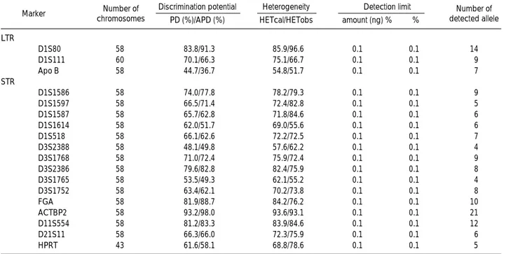
Results : The PCR sensivity of VNTR system tested, and detection limit of minor component in mixing experiment, were 100 pg and 0.1%, respectively. The most informative marker was ACTBP2 with 93.2% of PD, and 98.0% of actual PD (APD). The most informative test panel was ACTBP2, D3S2386 and D1S1768 loci-combination with 99.6% of PD and 100.0% of combined APD.
Conclusions :STRs, especially combination of ACTBP2, D3S2386, and D3S11768, were thought to be very useful screening markers for evaluating engraftment status in nonsibling allogeneic BMT. But most of allogeneic BMT are carried out between siblings, who have similar genetic mark- er each other, so further evaluation is need in sibling-BMT. (Korean J Clin Pathol 1999; 19: 258-65)
Key words :Bone marrow transplantation, Engraftment, Polymerase chain reaction, Variable number of tandem repeat
258
골수이식의 착상상태 평가를 위한 중합효소연쇄반응법에 의한 VNTR 표지자의 분석
Analysis of PCR-Based VNTR Markers to Evaluate Engraftment Status after Bone Marrow Transplantation
Hyeung Jong Lee, M.D., Gui Jeon Choi, M.D., Hyo Jin Chun, M.D., Dong Seok Jeon, M.D., and Jae Ryong Kim, M.D
Department of Clinical Pathology, Keimyung University, College of Medicine, Taegu, Korea 이형종∙최귀전∙전효진∙전동석∙김재룡
계명대학교 의과대학 임상병리학교실
서 론
골수이식은 재생불량성 빈혈 등의 골수기능부전이 있거나 백혈
병 등의 난치성 혈액질환 및 선천성 대사이상 질환의 치료로 이 용되고 있다. 이식후 환자의 골수가 공여자 세포로 완전히 치환 되는 완전 키메리즘(complete chimerism) 상태가 되면 성공적 인 골수이식이라고 볼 수 있다. 만약 숙주의 면역 혹은 암세포가 잔 존하여 혼합 키메리즘(mixed chimerism)의 상태가 되면 GVHD, 이식거부반응 및 질환의 재발 등의 합병증이 발생할 수 있다[1, 2]. 이러한 경우 대부분은 전형적인 임상양상과 혈액학적 소견을 보이지만 조기에 혼합 키메리즘 상태를 예측할 수 있다면 골수 258
258
접 수:1999년 2월 8일 접수번호:KJCP1261 수정본접수:1999년 4월 12일
교 신저 자:김 재 룡
우 700-712 대구광역시 중구 동산동 194 동산병원 임상병리과
전화 : 053-250-7221, Fax : 053-250-7275
이식후의 합병증을 미연에 방지하는데 도움이 될 것이다[3].
인간의 게놈성 DNA상에는 유전적 다형성 부위가 존재하는데 [4-7], 분자생물학적으로 공여자세포와 환자유래 세포사이의 차 이를 분석함으로써 이식골수의 착상상태를 평가할 수 있다. 다형 성 부위는 제한효소절편 길이다형성(restriction fragment length polymorphism, RFLP)[4], microsatellite 혹은 short tandem repeat (STR)[8, 9]와 minisatellite 혹은 long tandem repeat (LTR)[3, 6, 7, 10-12] 부위 등이 있는데, Mendel의 법칙에 따 라 유전되는 다양한 대립인자(allele)가 있어서 골수이식 착상상 태의 평가는 물론 친자감별 혹은 법의학적인 관계를 규명하는데 이용될 수 있다[13, 14]. STR과 LTR은 PCR법으로 간단히 검 사할 수 있다는 장점이 있다. 골수이식은 주로 혈연관계에서 이 루어지므로 골수 공여자와 환자사이에 대립인자의 양상이 유사한 경우가 많아 검사의 효율성을 높이기 위해서는 여러 표지자를 조 합하여 검사하는 것이 바람직하다[1, 2].
이 연구에서는 유전 표지자가 인종, 민족 혹은 지역에 따라 상 이할 수 있으므로, PCR-LTR 3종과 PCR-STR 15종에 대해 각 대립인자 프로파일(allelic profile)과 정보색인을 분석하여 한국인 의 골수이식 착상상태 평가를 위한 기초자료로 삼고자 하였으며, 표지자에 따른 검사 수행능력, 간편성, 경제성 등을 고려하여 선 별검사와 추가검사에 적절한 조합을 선정하고자 하였다.
재료 및 방법
1. 대상각 variable number of tandem repeat (VNTR) 표지자의 대립인자 출현빈도를 조사하기 위하여 건강한 성인 남, 여 각 15 명과 골수이식을 받은 2예의 환자를 대상으로 하였다. 환자중 1 예는 27세의 여자 환자로서 급성 림프구성 백혈병으로 진단받고 ABO형과 HLA형이 일치하는 친언니의 골수를 이식하였으나 8 개월 후 재발하였다. 항암요법을 실시한 후 GVHD가 속발하였고
골수이식 17개월 후에 PCR-VNTR 검사를 실시하였다. 다른 1 예는 22세의 여자 환자로서 중증 재생불량성 빈혈로 진단받고 ABO형과 HLA형이 일치하는 남동생의 골수를 이식받아 증세가 호전된 상태이며 골수이식 18개월 후에 검사하였다.
2. 방법
1) 게놈성 DNA의 분리
30명의 성인 남녀와 2명의 공여자와 각 환자의 골수이식 후의 말초혈액 20 mL을 채취하여 헤파린으로 처리한 다음 Ficoll- Hypaque 밀도 구배 원심분리법으로 단핵구를 분리하였다. 환자 의 골수 혹은 말초혈액 검체를 이식 전에 얻을 수 없었으므로 이 식 후에 각 환자의 모근을 10개씩 채취하였다. 말초혈액과 모근 은 phenol-chloroform법으로 DNA를 분리하였다.
2) PCR 반응
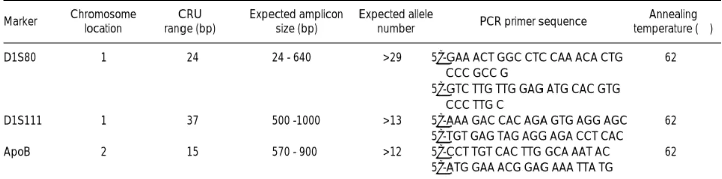
D1S80 (MCT118), D1S111 (33.6) 및 ApoB 등 LTR의 염 색체상의 위치, 공통반복단위(common repeat unit, CRU)의 크 기, 예상되는 PCR 증폭산물인 대립인자의 크기, 예상되는 대립 인자의 종류, 각 시발체의 염기서열 및 annealing 온도 등은 Table 1에 표시하였다. D1S1586, D1S1597, D1S1587, D1S1614, D1S518, D3S2388, D3S1768, D3S2386, D3S1765, D3S1752, FGA, ACTBP2, D11S554, D21S11 및 HPRT 등에 대해서는 Table 2에 표시하였다. 각 시발체는 바이오니아(주, 한국)에서 제조하여 사용하였다.
D1S80 PCR은 GeneAmp PCR Reagent Kit (Perkin Elmer, 미국)를 사용하여 다음과 같은 조건으로 시행하였다. 50 L의 PCR 반응액 [1 mM MgCl2, 0.14 mM의 dNTPs, 0.5 M 의 sense와 antisense 시발체, 2.5 단위의Taq DNA polymerase (Roche, 미국), 50 ng 표적 DNA]을 GeneAmp 9600 (Perkin Elmer, 미국) thermal cycler로 94℃에서 2분간 predenatura- tion하고, 94℃에서 20초 denaturation과 62℃에서 20초 anneal- ing하는 two-temperature 주기를 30회 반복하고 72℃에서 10분
Table 1. Characteristics of the human long tandem repeat markers used the in this study
Abbreviation: CRU, common repeat unit.
Marker Chromosome location
CRU range (bp)
Expected amplicon size (bp)
Expected allele
number PCR primer sequence Annealing temperature (℃) D1S80 1 24 24 - 640 >29 5’-GAA ACT GGC CTC CAA ACA CTG 62
CCC GCC G
5’-GTC TTG TTG GAG ATG CAC GTG CCC TTG C
D1S111 1 37 500 -1000 >13 5’-AAA GAC CAC AGA GTG AGG AGC 62 5’-TGT GAG TAG AGG AGA CCT CAC
ApoB 2 15 570 - 900 >12 5’-CCT TGT CAC TTG GCA AAT AC 62
5’-ATG GAA ACG GAG AAA TTA TG
간 postelongation하였다. ApoB와 D1S111 PCR은 PCR 반응액 (0.2 mM dNTPs, 2 mM MgCl2, 0.25 M sense와 antisense 시발체)을 D1S80과 동일한 조건으로 증폭하였다. D1S1586, D1S1597, D1S1587, D1S1614, D1S518, D3S2388, D3S1768, D3S2386, D3S1765, D3S1752, FGA, ACTBP2, D11S554, D21S11 및 HPRT 등 STR 표지자의 PCR은 50 L의 PCR 반 응액 (1.5 mM MgCl2, 0.2 mM dNTPs, 0.5 M sense와 antisense 시발체, 1단위Taq DNA polymerase, 50 ng 표적 DNA)를 94℃에서 2분간 predenaturation하고, 94℃에서 30초간 의 denaturation과 Table 2에 명시된 각 표지자의 온도에서 30초 간 annealing, 72℃에서 30초 elongation하는 two-temperature 주기를 30회 반복한 후 72℃에서 10분간 postelongation하였다.
LTR은 native polyacrylamide gel에서 Econo Sequencer 전 기영동기[바이오니아(주), 한국]로 분석하였다. 6% native polyacrylamide gel에 PCR product를 gel loading buffer (0.25% bromophenol blue, 0.25% xylene cyanol FF, 15%
Ficoll type 400)와 같이 loading하여 1×TBE 완충액으로 300
volt에서 5시간 혹은 200 volt에서 12시간 수직상태로 전기영동 하였다. STR는 8 M urea가 포함된 6% denaturing polyacry- lamide gel로 분석하였으며, PCR product를 formamide loading buffer[80% formamide, 10 mM EDTA (pH8.0), 1 mg/mL xylene cyanol FF, 1 mg/mL bromophenol blue]와 혼합하여 loading하고 1×TBE 완충액으로 1,200 volt에서 2시간내지 3시 간 수직상태로 전기영동하였다. 전기영동 후 DNA band는 Sil- ver Stain Kit [바이오니아(주), 한국]로 은염색하여 가시화 하 였다. VNTR 표지자의 대립인자 프로파일은 scan하여 분석하였다.
3) PCR 검출 예민도 조사 및 혼합실험 검출 한계
각 VNTR 표지자의 대립인자 검출 예민도를 보기 위하여 표 적 DNA 양을 10 ng, 1 ng, 100 pg, 10 pg, 1 pg, 0.1 pg 및 blank로 조절하여 PCR을 수행하였다. 또한 골수 공여자와 환자 유래 세포를 혼합하였을 때 VNTR 표지자의 minor component 에 대한 PCR 검출한계를 규명하기 위하여 혼합시험(mixing experiment)을 실시하였다. 실험방법은 PCR용액에 환자 DNA Table 2.Characteristics of the human short tandem repeat markers used the in this studyused the in this study
Abbreviation: See Table 1.
Marker Chromosome
location CRU Expected amplicon size (bp)
Expected allele
number PCR primer sequence Annealing temperature (℃) D1S1586 1 GTA 91-118 8 5’-AGA AAC AAA CAG TTC CAT TGC-3’ 55
5’-TCA AAG TTC TTT CTG TTT CAG TG-3’
D1S1597 1 TATC 155-179 6 5’-TTT ATT GAG ATA TAT TTG ACA TGC G-3’ 55 5’-AAG GAG GAA GCT TTT TGG A-3’
D1S1587 1 ATA 144-168 6 5’-TTG CTT GAA CCC AGA GGA C-3’ 57 5’-CAA GTC TCC CCA TGG TAG TG-3’
D1S1614 1 TTCC 268-293 8 5’-ATC TGA GGT GGA GAC CTT CC-3’ 65 TCCC 5’-CAC AAG ACA GAG TAG AGA AAC AGC-3’
D1S518 1 GATA 192-223 8 5’-TGC AGA TCT TGG GAC TTC TC-3’ 65 5’-AAA AAG AGT GTG GGC AAC TG-3’
D3S2388 3 GATA 101-125 4 5’-TCC ACT CAG TAC AGT CTG GG-3’ 62 5’-GAT CCT GTG GTT GTC GAT C-3’
D3S1768 3 GATA 186-206 7 5’-GGT TGC TGC CAA AGA TTA GA-3’ 60 5’-CAC TGT GAT TTG CTG TTG GA-3’
D3S2386 3 GATA 274-310 5 5’-AAG CTG TCC ATA CCA CAG CA-3’ 60 5’-TAC ATC ATC AGC TCT CTT GGG-3’
D3S1765 3 GATA 192-212 - 5’-TAT GGT GGT GCA TGA CTG TC-3’ 57 5’-TGC ACC CAG CCA TAA TAT CT-3’
D3S1752 3 ATC 189-209 - 5’-ACA GTC ACA TAC CTG GTA CTG-3’ 55 5’-CTG AGA AGC TGA GCC ACA TG-3’
FGA 4q28 TCTT 265 -297 11 5’-CCATAG GTT TTG AAC TCA CAG-3’ 55 5’-CTT CTC AGA TCC TCT GAC AC-3’
ACTBP2 6 AAAG 229 -337 34 5’-AAT CTG GGC GAC AAG AGT GA-3’ 60
D11S554 11p12 A3G/AAGG 177 - 281 8 5’-GGT AGC AGA GCA AGA CTG TC-3’ 55
-p11.2 5’-CAC CTT CAT CCT AAG GCA GC-3’
5’-ACA TCT CCC CTA CCG CTA TA-3’
D21S11 21q21 TCTA 197 -241 13 5’-GTG AGT CAA TTC CCC AAG-3’ 60
ATCT 5’-GTT GTA TTA GTC AAT GTT CTC C-3’
HPRT Xq26 ATCT 269 -297 44 5’-CTC TCC AGA ATA GTT AGA TGT AGG-3’ 55 5’-ATG CCACAG ATA ATA CAC ATC CCC-3’
를 100 ng으로 일정하게 조정하고 공여자의 DNA를 10 ng, 1 ng, 100 pg, 10 pg 및 1 pg되게 10배 연속 희석하여 혼합한 다 음 PCR을 수행하여 minor component 즉 공여자 세포의 검출 한계를 조사하였다.
3. VNTR 표지자의 정보색인 분석
각 VNTR 표지자의 공여자와 환자유래 세포의 키메리즘 상태 를 감별할 수 있는 능력을 보기 위하여, probability of discrimi- nation (PD), combined PD (PDcom), calculated heterozy- gosity (HETcal), observed heterozygosity (HETobs), actual power of discrimination (APD) 및 combined APD (APD- com) 등의 정보색인을 산출하였다[1, 15, 16].
결 과
1. VNTR 표지자의 PCR 검출 예민도와 검출 한계
Fig. 1과 Table 3에서와 같이 D3S1768의 검출 예민도는 100 pg이었으며, 기타 3종의 LTR과 14종 STR의 검출 예민도도 모 두 100 pg이었다. Fig. 2에서와 같이 D3S1768에 대해 공여자 세
포의 DNA 농도를 100 ng으로 일정하게 하고 환자 세포의 DNA를 혼합하였을 때 minor component인 환자 세포의 검출 한계는 0.1%이었으며, 기타 3종의 LTR과 14종 STR의 검출 한 계도 모두 동일하였다.
2. VNTR 표지자의 대립인자 분석 특성
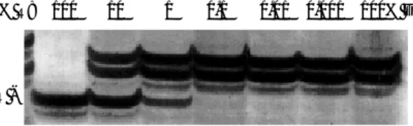
ACTBP2는 인척관계가 없는 29명의 한국인 남녀(남녀 각 15 명의 조사대상자 중 여자 1명은 PCR에서 DNA band가 증폭되 지 않아 제외시켰음), 즉 58개의 염색체를 분석한 결과 가장 많 은 21종의 대립인자가 관찰되었으며, 각 대립인자의 종류와 출현 빈도는 Fig. 3에 명시하였다. D3S2386은 9종의 대립인자가 관찰 되었으며, 각 대립인자의 종류 및 출현빈도는 Fig. 4에 명시하였 다. 기타 13종의 STR, 3종의 LTR의 대립인자 종류 및 HETcal, HETobs와 PD, APD는 Table 3에 요약하였다.
Fig. 1. Estimation of sensitivity of D3S1768 VNTR analysis. The DNA that was homozygote in polymorphic alleles at the D3S1768 polymorphism was used to demonstrate the potential sensitivity of this technique. DNA was serially diluted from 10 ng to 0.1 pg.
The D3S1768 VNTR was analyzed by PCR. After PCR, the prod- ucts were resolved using 6% polyacrylamide containing 8 M urea denaturing gel electrophoresis. The bands were visualized by silver staining.
10 ng 1 ng 100 pg 10 pg 1 pg 0.1 pg Blank
Fig. 3.PCR amplification of tetranucleotide (AAAG) repeat poly- morphism at the human (Korean) beta-actin related pseudogene ACTBP2 (H-beta-Ac-psi-2) on chromosome 6. The predicted length of amplifed sequence was expected to begin at base pair 176. The PCR products was analyzed by the method described in Fig 1. Various informative indices, which were estimated from 58 chromosomes of unrelated 29 Korean individuals, were as fol- lows: calculated heterozygosity (HETcal) = 93.6%, observed het- erozygosity (HETobs) = 93.1%, probability of discrimination (PD)
= 93.2%, actual power of discrimination (APD) = 98.0%. And number of observed alleles was 21, and allele types and fre- quencies were as follows: E (0.017), F (0.017), G (0.068), H (0.068), I (0.103), J (0.689), K (0.034), L (0.051), M (0.051), N (0.034), O (0.051), P (0.086), Q (0.034), R (0.017), S (0.017), T (0.068), U (0.103), V (0.034), W (0.034), X (0.034), and Y (0.017).
PR MT OS FN EK JQ IJ PV HW HI PU IJ GG KU IT HO IU QX LY MT UU PW GT GI OP MV JN LU HO Genotype
Fig. 4.PCR amplification of tetranucleotide (GATA) repeat poly- morphism at the human (Korean) D3S2386 gene on chromo- some 3. The genotypes are noted at the bottom of the figure. The predicted length of the amplified sequence was 274 - 310 bp.
The PCR products was analyzed by the method described in Fig. 1. Various informative indices, which were estimated from 58 chromosomes of unrelated 29 Korean individuals, were as fol- lows: HETcal = 82.4%, HETobs = 75.9%, PD = 79.6%, APD = 82.8%. And number of observed alleles was eight, and allele types and frequencies were as follows: H (0.0172), I (0.069), J (0.103), K (0.190), L (0.241), M (0.224), N (0.121) and O (0.034).
JM JM MM KL JN LL JN IL KM LL KM KN JM JN IL LO MM KL LN KL IN KL MM IL HO KL KN KK MM Genotype
Fig. 2. Estimation of sensitivity of D3S1768 VNTR analysis by mix- ing experiment. Two DNA specimens that differ in polymorphic alleles at the D3S1768 polymorphism were used to demonstrate the potential sensitivity of this technique. DNA from the recipient (R) is diluted in DNA from the donor (D) in the indicated propor- tions from 10% R to 0.001% R. The PCR products were resolved and visualizde by the method described in Fig. 1.
% R:
R -
100 10 1 0.1 0.01 0.001 100% D
3. 골수이식 착상상태 평가를 위한 VNTR 표지자의 선정
정보제공률은 STR인 ACTBP2가 가장 높았고, LTR인 D1S80, FGA, D11S554, D3S2386, D1S1586, D3S1768, D1S1597 및 D1S111의 순이었다. 이러한 VNTR 표지자가 골수 이식 착상상태평가를 위한 검사로 이용되기 위해서는 가능하면 검사조건이 서로 같고 검사결과의 판독이 용이해야 하며, 소수의 조합검사로 정보제공률이 높아야 한다. 이러한 조합을 Table 4에
요약하였다. ACTBP2가 정보제공률이 가장 높았고, 은염색 결과 DNA band의 판독성이 매우 좋았으므로 이 VNTR 표지자와 PCR 조건과 PCR 증폭산물의 분석조건이 동일한 D3S2386, D3S1768과의 조합이 일차 선별검사로 적합하다고 판단되었으며, PDcom/ APDcom은 99.6%/100.0%였다(Table 5). 일차검사 VNTR 표지자로 환자유래 세포와 공여자 세포를 감별할 수 없 을 때 실시하는 추가검사의 조합인 D1S80, D1S11과 ApoB의 PDcom/ APDcom은 97.2%/97.8%이었으며, D1S80, D1S111, ApoB, FGA와 D11S554의 PDcom/APDcom은 99.9%/100.0%
Table 3.Discrimination potential and other characteristics of VNTR markers
Abbreviations: PD, probability of discrimination; APD: actual power of discrimination; HETcal, calculated heterozygosity; HETobs, observed heterozy- gosity.
Marker Number of chromosomes
Number of detected allele Discrimination potential
PD (%)/APD (%) LTR
D1S80 58 83.8/91.3 85.9/96.6 0.1 0.1 14
D1S111 60 70.1/66.3 75.1/66.7 0.1 0.1 9
Apo B 58 44.7/36.7 54.8/51.7 0.1 0.1 7
STR
D1S1586 58 74.0/77.8 78.2/79.3 0.1 0.1 9
D1S1597 58 66.5/71.4 72.4/82.8 0.1 0.1 5
D1S1587 58 65.7/62.8 71.8/84.6 0.1 0.1 6
D1S1614 58 62.0/51.7 69.0/55.6 0.1 0.1 6
D1S518 58 66.1/62.6 72.2/72.5 0.1 0.1 7
D3S2388 58 48.1/49.8 57.6/62.2 0.1 0.1 4
D3S1768 58 71.0/72.4 75.9/72.4 0.1 0.1 9
D3S2386 58 79.6/82.8 82.4/75.9 0.1 0.1 8
D3S1765 58 53.5/49.3 62.1/55.2 0.1 0.1 4
D3S1752 58 63.4/62.1 70.2/73.8 0.1 0.1 8
FGA 58 81.9/88.7 84.2/76.2 0.1 0.1 10
ACTBP2 58 93.2/98.0 93.6/93.1 0.1 0.1 21
D11S554 58 81.2/83.3 83.9/84.6 0.1 0.1 12
D21S11 58 66.3/66.0 72.3/75.9 0.1 0.1 6
HPRT 43 61.6/58.1 68.8/78.6 0.1 0.1 5
Heterogeneity HETcal/HETobs
Detection limit amount (ng) % %
STR
ACTBP2 98.0 60 Denature gel Excellent FGA 88.7 55 Denature gel Good D11S554 83.3 55 Denature gel Good D3S2386 82.8 60 Denature gel Excellent D1S1586 77.8 55 Denature gel Good D3S1768 72.4 60 Denature gel Excellent D1S1597 71.4 55 Denature gel Good LTR
D1S80 91.3 62 Native gel Excellent D1S111 66.3 62 Native gel Good Locus APD
(%)
Annealing tempera- ture of PCR (℃)
Detection of PCR product EP condition Silver-stain quality Table 4. Characteristics of useful VNTR markers which can be selected as tests for evaluation of engraftment status of BMT
Abbreviations: APD, actual power of discrimination; EP: electrophore- sis.
Screening test
ACTBP2 93.2 98.0
ACTBP2 + D3S2386 98.7 99.8 ACTBP2 + D3S2386 + D3S1768 99.6 100.0 Additional test
D1S80 83.8 91.1
D1S80 + D1S111 95.0 97.5
D1S80 + D1S111 +ApoB 97.2 97.8 D1S80 + D1S111 +ApoB +FGA 99.5 99.5 D1S80 + D1S111 +ApoB +FGA +D11S55 499.9 100.0 Combination PDcom (%) APDcom (%) Table 5. Actual power of discrimination in combination of VNTR markers
Abbreviations: PDcom, combined probability of discrimination; APD- com: combined actual power of discrimination.
이었다(Table 5).
4. 골수이식환자의 착상상태 평가
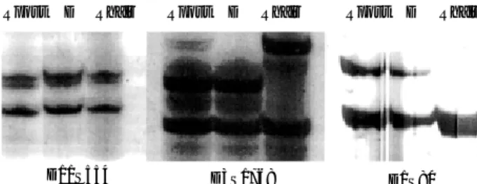
Fig. 5에서와 같이 D11S554는 공여자 세포(D), 골수이식 후 환자유래 세포(Rpost)와 환자의 모근세포(Rhair)가 모두 동일한 양상의 이형접합체로 키메리즘 상태의 평가가 불가능하였다.
D1S80와 D3S1768는 모두 Rhair와 D는 서로 다른 이형접합체 양상을 보여 이식 후 환자상태인 Rpost의 키메리즘 상태의 평가 가 가능하였는데, D1S80은 대립인자 band의 양상이 Rpost가 D 와 같이 완전 키메리즘의 양상을 보였으나 D3S1768 표지자에서 Rpost에 약한 Rhair의 대립인자 band가 관찰되어 혼합 키메리즘 으로 판정할 수 있었다. Fig. 6에서와 같이 재생불량성 빈혈 환자 는 D11S554, D1S80 및 D3S1768로써 골수이식 착상상태를 평가 하였다. 모든 표지자가 D와 Rhair의 band 양상이 모두 상이한 표지자였으며 Rpost의 대립인자 band의 양상이 D와 같아 완전 키메리즘으로 판정할 수 있었다.
고 찰
동종 골수이식은 백혈병과 재생불량성 빈혈 등 혈액학적 질환
에 유용한 치료법으로써 비교적 흔히 시도된다. 이식 후 환자의 골수는 이식세포의 착상상태에 따라 완전 키메리즘, 혼합 키메리 즘, 이식실패 등의 3가지 유형의 키메리즘 상태를 나타낸다[1].
혼합 키메리즘 상태가 반드시 이식이 실패하였다거나 백혈병의 재발을 뜻하지는 않지만, 완전 키메리즘 상태가 되면 환자의 예 후가 비교적 좋으며 성공적인 골수이식의 지표가 될 수 있다. 착 상상태를 평가할 수 있는 유전적 표지자로는 세포 유전학검사, 적혈구 형별 검사와 HLA 형별 검사 및 DNA 염기서열 다형성 분석 등이 있으며[2], 그 유형에 따라 예민도와 정보제공률에 차 이가 있을 수 있다[1, 5, 7, 17].
게놈성 다형성을 보이는 DNA 표지자로는 RFLP 표지자와 VNTR 표자자가 있다. RFLP 표지자는 사람의 게놈성 DNA를 제한효소로 처리하였을 때 DNA의 절편 크기가 다형성을 보이는 것으로 southern blot법으로 가시화 할 수 있다. 따라서 검사가 번거로우며, 혼합실험에서 minor component의 검출 한계는 비 교적 낮은 1%에서 10%정도로서 약 106개의 세포에 해당한다 [4]. VNTR 표지자는 동일한 DNA 염기서열이 반복되는 구조 이며, 반복회수에 따라 다양한 대립인자 양상을 보이며, 반복구조 CRU의 염기수에 따라 microsatellite 혹은 STR과 minisatellite 혹은 LTR로 구별할 수 있다. STR은 CRU의 반복 염기수는 2-5 개, LTR의 경우 8-70개이며[8, 9, 11, 13, 18-20], 여러 염색체 에 다양하게 분포되어 있다. VNTR 대립인자는 30여종 이상 고 도의 다형성을 보이는 경우도 있으며, PCR법으로 검사가 가능하 다[1]. 그러므로 250 ng 정도의 적은 량의 DNA로도 검사가 가 능하며 제한효소의 처리가 필요 없어 RFLP법에 비해 신속하고, 예민도는 선택되는 표지자의 종류에 따라 상이할 수 있지만, minor component의 검출 한계는 대체로 0.05%에서 1.0%의 수 준이며, 이 때 표적 DNA의 양은 25에서 50 ng이다[1, 2, 5, 12].
본 연구에서 골수이식 착상상태를 평가를 위해 분석한 표지자 는 LTR 3종과 STR 15종을 PCR법으로 증폭하고 은염색법에 의해 각 대립인자의 DNA band를 확인하였다. 각 표지자의 PCR 검출 예민도는 표적 DNA의 농도로 볼 때 100 pg이었으며, 혼합실험에서 minor component의 검출 한계는 0.1%로서 다른 보고[1, 2, 5]와 거의 일치하였다. 또한 각 VNTR의 대립인자 프로파일 양상과 정보색인은 백인을 주 대상으로 조사한 다른 보 고[1, 2, 5]와 비교해 볼 때 약간의 차이가 있었다. 백인에 있어 서는 ApoB가 82.1%의 APD값을 나타내어 유용성이 높은 표지 자로 평가되었으나[5], 본 연구에서는 36.7%로서 유용성이 매우 낮은 표지자로 평가되었다. 그리고 백인의 경우 FGA, ACTBP2, D11S554, D21S11 및 HPRT의 PD가 각각 95.3%, 99.4%, 98.8%, 94.3% 및 89.9%이었으며 allele이 각각 10, 44, 34, 13 및 8개이었는데, 본 연구에서는 PD가 각각 81.9%, 93.2%, 81.2%, 66.3% 및 61.6%이었으며 allele이 각각 10, 21, 12, 6 및 5개로서 ACTBP2를 제외한 다른 표지자들의 정보제공률은 백인 에 비해 비교적 낮음을 알 수 있었다. 이는 각 대립인자의 특성이 민족 혹은 지역에 따른 차이일 수도 있겠으나, 본 연구의 조사 대 Fig. 5.DNA typing of patient with acute lymphoblastic leukemia
(ALL), after 17 months of BMT. Selected markers were shown, in which D11S554 was not informative, D1S80 was represented by complete chimerism, and D3S1768 was represented by mixed chimerism.
Abbreviations: Rpost, recipient peripheral blood after BMT; D, donor peripheral blood; Rhair, recipient hair follicle after BMT.
Rpost
D11S554 D3S1768 D1S80
Rpost Rpost
D Rhair D Rhair D Rhair
Fig. 6.DNA typing of patient with aplastic anemia, after 18 months of BMT. Selected informative markers were shown such as D11S554, D3S1768 and D1S80.
Abbreviations: See Fig. 5.
Rpost
D11S554 D3S1768 D1S80
Rpost Rpost
D Rhair D Rhair D Rhair
상자 수가 적었기 때문일 수 있으므로 보다 많은 대상자를 조사 할 필요가 있다고 생각된다.
각 VNTR 표지자의 정보색인은 표지자에 따라 다양한 양상을 보이고 있다. 비혈연관계의 법의학적인 데이터베이스에서 추출한 각 VNTR 표지자의 대립인자 출현빈도의 양상을 볼 때 RFLP 법에 비해 구별능력이 낮음을 알 수 있으며[1], RFLP법과 동등 한 정보제공률을 나타내기 위해서는 여러 PCR VNTR 표지자를 조합하여 검사할 필요가 있으며, 효율을 극대화시키기 위해 선별 검사와 추가검사로 구분하여 적절한 표지자를 선정해야 될 필요 성이 있다. 본 연구에서는 ACTBP2, FGA, D11S554, D3S2386, D1S1586, D3S1768 및 D1S1597 등의 STR과 LTR인 D1S80과 D1S111이 골수이식 착상상태를 평가하는데 있어서 정보제공률이 높 은 표지자들이었다. 이러한 VNTR 표지자 중 ACTBP2, D3S2386, D21S11의 STR 조합이 PDcom/APDcom이 99.6%/100.0%로 정보제공률이 매우 높았으며 PCR 분석조건이 동일하고 은염색 결과 DNA band의 판독성이 좋아 골수이식 착상상태 평가를 위 한 선별검사로 가장 바람직한 것으로 판단되었다. 이차 혹은 확 인검사로서는 D1S80, D1S111, ApoB LTR조합이나 D1S80, D1S111, ApoB, FGA, D11S554 조합이 우수한 것으로 평가되 었다.
혼합 키메리즘을 증명하기 위해서는 일정한 시간간격을 두고 연속적으로 DNA 표지자 검사를 하는 것이 가장 바람직하지만, 환자의 골수이식전 가검물이나 공여자의 가검물을 구할 수 없는 경우와 같이 임상적인 여건상 single-point 검사가 필요한 경우도 있다[4]. 법의학적 검사나 친자감별검사는 두 검체가 서로 동일 하다는 것을 증명하는 것이지만, 골수이식에서 키메라즘 상태를 규명하는 것은 두 검체가 서로 다르다는 것을 증명하는 것이므로 비교의 지표가 되는 환자의 골수이식전 가검물이나 공여자의 가 검물이 없으면 감별이 힘들며, 특히 공여자와 환자의 성별이 동 일한 골수이식에서 그러하다. 골수이식 전과 이식 후에 환자의 골수 혹은 말초혈액을 확보해 두는 것이 가장 바람직하지만, 많 은 경우의 골수이식이 DNA 표지자 분석에 대한 계획이 없이 시 행되고 있다. 그러므로 검사실에서는 환자의 이식 전 혈액도말 슬라이드이나 이식 전 환자의 가검물로 대신할 수 있는 체세포 가검물을 확보하여야 한다. 체세포로서 검체의 채취가 쉬운 것으 로는, 피부생검조직[21], 구강탈락세포[22] 혹은 모근(hair folli- cle)[1] 등이 있으며, 피부생검조직이나 구강탈락세포는 환자의 골수가 혼합 키메리즘을 보일 때 동일한 양상의 혼합 키메리즘을 나타내 이식전 환자의 골수를 대신할 수 없는[1, 21, 22] 반면, 모근은 이와 같은 현상을 보이지 않으며 충분한 양의 DNA를 비 교적 쉽게 확보할 수 있다[1]. 본 연구에서도 급성 림프구성 백 혈병 및 재생불량성 빈혈 환자의 골수이식전 가검물을 확보하지 못하여 환자의 모근을 이용하였는데, 10개 정도의 모발을 채취하 였을 때 700에서 1,000 ng 정도의 충분한 DNA를 확보할 수 있 었다.
환자가 혼합 키메리즘 상태일 때 표지자에 따라서는 minor
component를 검출하지 못하는 경우가 있다. 이러한 이유는 첫째, RFLP법의 경우 DNA band를 관찰하기 위해 표지자의 종류에 따라 방사선 동위원소의 노출시간을 조절해야 되는데, 노출시간 이 짧은 경우 minor component를 검출하지 못할 수 있다. 둘째, DNA band 크기의 차이가 뚜렷하지 않는 경우 구별이 어렵다.
셋째, PCR의 경우 표적 DNA의 크기에 따라 증폭효율의 차이가 있을 수 있는데, minor component의 표적 DNA의 크기가 major component에 비해 클 경우 증폭되지 않을 수 있다. 본 연구에서 도 혈연관계에서 동종 골수이식이 실시된 급성 림프구성 백혈병 환자의 경우 D1S80와 D3S1768로써 골수이식의 착상상태를 평가 한 결과 D3S1768에서만 minor component을 관찰할 수 있었다.
이런 현상을 방지하기 위해서 표적 DNA의 농도를 낮추거나 크 기가 다른 표적 DNA의 증폭효율을 조절하기 위해 화학물질을 PCR 반응에 첨가할 수 있지만 그 효과는 뚜렷하지 않다.
요 약
배경 : 골수이식에 있어서 이식한 골수의 착상상태를 평가하기 위한 검사로서 variable number of tandem repeat (VNTR) 표지자가 유용성이 높다. 그러나 VNTR 표지자의 특성은 민족 혹은 지역에 따라 상이할 수 있으므로 이 연구에서는 PCR법으로 3종의 long tandem repeat (LTR)과 15종의 short tandem repeat (STR)에 대해 각 VNTR 표지자의 대립인자 프로파일 (alleleic profile)과 정보색인(informative index)을 분석하고 한 국인의 골수이식 착상상태를 평가하기에 적절한 검사방법을 모색 하고자 하였다.
방법 : 연구대상은 정상성인 남녀 각 15명과 골수이식을 시행 한 27세의 급성 림프구성 백혈병 환자 1명과 22세의 중증 재생불 량성 빈혈 환자 1명으로 하였다. PCR은 LTR 3종과 15종의 STR 표지자에 대해 실시하였다. PCR 증폭산물의 분석은 poly- acrylamide gel electrophoresis법으로 분리하고 은염색으로 각 DNA 분획을 가시화 하였다.
결과 : 각 VNTR 표지자의 PCR 검출 예민도는 게놈성 DNA 100 pg이었으며, 혼합실험에 의한 minor component의 검출 한 계는 major component의 0.1%였다. 대립인자 프로파일을 분석 한 결과 STR인 ACTBP2가 HETcal/HETobs가 93.6%/93.1%, PD/APD가 93.2%/98.0%로서 정보제공률이 가장 높았으며, ACTBP2, D3S2386과 D3S1768의 STR 조합이 PDcom/APD- com이 99.6%/100.0%로 정보제공률이 매우 높아, 골수이식 착상 상태 평가를 위한 일차 혹은 선별검사로 적합한 panel인 것으로 판단되었다.
결론 : ACTBP2, D3S2386, D3S1768 등의 VNTR 표지자 들은 한국인에 있어서 비혈연관계의 골수이식 착상상태 평가와 친자감별이나 법의학적 조사에 유용성이 있을 것으로 생각된다.
그러나 우리나라에서의 골수이식이 주로 혈연관계에서 실시되므
로 상기 VNTR 표지자들의 혈연간 골수이식에 있어서의 유용성 에 대한 조사가 이 연구를 바탕으로 이루어져야 할 것으로 사료 된다.
참고문헌
1. Leclair B, Freau CJ, Aye MT, Fourney RM. DNA typing for bone marrow engraftment follow-up after allogeneic transplant: a comparative study of current technologies. Bone Marrow Transplant 1995; 16: 43-55.
2. Frankel W, Chan A, Corringham RET, Shepherd S, Rearden A, Wang-Rodriguez J. Detection of chimerism and early engraftment after allogeneic peripheral blood stem cell or bone marrow transplantation by short tandem repeats. Am J Hematol 1996; 52: 281-7.
3. O’Reilly J, Meyer B, Stoner M, Erber W, Herrmann R, Davies J.
Very early analysis of graft establishment after allogeneic bone marrow transplantation using the polymerase chain reaction. Br J Haematol 1993;
85: 169-72.
4. Ginsburg D, Antin JH, Smith BR, Orkin SH, Rappeport JM. Origin of cell populations after bone marrow transplantation: analysis using DNA sequence polymorphims. J Clin Invest 1985; 75: 596-603.
5. Ugozzoli L, Yam P, Petz LD, Ferrara GB, Champlin RE, Forman SJ, et al. Amplification by the polymerase chain reaction of hypervariable regions of the human genome for evaluation of chimerism after bone mar- row transplantation. Blood 1991; 77: 1607-15.
6. MacKinnon S, Barnett L, Heller G. Polymerase chain reaction is highly predictive of relapse in patients following T cell-depleted allogeneic bone marrow transplantation for chronic myeloid leukemia. Bone Marrow Transplant 1996; 17: 643-7.
7. Roux E, Helg C, Chapuis B, Jeannet M, Roosnek E. Evolution of mixed chimerism after allogeneic bone marrow transplantation as deter- mined on granulocytes and mononuclear cells by the polymerase chain reaction. Blood 1992; 79: 2775-83.
8. Armour JA and Jeffreys AJ. Recent advances in minisatellite biology (Lett). FEBS 1992; 307: 113-5.
9. Edwards A, Hammond HA, Jin L, Caskey CT, Chakraborty R.
Genetic variation at five trimeric and tetrameric tandem repeat loci in four human population groups. Genomics 1992; 12: 241-53.
10. Budowle B, Chakraborty R, Giusti AM, Eisenberg AJ, Allen RC.
Analysis of the VNTR locus D1S80 by the PCR followed by high-resolu- tion PAGE. Am J Hum Genet 1991; 48: 137-44.
11. Beckmann JS and Weber JL. Survey of human and rat microsatellites.
Genomics 1992; 12: 627-31.
12. Van Leeuwen JEM, van Tol MJD, Bodzinga BG, Wijnen JT, van der Keur M, Joosten AM, et al. Detection of mixed chimerism in flow-sorted cell subpopulations by PCR-amplified VNTR markers after allogeneic bone marrow transplantations. Br J Haematol 1991; 79: 218-25.
13. Bever RA. Characterization of five VNTR loci by Hae III RFLP analysis:
application to paternity testing, In: Britten C, ed. Proc 2nd Intern Sympos Hum Identif Promega, edition New York; Madison W1 USA, 1991: 103-28.
14. Sajantila A, Makkonen K, Ehnholm C, Peltonen L. DNA profiling in a genetically isolated population using three hypervariable DNA markers.
Hum Hered 1992; 42: 372-9.
15. Jones DA. Blood samples: probability of discrimination. J Forens Sci Soc 1972; 12: 355-9.
16. Budowle B, Giusti AM, Waye JS, Baechtel FS, Fourney RM, Adams DE, et al. Fixed-bin analysis for statistical evaluation of continuous distri- butions of allelic data from VNTR loci, for use in forensic comparisons.
Am J Hum Genet 1991; 48: 841-55.
17. MacKinnon S, Barnett L, Bourhis JH, Black P, Heller G, O’Reilly RJ.
Myeloid and lymphoid chimerism after T-cell-depleted bone marrow transplantation: evaluation of conditioning regimens using the poly- merase chain reaction to amplify human minisatellite regions of genomic DNA. Blood 1992; 80: 3235-41.
18. Kasai K, Nakamura Y, White R. Amplification of a variable number of tandem repeats (VNTR) locus (pMCT118) by the polymerase chain reac- tion (PCR) and its application to forensic science. J Forensic Sci 1990; 35:
1196-200.
19. Wu S, Senio S, Bell GI. Human collagen, type II, alpha 1 (Col2A1) gene:
VNTR polymorphism detected by gene amplification. Nucleic Acids Res 1990; 18: 3102.
20. Fregeau CJ and Fourney RM. DNA typing with fluorescently tagged short tandem repeats: a sensitive and accurate approach to human identifi- cation. BioTechniques 1993; 15: 100-9.
21. Hansen-Hagge TE, Yokota S, Bartram CR. Detection of minimal residual disease in acute lymphoblastic leukemia by in vitro amplification of rearranged T-cell receptor chain sequences. Blood 1989; 74: 1762-7.
22. Roth MS, Antin JH, Bingham EL, Ginsburg D. Use of polymerase chain reaction-detected sequence polymorphisms to document engraft- ment following allogeneic bone marrow transplantation. Transplantation 1990; 49: 714-20.