Assessment of Clinical, Electrocardiographic, and Physiological Relevance of Diagonal Branch in Left Anterior Descending Coronary Artery Bifurcation Lesions
Bon-Kwon Koo, MD, P
HD,* Seung-Pyo Lee, MD,* Ju-Hee Lee, MD,*
Kyung-Woo Park, MD, P
HD,* Jung-Won Suh, MD, P
HD,† Young-Seok Cho, MD, P
HD,†
Woo-Young Chung, MD, P
HD,‡ Joon-Hyung Doh, MD, P
HD,§
Chang-Wook Nam, MD, P
HD,储 Cheol Woong Yu, MD, P
HD,¶ Bong-Ki Lee, MD, P
HD,#
Dobrin Vassilev, MD,** Robert Gil, MD,†† Hong-Seok Lim, MD, P
HD,‡‡
Seung-Jea Tahk, MD, P
HD,‡‡ Hyo-Soo Kim, MD, P
HD*
Seoul, Gyeonggi-do, Daegu, Bucheon, and Kangwo˘n-do, Korea; Sofia, Bulgaria; and Warsaw, Poland
Objectives This study sought to investigate the clinical, electrocardiographic, and physiological rele- vance of main and side branches in coronary bifurcation lesions.
Background Discrepancy exists between stenosis severity and clinical outcomes in bifurcation le- sions. However, its mechanism has not been fully evaluated yet.
Methods Sixty-five patients with left anterior descending coronary artery (LAD) bifurcation le- sions were prospectively enrolled. Chest pain and 12-lead electrocardiogram were assessed after 1-min occlusion of coronary flow and coronary wedge pressure (Pw) was measured using a pressure wire.
Results ST-segment elevation was more frequent during LAD occlusion (92%) than during diagonal branch occlusion (37%) (p ⬍ 0.001). Pain score was also higher with the occlusion of LAD than with the diagonal branch (p ⬍ 0.001). However, both Pw and Pw/aortic pressure (Pa) were lower in the LAD than in diagonal branches (Pw: 21.0 ⫾ 6.5 vs. 26.7 ⫾ 9.4, p ⬍ 0.0001; Pw/Pa: 0.22 ⫾ 0.07 vs.
0.27 ⫾ 0.08, p ⫽ 0.001). The corrected QT interval was prolonged with LAD occlusion (435.0 ⫾ 39.6 ms to 454.0 ⫾ 45.4 ms, p ⬍ 0.0001) but not with diagonal branch occlusion. There was no differ- ence in vessel size between the diagonal branches with and without ST-segment elevation during occlusion. Positive and negative predictive values of vessel size ( ⱖ2.5 mm) to determine the pres- ence of ST-segment elevation were 48% and 72%, respectively.
Conclusions Diagonal branch occlusion caused fewer anginas, less electrocardiogram change, less arrhythmogenic potential, and higher Pw than did a LAD occlusion. These differences seem to be the main mechanism explaining why aggressive treatment for side branches has not translated into clinical benefit in coronary bifurcation lesions. (Comparison Between Main Branch and Side Branch Vessels; NCT01046409) (J Am Coll Cardiol Intv 2012;5:1126 –32) © 2012 by the American College of Cardiology Foundation
From the *Department of Internal Medicine, Seoul National University Hospital, Seoul, Korea; †Department of Internal Medicine, Bundang Seoul National University Hospital, Seongnam, Gyeonggi-do, Korea; ‡Department of Internal Medicine, Seoul National University Boramae Medical Center, Seoul, Korea; §Department of Internal Medicine, Inje University Ilsan Paik Hospital, Gyeonggi-do, Korea;储Department of Internal Medicine, Keimyung University Dongsan Medical Center, Daegu, Korea;
¶Department of Cardiology, Sejong Hospital, Bucheon, Korea; #Department of Internal Medicine, Kangwon National University Hospital, Chuncheon, Kangwo˘n-do, Korea; **Department of Cardiology, National Heart Hospital, Sofia, Bulgaria; ††Department of Cardiology, Central Hospital of the Internal Affairs and Administration Ministry, Warsaw, Poland; and the ‡‡Department of
Despite the outstanding outcomes of drug-eluting stents, their effectiveness in the treatment of side branches of bifurcation lesions is still controversial (1–5). In most of the recent randomized clinical trials for bifurcation lesions, routine 2-stenting strategy did not prove to be better than the provisional side branch intervention strategy (1–3).
However, the mechanism of these disappointing results of revascularization has not been fully investigated yet.
Previous studies revealed the discrepancy between angio- graphic stenosis severity and the presence of ischemia assessed by fractional flow reserve (FFR) in jailed side branches (6 – 8). However, the presence of ischemia is not enough and a certain threshold of inducible ischemia is required to prove the benefit of revascularization over medical treatment (9,10). As the ischemic burden is deter- mined by the amount of myocardium supplied by a target lesion and collateral supply as well as the severity of a stenosis (11), understanding the clinical relevance of bifurcation lesions in terms of myocardial territory and collateral recruitability is necessary to select the adequate treatment strategy for each different side branch.
We performed this study to investigate the differences in clinical, electrocardiographic, and physiological properties of main and side branches in coronary bifurcation lesions and to define the angiographic characteristics of clinically relevant side branches.
Methods
Patient population. Patients with de novo proximal or mid left anterior descending coronary artery (LAD)-diagonal branch true bifurcation lesions (1/1/1, 1/0/1, or 0/1/1 by Medina classification (12)) with a planned diagonal branch intervention were prospectively and consecutively enrolled from 6 centers. To be included, the size of a diagonal branch was to be ⬎2.25 mm and vessel length ⬎40 mm by visual estimation. Patients were excluded if any of the following was present: ST-segment elevation myocardial infarction, left main stenosis, main or side branch TIMI (Thrombolysis In Myocardial Infarction) flow grade ⬍3, total occlusion of other vessels with collateral flow from the target bifurcation, regional wall motion abnormalities of the LAD or diagonal branch territory, left ventricular ejection fraction ⬍40% or serum creatinine ⱖ2 mg/dl. The study protocol was ap- proved by the institutional review board at each participat- ing center, and all patients provided written, informed consent.
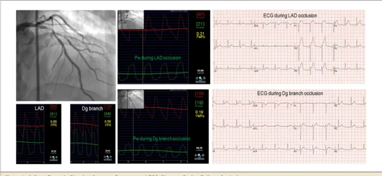
Study procedures. Coronary intervention was performed using standard intervention techniques. Coronary interven- tion strategy for bifurcation lesions and the sequence of the procedures, including balloon occlusion of main and side branches were selected at the operator’s discretion. During the procedures, blood flow to each branch was completely occluded by the angioplasty balloon. The occurrence of anginal symptoms and 12-lead electrocardiogram (ECG) were checked after balloon occlusion for 1 min. Complete occlusion of the vessel was confirmed by contrast injection (Fig. 1). The severity of chest pain was assessed by a visual analogue scale pain score (range 0 to 10).
A physiological substudy was performed in patients enrolled from 4 centers. In these patients, the physiological evaluation of both branches was performed after diagnostic angiography. FFR was measured using an intracoronary pressure wire (PressureWire, St. Jude, St. Paul, Minnesota;
ComboWire, Volcano Corporation, San Diego, California).
Maximal hyperemia was induced by intravenous adenosine infusion (140 g/kg/min). The
coronary wedge pressure (Pw) of the LAD or diagonal branch was measured using the intracoro- nary pressure wire after balloon inflation of each vessel for 1 min (13). During balloon oc- clusion, Pw and aortic pressure (Pa) were simultaneously re- corded (Fig. 1). In 10 patients without ST-segment elevation during side branch balloon oc- clusion, the presence of local ischemia was monitored using intracoronary ECG as previously described (14).
Quantitative coronary angiography, side branch scoring, and ECG analysis. All angiographic images, pressure tracings, and ECG recordings were collected and analyzed by an independent core laboratory at Seoul National University Hospital. Each dataset was analyzed in a blinded fashion.
Quantitative coronary angiography was performed by an experienced operator in 3 segments (proximal main branch, distal main branch, and side branch) using a dedicated bifurcation quantitative coronary angiography software (CAAS 5, Pie Medical Imaging, Maastricht, the Nether- lands). To better estimate the mass of myocardium at risk, a scoring system (SNuH score) for a diagonal branch was developed by incorporating size, number, and distribution
Internal Medicine, Ajou University Hospital, Suwon, Gyeonggi-do, Korea. This study was supported by grants from the Korean Society of Interventional Cardiology, Seoul, Republic of Korea (2009), Innovative Research Institute for Cell Therapy, the Clinical Research Center for Ischemic Heart Disease (no. 0412-CR02-0704-0001) sponsored by the Ministry of Health and Welfare, Republic of Korea and the Seoul
National University Hospital Research Fund (no. 03-2010-0270). The authors have reported that they have no relationships relevant to the contents of this paper to disclose.
Manuscript received February 13, 2012; revised manuscript received May 1, 2012, accepted May 27, 2012.
Abbreviations and Acronyms
ECGⴝ electrocardiogram(s) IQRⴝ interquartile range LADⴝ left anterior descending coronary artery Paⴝ aortic pressure Pwⴝ coronary wedge pressure
QTcⴝ QT Interval corrected for heart rate
TIMIⴝ Thrombolysis In Myocardial Infarction
of diagonal branches (Table 1). The SNuH score was calculated as the sum of each factor, which ranged from 0 to 3. For this scoring, only branches with ⬎1.5 mm in diameter by visual estimation were included.
All ECG were analyzed by an independent cardiologist.
ST-segment elevation was considered significant when the elevation was ⱖ1 mm compared to the baseline ECG. QT interval corrected for heart rate (QTc) and QTc dispersion were also measured both before and after balloon occlusion of the LAD or diagonal branch. The QTc interval was calculated from the Bazett formula (15) and the QTc dispersion, as the difference between the maximal and minimal QTc intervals.
Statistical analysis. Data are presented as mean ⫾ SD for continuous variables and frequency for categorical variables.
Comparison of continuous variables was performed with the Student t test or paired t test. Analysis of discrete variables was performed with the chi-square test. McNemar test was used for paired comparison of binary values. Variables that do not have normal distribution were reported as medians and interquartile ranges (IQR) and were compared by nonparametric tests. All statistical analyses were performed
with SPSS (version 11.0, SPSS, Inc., Chicago, Illinois), and a probability value of ⬍0.05 was considered statistically significant.
Results
Between May 2010 and May 2011, 73 consecutive patients were enrolled. After excluding 8 patients (diagonal branch wiring failure in 3, LAD TIMI flow grade 2 in 1, collateral feeding LAD in 2, and failure to achieve occlusion ECG in 2), 65 patients were finally included in this study. Among them, 47 patients underwent the physiological substudy.
There were no complications during any of the procedures.
Baseline characteristics. Clinical, angiographic, and proce- dural characteristics of the patients and lesions are shown in Table 2. Thirty-six lesions (55%) were classified as Medina 1,1,1 bifurcation lesion and most lesions (89%) were Y-type bifurcations. The reference diameter and percentage of diameter stenosis of diagonal branches were 2.4 ⫾ 0.3 mm and 66.1 ⫾ 15.2%, respectively.
Different responses to balloon occlusion between LAD and diagonal branches. There were differences in clinical, elec- trocardiographic, and physiological parameters between LAD and diagonal branches (Table 3). ST-segment eleva- tion was shown in 60 cases (92%) during LAD occlusion and in 23 cases (35%) during diagonal branch occlusion (p ⫽ 0.001). Pain score was also higher during LAD occlusion than in diagonal branch occlusion (median [IQR]:
Figure 1.A Case Example Showing Coronary Pressure and ECG Changes During Balloon Occlusion
A case example showing coronary pressure and electrocardiogram (ECG) changes during left anterior descending (LAD) and diagonal (Dg) branch (SNuH score 3) occlusion. Fractional flow reserve (FFR) before intervention was 0.65 and 0.56 for LAD and Dg branch, respectively. After 1-min occlusion, coronary wedge pres- sure (Pw) and Pw/aortic pressure (Pa) were 21 mm Hg and 0.21 for LAD and 19 mm Hg and 0.19 for Dg branch. The 12-lead ECG showed ST-segment elevation in precordial leads with LAD occlusion and in lateral leads with Dg branch occlusion.
Table 1.A Diagonal Branch Scoring System (SNuH Score)
Variables Description Score
Size (S) Vessel diameterⱖ2.5 mm 1
Number (Nu) Number of diagonal branchesⱕ2 1
Highest (H) No branch below the target branch 1
5 [0 to 7] vs. 2 [0 to 4], p ⬍ 0.0001). Mean baseline QTc interval and QTc dispersion were 435.0 ⫾ 39.6 ms and 67.3 ⫾ 33.7 ms, respectively. Mean QTc interval was prolonged with LAD occlusion (454.0 ⫾ 45.4 ms, p ⬍ 0.0001), but not with diagonal branch occlusion (440.4 ⫾ 35.7 ms, p ⫽ 0.213). Mean QTc dispersion after 1-min occlusion was greater (83.8 ⫾ 39.2 vs. 70.7 ⫾ 28.5 ms, p ⫽ 0.04) with LAD occlusion than with diagonal branch occlusion. In patients who underwent physiological evalua- tions, pre-intervention FFR was lower in the LAD than in the diagonal branches (0.67 ⫾ 0.10 vs. 0.71 ⫾ 0.11, p ⫽ 0.02). Both Pw and Pw/Pa were lower in the LAD than in the diagonal branches (Pw: 21.0 ⫾ 6.5 vs. 26.7 ⫾ 9.4, p ⬍ 0.0001; Pw/Pa: 0.22 ⫾ 0.07 vs. 0.27 ⫾ 0.08, p ⫽ 0.001). In 10 patients without ST-segment elevation by 12-lead sur- face ECG, all patients showed ST-segment elevation by intracoronary ECG during diagonal branch occlusion.
Differences in diagonal branches with and without ST- segment elevation during occlusion. When the diagonal branches were divided according to ST-segment elevation during occlusion, there was no difference in clinical and angiographic findings between the 2 groups (Table 4).
However, SNuH score was higher in branches with ST- segment elevation than in those without ST-segment ele- vation (median [IQR]: 3 [2 to 3] vs. 2 [1 to 3], p ⫽ 0.005).
There was a trend toward lower Pw and Pw/Pa in patients with ST-segment elevation during balloon occlusion than in those without. Among the 13 diagonal branches with Pw/Pa ⬎0.3, 2 lesions (15.4%) showed ST-segment eleva- tion during balloon occlusion. In 34 branches with Pw/Pa ⱕ0.3, 15 branches (44.1%) showed ST-segment elevation.
Diagnostic performance of angiographic parameters to predict ST-segment elevation during balloon occlusion. The crite- rion of vessel size (ⱖ2.5 mm) had sensitivity, specificity, and positive and negative predictive values of 58%, 63%, 48%, and 72%, respectively (Fig. 2). The sensitivity of SNuH score for the prediction of ST-segment elevation was 83%
and negative predictive value was 83%. However, its spec- ificity and positive predictive value were 49% and 49%, respectively. Diagnostic accuracy of both vessel size and SNuH score was 62%. The positive and negative predictive values of the variable “highest” (no branch below the target branch) were 49% and 86%, respectively.
Discussion
By comprehensive analysis of patients with true LAD bifurcation lesions, this study demonstrated that: 1) the clinical, electrocardiographic, and physiological responses to coronary artery occlusion were different between LAD and diagonal branches; 2) diagonal branch occlusion caused less pain, less frequent ST-segment elevation, and less arrhyth-
Table 2.Clinical, Angiographic, and Procedural Characteristics Patient characteristics
Age, yrs 63⫾ 11
Male 47 (72)
Diabetes 21 (32)
Stable angina 33 (51)
Unstable angina 15 (23)
Previous revascularization 4 (6)
LV ejection fraction, % 63⫾ 8
Angiographic and procedural characteristics Medina classification
1,1,1 36 (55)
1,0,1 12 (18)
0,1,1 17 (26)
T type (distal angleⱖ70°) 7 (11)
Left anterior descending artery stent
Promus/Xience/Endeavor 16/14/13
Diameter/length, mm 3.1⫾ 0.3/29.2 ⫾ 10.6
Diagonal branch balloon
Diameter/length, mm 2.1⫾ 0.3/15.5 ⫾ 2.8
Quantitative coronary angiography Proximal main branch
Reference diameter, mm 3.0⫾ 0.4
% diameter stenosis 57.9⫾ 15.5
Lesion length, mm 12.8⫾ 8.0
Distal main branch
Reference diameter, mm 2.7⫾ 3.5
% diameter stenosis 55.3⫾ 16.8
Lesion length, mm 12.5⫾ 8.2
Side branch
Reference diameter, mm 2.4⫾ 0.3
% diameter stenosis 66.1⫾ 15.2
Lesion length, mm 12.9⫾ 9.4
Values are mean⫾ SD or n (%). The Promus stent is a product of Boston Scientific (Natick, Massachusetts; the Xience stent is a product of Abbott Vascular (Abbott Park, Illinois); and the Endeavor stent is a product of Medtronic (Minneapolis, Minnesota).
LV⫽ left ventricle.
Table 3.Clinical, Electrocardiographic, and Hemodynamic Responses to 1-Min Balloon Occlusion Between LAD Artery and Diagonal Branches
LAD Diagonal p Value
Chest pain and ECG parameters, n⫽ 65
VAS pain score 5 (0–7) 2 (0–4) ⬍0.0001
ST-segment elevationⱖ1 mm 60 (92.3) 23 (35.4) 0.001 QTc interval, ms 454.0⫾ 45.4 440.4⫾ 35.7 0.07 QTc dispersion, ms 83.8⫾ 39.2 70.7⫾ 28.5 ⬍0.0001 Coronary hemodynamic
parameters, n⫽ 47
Pre-intervention FFR 0.67⫾ 0.10 0.71⫾ 0.11 0.02
Pw, mm Hg 21.0⫾ 6.5 26.7⫾ 9.4 ⬍0.0001
Pw/Pa 0.22⫾ 0.07 0.27⫾ 0.08 0.001
Values are median (interquartile range), n (%), or mean⫾ SD.
ECG⫽ electrocardiogram; FFR ⫽ fractional flow reserve; LAD ⫽ left anterior descending coronary artery; Pa⫽ aortic pressure; Pw ⫽ coronary wedge pressure; QTc ⫽ QT interval corrected for heart rate; VAS⫽ visual analogue scale.
mogenic potential than did LAD occlusion; 3) collateral recruitability of diagonal branch was better than LAD; and 4) angiographic parameters could not reliably predict the clinical relevance of a diagonal branch.
In the history of percutaneous coronary intervention, bifurcation lesions are unique, as most studies have failed to show the benefit of routine stenting over provisional inter- vention strategy for side branch lesions (1–3). Moreover, even balloon angioplasty was not better than a no-treatment strategy for side branches in a recent randomized trial (16).
Therefore, a gap exists between the lesion severity and clinical outcomes of revascularization in coronary bifurca- tion lesions.
Better assessment using FFR may reduce this gap (6 – 8) as the presence of ischemia is essential to prove the benefit of revascularization over medical treatment. However, the amount of ischemia that is determined by myocardial mass at risk and collateral recruitability is the other key element in this regard (9,10) and should also be considered. In this study, we assessed the clinical relevance of the bifurcation lesion using 1-min occlusion of coronary blood flow. Myocar- dial mass at risk was indirectly assessed by ECG changes during coronary occlusion and collateral recruitability by Pw.
ST-segment elevation or changes in QTc interval in standard 12-lead ECG are regarded as the clinically useful surrogate marker of infarct size or clinical outcomes (17–20). In our study, LAD occlusion caused more pain and electrocardiographic changes (ST-segment elevation, QTc interval, and QTc dispersion) than did diagonal branch occlusion. These differences explain why main branch–
related clinical events are more frequent and more severe than side branch–related events and why the previous studies failed to prove the benefit of routine stenting for side
branches in bifurcation lesions. Revascularization for clini- cally insignificant side branches cannot be translated into clinical benefit and may even be harmful (21–23). In this regard, future studies designed to show the clinical benefit of a new device or treatment strategy in bifurcation lesions should include only the clinically relevant side branches.
An interesting finding in our study was that Pw and Pw/Pa were lower in the LAD than in the diagonal branches despite more severe pre-intervention ischemia (lower FFR) in the LAD. Previous studies showed that high collateral flow index is associated with better collateral recruitability and better outcomes (17,24). This difference in Pw between the LAD and diagonal territories may reflect the difference in collateral recruitability and might be one possible mechanism in how the side branch territory can be protected from clinical events. Although the mechanism of this difference is beyond the scope of this study, there is some evidence suggesting regional differences in the protec- tion from and development of infarction within the LAD territory (25) and within the whole myocardium (19).
Vessel size is the most commonly used parameter to define the clinical significance of a side branch. However, in our study, among 29 branches with reference diameter ⱖ2.5 mm, ST-segment elevation was found in 14 branches (48%) and 28% of the branches with ⬍2.5 mm showed ST- segment elevation during 1-min balloon occlusion. To overcome this limitation for vessel size, we developed a scoring system (SNuH score) that incorporates the number and distribution of branches as well as the size to better reflect the myocardial mass at risk. Although SNuH score was higher in branches with ST-segment elevation than in those without and showed the negative predictive value of 83%, its positive predictive value to determine the presence of ST-segment elevation during balloon occlusion was just 49%. This low positive predictive value represents the limitation of angiographic parameters in the assessment of
Figure 2.Diagnostic Performance of Vessel Diameter and SNuH Score Diagnostic performance of vessel diameter (ⱖ2.5 mm) and SNuH score (ⱖ2) in the predication of ST-segment elevation during 1-min balloon occlusion of diagonal branch. NPV⫽ negative predictive value; PPV ⫽ pos- itive predictive value; SB⫽ side branch.
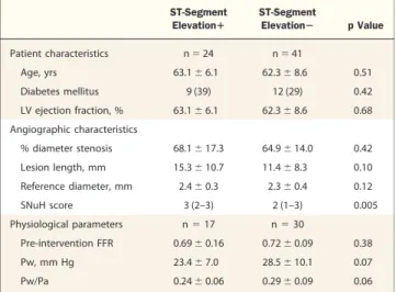
Table 4.Differences in Clinical, Angiographic, and Physiological Parameters Between Diagonal Branches With and Without ST-Segment Elevation During 1-Min Balloon Occlusion
ST-Segment Elevationⴙ
ST-Segment
Elevationⴚ p Value
Patient characteristics n⫽ 24 n⫽ 41
Age, yrs 63.1⫾ 6.1 62.3⫾ 8.6 0.51
Diabetes mellitus 9 (39) 12 (29) 0.42
LV ejection fraction, % 63.1⫾ 6.1 62.3⫾ 8.6 0.68 Angiographic characteristics
% diameter stenosis 68.1⫾ 17.3 64.9⫾ 14.0 0.42
Lesion length, mm 15.3⫾ 10.7 11.4⫾ 8.3 0.10
Reference diameter, mm 2.4⫾ 0.3 2.3⫾ 0.4 0.12
SNuH score 3 (2–3) 2 (1–3) 0.005
Physiological parameters n⫽ 17 n⫽ 30
Pre-intervention FFR 0.69⫾ 0.16 0.72⫾ 0.09 0.38
Pw, mm Hg 23.4⫾ 7.0 28.5⫾ 10.1 0.07
Pw/Pa 0.24⫾ 0.06 0.29⫾ 0.09 0.06
Values are mean⫾ SD, n (%), or median (interquartile range).
Abbreviations as inTables 2and3.
the clinical significance of a diagonal branch and the difference in collateral recruitability. In our study, 11 of 13 diagonal branches with Pw/Pa ⬎0.3 did not show ST- segment elevation during balloon occlusion. Considering these limitations of angiographic parameters, 1-min balloon occlusion while monitoring the severity of pain and ECG changes can be helpful in defining the clinical relevance of diagonal branches when the vessel is large or the SNuH score is high. Diagnostic occlusion of the coronary artery with low-inflation pressure to measure Pw is reported to be safe (26). However, considering the possible injury during balloon occlusion, this assessment should be performed in braches with a planned intervention.
Study limitations. First, the number of patients included in this study was relatively small. Second, as this study in- cluded only LAD/diagonal bifurcation lesions, the results cannot be applied to other bifurcation lesions. Third, the primary endpoint of this study does not include the clinical outcomes. For the validation of these results, further studies with a larger population is needed. Fourth, collateral flow index could not be accurately calculated in this study as the central venous pressure was not measured. However, con- sidering the possibility that the LAD occlusion could increase left ventricular end-diastolic pressure more than diagonal branch occlusion could, the difference in collateral flow index between LAD and diagonal branches could have been greater than the difference in Pw/Pa in our study.
Conclusions
Diagonal branch occlusion caused fewer symptoms, fewer ECG changes, less arrhythmogenic potential, and better collateral recruitability than LAD occlusion did. These differences seem to be the main cause why aggressive treatment of side branches does not translate into clinical benefit in the percutaneous treatment of coronary bifurca- tion lesions involving the LAD and diagonal branches.
Reprint requests and correspondence: Dr. Hyo-Soo Kim, De- partment of Internal Medicine, Seoul National University Hospi- tal, Yongon dong 28, Jongno-gu, Seoul, Republic of Korea, 110-744. E-mail: [email protected].
REFERENCES